
1. Background
The liver is considered an essential organ for body metabolism. Diseases that can create masses or cysts in the liver include primary hepatic malignancies and benign lesions like hemangioma, hepatocellular nodules, hepatic adenoma, hepatic cysts, and abscess (1). Hepatic space-occupying lesions are among liver disorders due to growth and development, neoplasm, inflammation, and other complications. Although the diagnosis of hepatic lesions based on imaging alone is difficult in some cases, focal hepatic lesions can be diagnosed with ultrasonography, computed tomography (CT), and magnetic resonance (MR) (2). Ultrasonography is a non-invasive technique, usually welcomed by patients for its relatively low cost and ability to determine focal liver lesions, which uses probes with 2.5 to 7.5 megahertz (MHz) frequencies (3, 4).
Space-occupying liver lesions result from various diseases with or without clinical symptoms (5), which are divided into two sets, benign and malignant (6). Benign hepatic lesions usually represent pain in the upper quarter of the right side of the abdomen and/or a mass touchable in examination or are discovered by chance during imaging and laparotomy for other problems. In addition, malignant hepatic lesions can also be diagnosed by jaundice, blood ascites, hepatic bruit, loss of appetite, and unjustifiable weight loss (3). The most frequent liver lesions are hemangiomas, hepatic cysts, focal nodular hyperplasia (FNH), and hepatocellular adenomas as benign lesions (7-9). Sometimes, atypical hemangiomas are difficult to distinguish from other more worrying hepatic lesions such as metastases and hepatocellular carcinoma. Furthermore, some malignant lesions can display features that simulate hemangiomas (10). Also, the epidemiology of hepatic hemangioma has not been evaluated well.
The prevalence of hepatic hemangioma ranges from 0.4% to 20% (11). A retrospective study on patients referred for transabdominal ultrasonography to the Ultrasound Unit of Firoozgar hospital, Tehran, Iran, reported that the prevalence of hemangioma was 2.04% in the study population, with women predominance (12).
2. Objectives
As no comprehensive study has been performed in the northern population of Iran, we aimed to evaluate the prevalence of hepatic space-occupying lesions by sonography in Guilan Cohort Center participants.
3. Methods
3.1. Participants
This cross-sectional study was conducted within the prospective epidemiological research studies of Iranian adults (PERSIAN) cohort study (13), started in September 2014 in Sowme'eh Sara (GPS coordinator latitude: 37.308003 and longitude: 49.315022), Guilan, northern Iran, engaging both genders aged 35 - 60 years, being followed for 10 years to determine new diseases and underlying genetic factors for chronic diseases. We selected 960 out of 10,520 individuals based on a non-randomized simple sampling method. Guilan cohort profile was published in detail previously (14). Written consent was taken after informing each participant of the purpose and importance of the study. To ensure the confidentiality of participants’ information, we used codes to omit the participants' names and identifiers in the questionnaires. This study was approved by the Ethics Committees of the Ministry of Health and Medical Education and the Guilan University of Medical Sciences (P/3/132/215). Informed consent was obtained from all participants.
3.2. Data Collecting
Demographic data and clinical characteristics of all individuals were recorded in questionnaires, including age, sex, marital status, education level, occupation, body mass index (BMI; as low weight < 18.5 kg/m2, normal weight 18.5 - 24.99 kg/m2, overweight 25 - 29.9 kg/m2, and obese ≥ 30 kg/m2), history of smoking, alcohol consumption, and diabetes. Ultrasonography was carried out by a radiologist after at least 12 hours of fasting, using an Ultrasonic Device of Sonix SP type with 3.5 MHz to 5 MHz probes to determine the hepatic space-occupying lesions. The exclusion criteria were acute and chronic hepatic diseases like B and C viral hepatitis, known hepatic and biliary diseases, cancer, and pregnancy.
3.3. Statistical Analysis
Continuous variables were expressed as percentage and frequency. The normality was evaluated using the Shapiro-Wilk normality test. Absolute and relative frequency, mean, and standard deviation were used to describe the data, and the chi-Square test was used to determine the relationship between variables. Statistical analysis was undertaken using SPSS for Windows (version 16.0) at the significance level of 0.05.
4. Results
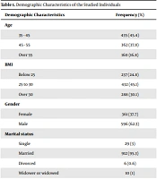
From 2017 to 2018, the presence of liver lesions was examined by ultrasound in 950 individuals, of whom 37.7% (n = 361) were female, and 62.3% (n = 596) were male. About 83.2% (n = 797) of the individuals were aged 35-55 years and 16.8% (n = 160) were over 55 years. Most participants were married (95.3%, n = 912), had a diploma degree (56.7%, n = 543), with no history of smoking (69.3%, n = 663) or alcohol consumption (95.6%, n = 915), without diabetes (90.3%, n = 864) (Table 1).
Table 1.
Demographic Characteristics of the Studied Individuals
| Demographic Characteristics | Frequency (%) |
|---|---|
| Age | |
| 35 - 45 | 435 (45.4) |
| 45 - 55 | 362 (37.8) |
| Over 55 | 160 (16.8) |
| BMI | |
| Below 25 | 237 (24.8) |
| 25 to 30 | 432 (45.1) |
| Over 30 | 288 (30.1) |
| Gender | |
| Female | 361 (37.7) |
| Male | 596 (62.3) |
| Marital status | |
| Single | 29 (3) |
| Married | 912 (95.3) |
| Divorced | 6 (0.6) |
| Widower or widowed | 10 (1) |
| Education level | |
| Illiterate | 64 (6.4) |
| Primary school | 207 (21.6) |
| Diploma | 543 (56.7) |
| University degrees | 143 (15.3) |
| Occupation | |
| Farmer | 109 (11.4) |
| Employee | 164 (17.1) |
| Worker | 133 (13.9) |
| University student | 6 (0.6) |
| Housewife | 308 (32.2) |
| Self-employed | 237 (24.8) |
| Smoking | |
| Nonsmoker | 663 (69.3) |
| Smoker | 294 (30.7) |
| Alcohol use | |
| Nonalcoholic | 915 (95.6) |
| Alcoholic | 42 (4.4) |
| Diabetes | |
| Nondiabetic | 864 (90.3) |
| Diabetic | 93 (9.7) |
Abbreviation: BMI, body mass index.
The total frequency of liver lesions was 2.3%; the most frequent was hemangioma with 1.1% (n = 11) while liver cyst was 0.4% (n = 4) and others (not defined) were 0.8% (n = 8) (Table 2).
Table 2.
Frequency of Hepatic Lesions in the Studied Individuals
| Lesion | Frequency (%) |
|---|---|
| Hemangioma | 11 (1.1) |
| Cyst | 4 (0.4) |
| Others | 8 (0.8) |
| No lesion | 934 (97.6) |
Females had a higher frequency of hemangioma (63.3%, n = 7) than males (36.4%, n = 4). Compared to females, males had a higher frequency of liver cysts (25%, n = 1 vs. 75% n = 3). Statistical analysis revealed that the presence of liver lesions was slightly higher in females than in males (3.6%, n = 13 vs. 1.7%, n = 17; P = 0.05) (Table 3). Other variables had no statistically significant relationship with the presence of liver lesions (Table 3).
Table 3.
Hepatic Lesions by Demographic Characteristics of the Studied Individuals
| Demographic Characteristics | Hepatic Lesion | P Value | |
|---|---|---|---|
| Negative | Positive | ||
| Age | 0.421 | ||
| 35 - 44 | 428 (98.4) | 7 (1.6) | |
| 45 - 54 | 353 (97.5) | 9 (2.5) | |
| Over 55 | 153 (95.6) | 7 (4.4) | |
| BMI | 0.117 | ||
| Below 25 | 231 (97.5) | 6 (2.5) | |
| 25 to 30 | 418 (96.8) | 14 (3.2) | |
| Over 30 | 285 (99) | 3 (1) | |
| Gender | 0.050 | ||
| Female | 348 (96.4) | 13 (3.6) | |
| Male | 586 (98.3) | 10 (1.7) | |
| Marital status | 0.711 | ||
| Single | 28 (100) | 1 (0) | |
| Married | 890 (97.5) | 22 (2.5) | |
| Divorced | 6 (100) | 0 (0) | |
| Widowed or widower | 10 (100) | 0 (0) | |
| Education level | 0.613 | ||
| Illiterate | 59 (96.7) | 5 (3.4) | |
| Primary school | 200 (96.6) | 7 (3.4) | |
| Diploma | 532 (98) | 11 (2) | |
| Academic degree | 143 (97.9) | 3 (2.1) | |
| Occupation | 0.251 | ||
| Farmer | 107 (98.2) | 2 (1.8) | |
| Employee | 157 (95.7) | 7 (4.3) | |
| Worker | 131 (98.5) | 2 (1.5) | |
| University student | 6 (100) | 0 (0) | |
| Housewife | 298 (96.8) | 10 (3.2) | |
| Self-employed | 235 (99.2) | 2 (0.8) | |
| Smoking | 0.110 | ||
| Smoker | 290 (98.6) | 4 (1.4) | |
| Nonsmoker | 644 (97.1) | 19 (2.9) | |
| Alcohol use | 0.711 | ||
| Alcoholic | 41 (97.6) | 1 (2.4) | |
| Nonalcoholic | 893 (97.6) | 22 (2.4) | |
| Diabetes | 0.623 | ||
| Diabetic | 91 (97.8) | 2 (2.2) | |
| Nondiabetic | 843 (97.6) | 21 (2.4) | |
Abbreviation: BMI, body mass index.
5. Discussion
Recently, there has been a significant increase in incidentally detected focal liver lesions. These lesions are found in up to one-third of individuals screened with cross-sectional imaging without any liver-related diseases or symptoms (15, 16).
Focal hepatic lesions have various benign and malignant causes, and many are not readily characterizable in imaging studies, particularly when smaller than 1 cm. These lesions can usually be evaluated with serial follow-up imaging tests in patients without known cancer because nearly all will be benign (17). In cancer patients, however, prompt determination of the cause (or likely cause) of such lesions may be pivotal for defining prognosis and therapy (18). Despite extensive complete diagnostics, the precise diagnosis of focal hepatic lesions remains unclear in up to 50% of cases (19-21). Semaan et al. report a rate of merely 64% of correctly diagnosed lesions by CT and MRI. Studies concerning the prevalence of benign focal liver lesions present a quite heterogeneous picture due to population size, and investigation method. It is, therefore, difficult to compare different study results and apply them to routine ultrasound primary diagnostics (22). The prevalence of liver lesions in our study was 2.3%. In the same way, Kaltenbach et al. studied the sonographic prevalence of benign focal liver lesions in hospital patients in Germany. They indicated about 15.1% of the individuals with at least one hepatic lesion (23). The prevalence of hepatic hemangioma in various studies ranges from 0.1% to 20.0% (24-27). Our prevalence of 1.1% for hemangioma lies in the mid-range compared to the results of ultrasound-based studies (26-29). Compared to CT, MRI, and autopsy studies, which show a far higher prevalence range, our figure is in the lower third (24, 25, 30, 31).
Concerning hemangioma, there are a comparatively large number of prevalence studies for hepatic cysts, but they also differ in study size, patient population, and diagnostic techniques. Retrospective and prospective studies based on ultrasound have reported the prevalence of hepatic cysts between 0.1% and 11.3% (27, 29, 32, 33). We are also in the mid-range with our determined prevalence of 0.8%.
In our collective, hemangioma was the highest diagnostic hepatic lesion in ultrasonography examinations, with a higher frequency in females. Similarly Rungsinaporn and Phaisakamas reported a higher prevalence of hepatic hemangioma in women, while some other studies reported a higher prevalence of hemangioma in middle-aged or elderly patients, which was contrary to our results (32-34). On the contrary, Jacob et al. reported no statistically significant relationship between gender and liver lesions, which is inconsistent with the present study (35). Studies reported an increasing prevalence of hepatic cysts with age (7, 36-38). Most studies have also found a gender-dependent relationship, with a higher prevalence of hepatic cysts in women (32, 36-38). On the contrary, the presence of liver lesions was not significantly related to age in our results. In line with our results, some studies in France and Japan indicated no statistically significant relationship between age and the presence of liver lesions (39, 40).
In the present study, there was no statistically significant relationship between BMI and the presence of liver lesions. Hussain and Semelka in the Netherlands and Jacob et al. in England obtained similar results consistent with the present study (2, 35). CT, MRI, or autopsy studies report much broader prevalence rates than ultrasound-based studies for liver lesions (24, 41-43). Our prevalence is in the mid to lower third of the range compared to CT, MRI, and autopsy studies. CT studies have reported the highest prevalence rates (43). Ultrasound is a high-resolution, non-invasive imaging technique with high accuracy, low cost, and without side effects, which is well accepted by patients in many diagnostic situations (3). Benign masses, on the other hand, are relatively common in the general population. Most of these lesions are cystic, and some are hemangiomas. Focal nodular hyperplasia and adenomas are rare but can produce an appearance quite similar to malignant masses, requiring careful ultrasound examination (44).
This virtual epidemic of hepatic incidentalomas is mainly based on the constant improvement and widespread availability of radiologic technologies and their inflationary use in routine clinical work. This progress holds both opportunities and risks. On the one hand, it may result in the detection of a life-threatening lesion at an early, curable stage. On the other hand, detecting focal liver lesions may result in a harmful over-treatment of patients for whom merely follow-up or no treatment is adequate (45).
Despite recent technical advances in modern radiology settings, a correct preoperative diagnosis of asymptomatic focal liver lesions remains challenging. This was a cross-sectional study; hence, further research is suggested, including the group and prospective studies characterized by long follow-up periods and long-term intervals that can be instrumental in identifying more variables influencing hepatic lesions. Hence, along with the determination of accuracy and justification of ultrasound efficiency, more comprehensive research concerning some variables, such as the history of oral contraceptive pills (OCP), is warranted. It is also suggested that other diagnostic tools be used along with ultrasonography to confirm diagnostic findings. As our study was conducted on outpatients with no acute or chronic hepatic diseases, like B and C viral hepatitis, biliary diseases, and cancer, the results can be powerfully generalized to the general population.
5.1. Conclusions
Hemangioma was the most common liver lesion diagnosed by ultrasound, which was reported marginally higher in females. Also, sonography is not recommended for the general population but is advisable in risk groups.