1. Background
Hepatitis C virus (HCV) is a blood-borne pathogen which causes acute and chronic hepatitis infection. In the second half of the 20th century, HCV transmission occurred frequently due to unsafe blood transfusion (1). Routine screening for blood donation has been subsequently proven effective in minimizing the residual risk of transfusion-transmitted HCV. With the implementation of enhanced sensitive serological tests and nucleic acid tests (NAT), the risk has reduced. In developed countries, it was as low as below 1:1 million for HCV in the last decade (2-4). Therefore, blood transfusion has currently not been considered as a major transmission route.
In China, legislation regulating blood donation was enacted in 1998, followed by mandatory transition from paid donors or family members to voluntary blood donors (VBD). Additionally, the implementation of improved screening methods for HCV has been performed. A systematic review covering the period 1990 - 2010 reported a rapid decrease in the prevalence of HCV after 1998 (pooled prevalence: 1.7%) compared to before 1998 (pooled prevalence: 12.9%) in Chinese blood donors (5). However, the HCV prevalence was still higher in China than that in developed countries and even some developing countries. Based on recent studies in different areas of China, the prevalence of anti-HCV antibody in VBD varied from 0.25% to 0.86% (2005 - 2012) (6-9), whereas it was 0.13% in Iran (2004 - 2007) (10) and 0.19% in Brazil (2007) (11). Thus, it remains a crucial challenge to Chinese VBD and blood supply safety.
Currently, HCV has been classified into 7 confirmed genotypes and a number of identified subtypes (12, 13). Diverse genotypes and subtypes make it difficult to thoroughly understand the transmission. A previous study based on the VBD from 17 provinces in China revealed that 5 major genotypes (1b, 2a, 3a, 3b, and 6a) displayed totally different patterns of transmission (14). Direct-acting antiviral agents amazingly revolutionized the treatment for HCV infection; however, treatment failures have been observed in many patients due to individual variability of efficacy such as the cases infected with non-genotype-1 HCV (15). In this study, we determined the prevalence and genotype of HCV in VBD in Shanghai, China. Then, we conducted phylogenetic analysis to determine potential transmission of HCV between VBD and local intravenous drug users (IDU).
2. Methods
2.1. Study Subjects
In China, pre-donation screening includes a health history questionnaire, a physical examination, and a rapid test for hepatitis B virus (HBV) surface antigen (HBsAg). Candidates who meet the following criteria are permanently excluded from donation: past diagnosis of syphilis, HIV infection, other sexually transmitted diseases, or viral hepatitis; reported history of drug use, paid blood donation, or the involvement of commercial sex; reactivity to HBsAg (by the rapid test). In this study, a total of 38952 VBD aged 18-55 years were enrolled in Pudong New Area between January 1st and December 31st, 2014, after the pre-donation screening. Demographics were collected from the health history questionnaire, including gender, age, ethnicity, and donation status (first, second, or regular donor; regular donor was defined as those made donation in the current month and made donations ≥ 3 times within past 36 months). The study protocol was reviewed and approved by Ethics Committee of Shanghai Pudong New Area Center for Disease Control and Prevention.
2.2. Examination of Anti-HCV Antibody and Viral Nucleic Acid
The donated blood specimens underwent two rounds of serological test for anti-HCV antibody. Two different commercial ELISA kits, one domestic (EIA Kit for the Detection of Anti-HCV, Ke-Hua BIO-Engineering Co., Ltd., Shanghai, China; or Diagnostic Kit for Antibody to HCV, InTec Products, Inc., Xiamen, China) and one imported (Ortho HCV 3.0, Ortho-Clinical Diagnostics, Inc., USA; or Murex anti-HCV, DiaSorin, Italy), were employed to test anti-HCV according to the instructions of the kits. Blood specimens were determined as positive for anti-HCV if either test showed reactive. Specimens negative for anti-HCV were subsequently examined to determine the presence of viral nucleic acid (HBV DNA/HCV RNA/HIV-1 RNA) by NAT (Procleix Ultrio Assay on the Tigris System, Novartis Vaccines & Diagnostics, Emeryville, CA).
2.3. Phylogenetic Analysis
In the blood specimens positive for anti-HCV or positive by NAT, a 377 nucleotide (nt) partial sequence in HCV NS5B region corresponding to 8256-8632 nt on the strain H77 (GenBank accession no.AF009606) was amplified with nested reverse transcription PCR (RT-PCR). PCR primers and thermal profile were described elsewhere (16). Duplicate tests were performed to ensure the accuracy of positive results. The amplified 377-nt NS5B partial sequence has been deposited in the GenBank under accession nos.KU707094-KU707169.
HCV genotype was determined with BLAST search using software MEGA 7.0. Ten sequences in the GenBank that were most phylogenetically identical to each amplified sequence in VBD were determined. If some sequences belonged to same subtypes and were isolated in the same area/year in the same study, only the most identical one was included in the study. Highly similar cloned sequences were excluded. Additionally, 39 HCV NS5B 377-nt partial sequences isolated in IDU visiting methadone clinics in Pudong New Area during 2013 - 2014 were included for comparison. Genetic distances between pairwise sequences were calculated using the Kimura-2-parameter method. A phylogenetic tree was reconstructed using maximum likelihood method with bootstrap tests of 500 replications in MEGA 7.0 (17).
2.4. Statistical Analysis
Association between demographic characteristics and prevalence of anti-HCV antibody was assessed with chi-square test and Cochran-Armitage trend test. Statistical analysis was performed using SAS 9.2 for Windows (Cary, NC, USA). A P-value of <0.05 was considered statistically significant.
3. Results
3.1. Prevalence of Anti-HCV Antibody and Viral Nucleic Acid
Majority of VBD were male (69.7%), 21 - 40 years of age (78.0%), Han Chinese (97.7%), and first donor (72.5%) (Table 1). Overall prevalence of anti-HCV was 0.46%. The prevalence was not significantly different in gender (P = 0.91) or in ethnicity (P = 0.34). Among age groups, the prevalence decreased substantially with age (P = 0.021). It was highest in the age group of 18-20 years old and lowest in the 41 - 50 years old. Additionally, the prevalence in the first and second donors was 4.3 times and 2.6 times higher than that in regular donors, respectively (P < 0.001).
In the VBD negative for anti-HCV, the prevalence of HBV DNA/HCV RNA/HIV-1 RNA by NAT was 0.25% (Table 1). Of these donors, 62 were positive for HBsAg, 8 were positive for anti-HIV-1 antibody, and 28 were negative for both HBsAg and anti-HIV-1, indicated by further screening of HBsAg and anti-HIV-1.
3.2. HCV Genotyping and Phylogenetic Analysis
A 377-nt NS5B partial sequence was successfully amplified in 37 out of 179 specimens positive for anti-HCV, whereas it was not amplified in any specimens negative for anti-HCV while positive by NAT. Genotype was classified as 1a (n = 4), 1b (n = 18), 2a (n = 5), 3a (n = 5), 3b (n = 2), 6a (n = 1), and 6n (n = 2). Additionally, thirty nine 377-nt partial sequences isolated in the local IDU were included in the study as follows: 1b (n = 7), 3a (n = 14), 3b (n = 13), 6a (n = 4), and 6n (n = 1). Within genotype 1b, 3a, 3b, 6a, and 6n, VBD and IDU strains shared the sequence identities of 91.3% - 94.7%, 94.6% - 96.8%, 95.3% - 97.5%, 94.1% - 96.9%, and 97.4% - 99.6%, respectively.
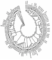
With BLAST search, a total of 179 sequences that were most phylogenetically identical to VBD strains were retrieved in the GenBank (Supplementary Material). Phylogenetic tree was reconstructed after alignment of 37 VBD strains and 39 IDU strains isolated in Pudong New Area. In genotype 1b, the retrieved sequences most phylogenetically identical to VBD strains were isolated in the different areas of China, whereas in genotypes 3a, 3b, 6a, and 6n, most of these sequences were isolated in southern China and Southeast Asian countries (Supplementary Material). In genotypes 1b, 3a, 3b, and 6n, some VBD strains clustered more closely with IDU strains (indicated by a bar) than the retrieved sequences (of 130 retrieved sequences, 55 were previously isolated in VBD in other studies) (Figure 1). In genotypes 1a and 2a, VBD strains were more identical to the sequences isolated in foreign countries than those in China.
Table 1.
Detection of Anti-HCV Antibody, Viral Nucleic Acid, and HCV 377-nt Partial Sequence by Demographic Lines (n = 38952)
| No. Detected | Proportion, % | No. Positive for Anti-HCV (No NAT) | No. Negative for Anti-HCV and Positive by NAT | No. Negative for Anti-HCV and Negative by NAT | |
|---|---|---|---|---|---|
| Gender | |||||
| Male | 27142 | 69.7 | 124 | 75 | 26943 |
| Female | 11810 | 30.3 | 55 | 23 | 11732 |
| Age, y | |||||
| 18 - 20 | 2599 | 6.7 | 14 | 3 | 2582 |
| 21 - 30 | 19179 | 49.2 | 102 | 31 | 19046 |
| 31 - 40 | 11190 | 28.7 | 44 | 30 | 11116 |
| 41 - 50 | 5225 | 13.4 | 16 | 33 | 5176 |
| 51 - 55 | 759 | 1.9 | 3 | 1 | 755 |
| Ethnicity | |||||
| Han | 38064 | 97.7 | 173 | 95 | 37796 |
| Minority | 888 | 2.3 | 6 | 3 | 879 |
| Donation status | |||||
| First | 28240 | 72.5 | 151 | 90 | 27999 |
| Second | 6658 | 17.1 | 24 | 7 | 6627 |
| Regular | 4054 | 10.4 | 4 | 1 | 4049 |
| Total | 38952 | 100 | 179 | 98 | 38675 |

Figure 1.
The bootstrap test values of > 70% were shown at the respective nodes. The circle indicated 37 strains isolated from voluntary blood donors (VBD) and the triangle indicated 39 strains from intravenous drug users (IDU) in the study. Black bar indicated the VBD strains clustering closely with IDU strains. 179 sequences were retrieved in the GenBank.
4. Discussion
In our study, the prevalence of anti-HCV antibody was determined to be 0.46% in post-donation screening in the VBD in Pudong New Area, Shanghai. It was consistent with recent studies in other areas of China (6-9). So far, China has achieved almost 100% voluntary blood donation and subsequently a rapid decrease of transfusion-transmitted infection (18). Chinese VBD is characterized by a healthier population who has fewer risk factors for HCV infection than other populations. However, through a nationwide survey in 2006, the prevalence of anti-HCV antibody in the general population aged 1 - 59 years old was 0.43% (19), which was similar to that in Chinese VBD, suggesting that current pre-donation screening failed to identify some high-risk donors. Thus, risk factors for HCV transmission in Chinese VBD may not be well understood, posing a public health concern that VBD should be given particular attention.
Our study suggested that younger donors aged 18 - 30 years were likely to have higher HCV prevalence. Previous studies in China reported conflicting association between age and HCV prevalence in VBD. Younger donors may have more high risk factors whereas older donors may have accumulated exposure (6-9). It depends on the demographics of participants. Shanghai is a highly developed metropolitan in China with a permanent population of about 25 million. Pudong New Area is the largest administrative unit of Shanghai, which contains about one-fifth of permanent population of Shanghai and comprises urban and suburb districts. There is frequent population migration that may increase the risk of HCV transmission in local younger donors. Additionally, the prevalence of anti-HCV was significantly higher in the first and second donors compared to the regular donors, whereas it was not significantly different between men and women, Han Chinese and minorities in the study, which was very similar to previous studies (6-9).
In our study, a 377-nt HCV NS5B partial sequence was successfully amplified in the 20.7% of blood specimens positive for anti-HCV. The low detection rate could be generally interpreted by different dynamics of antibody and viral nucleic acid, such as spontaneous clearance of HCV or resolved HCV RNA in blood specimens (20-23). However, no partial sequence was amplified in the specimens negative for anti-HCV while positive by NAT. Majority of these specimens (71.4%) were positive for HBsAg or anti-HIV, suggesting probable existence of HBV DNA or HIV-1 RNA other than HCV RNA. Also, higher sensitivity of NAT than that of PCR may account for the zero detection of the partial sequence in the rest 28 specimens negative for both HBsAg and anti-HIV.
Further phylogenetic analysis suggested that HCV genotype 1b was most predominant in VBD, whereas 3a and 3b were dominant in IDU. In genotypes 3a, 3b, 6a, and 6n, VBD and IDU strains shared high sequence identities and clustered together in the phylogenetic tree; however, in genotype 1b, some VBD strains clustered closely with IDU strains while others did not. It was consistent with previous studies, which reported that increasing HCV strains including 3a, 3b, 6a, and some 1b strains in Chinese VBD may preferentially be associated with transmission via IDU network (14). Five genotype 1b sequences that were reported to be infected through IDU in that study were identified as most phylogenetically identical to VBD strains in our study (1b.KF585861.China, designated as XA03 in the original article; 1b.KF585765. China, SH23; 1b.KF585515.China, BJ16; 1b.KF585842.China, WL 43; 1b.KF585846.China, WL47). In our study, all the VBD denied the involvement of IDU or paid blood donation in the pre-donation screening. One explanation is that the HCV-infected VBD may be infected by non-IDU means like invasive medical treatment and the original reservoir was IDU. The identification of genotypes 3 and 6 in VBD that were proven to be mainly circulated in IDU (24) indicated possible transmission from high-risk population to general population. Another possibility is that the VBD did not disclose their risk factors and in fact they belonged to a high-risk population.
There were some limitations in the study. In the blood specimens negative for anti-HCV while positive by NAT, we did not conduct HBV/HCV/HIV-1 discriminatory test due to limited volume of blood sample and routine protocol in blood centers. Generally, blood samples positive by NAT will be discarded immediately regardless of which viral DNA/RNA is positive. Consequently, we could not determine the presence of HCV RNA in these specimens and may attain an underestimate of HCV prevalence. In contrast, the blood specimens positive for anti-HCV were not confirmed by NAT as they had been labelled as “positive for HCV”, possibly resulting in false positivity. Additionally, we employed RT-PCR to amplify HCV partial sequence for phylogenetic analysis. Compared to NAT, lower sensitivity of RT-PCR could not guarantee the amplification in the real HCV-infected VBD. Subsequently, we could not obtain all HCV strains and genotypes in the VBD, which may bias the findings of the phylogenetic analysis.
4.1. Conclusions
Our study suggested that HCV prevalence in VBD in Shanghai remained low. However, there were diverse genotypes of HCV in VBD, including 1b, 2a, 3a, 1a, 3b, 6n, and 6a. The identification of genotypes 3 and 6 in VBD that are mainly circulated in IDU suggested that HCV transmission from high-risk population to general population is likely to occur.
4.2. Implication for Health Policy Makers/Practice/Research/Medical Education
In Chinese voluntary blood donors, diverse HCV genotypes were identified, including those were mainly circulated in intravenous drug users. The strains isolated from these two populations shared close phylogenetic relationship, suggesting possible HCV transmission from high-risk population to general population.