1. Background
Chronic hepatitis B (CHB) is a heavy-burden disease around the world. According to the World Health Organization, about 296 million people lived with CHB in 2019. Also, about 1.5 million people get infected with the hepatitis B virus (HBV) annually despite the safe and effective HBV vaccine being available in most countries (1). About 90 million people in China are chronically infected with HBV (2). If not timely and effectively treated, CHB can progress to cirrhosis, hepatocellular carcinoma, and even death. The short-term goal of anti-HBV treatment is to achieve sustained suppression of HBV replication and, ideally, a functional cure, which refers to the loss of serum hepatitis B surface antigen (HBsAg). The ultimate goal is to reduce the risk of CHB-related complications (3). There are several drugs in development that target HBV itself or the host immune system, but currently, only nucleos(t)ide analogs (NAs) and PEGylated interferon have been approved (4). Guidelines from AASLD, EASL, and APASL all recommend entecavir, TDF, and TAF as first-line therapy for CHB patients because of their high efficacy and resistance barrier (5). PEGylated interferon is also a recommended drug. However, considering its limited ability to control HBV replication and multiple adverse effects, it was not as widely used as NAs in clinical practice.
An oral phosphonate prodrug of tenofovir, TAF is a reverse transcriptase nucleotide inhibitor. When ingested by peripheral blood mononuclear cells and liver cells, TAF can eventually be transformed into its active form, tenofovir (TFV) (6). Moreover, after TFV is phosphorylated, it can be incorporated into the viral DNA by HBV reverse transcriptase, inhibiting HBV replication and ultimately leading to the eradication of the virus. A systematic review of 28 articles with 8624 patients found that CHB patients treated with TDF had a better virologic response than patients treated with ETV during a 96-week follow-up period (7). Meanwhile, TAF was as effective as TDF in suppressing HBV replication, but with a higher normalization rate of ALT and fewer adverse effects on the kidney and bone (8), although the exact mechanism behind it is still not completely understood.
As mentioned above, TAF has become the mainstream drug among NAs due to its potent antiviral ability and safety. However, research data are mainly from countries other than China. Abroad, TAF was first brought to market in the United States in November 2016, followed by Japan, Europe, South Korea, and other regions. However, TAF was just approved to treat HBV infection in China in November 2018. China has the most significant number of people infected with HBV. In order to achieve the goal of hepatitis B elimination by 2030 (9), potent anti-HBV drugs are becoming an impending demand. However, due to the limited marketing time of TAF in China, there is relatively little data available to demonstrate its effectiveness and safety in real-world settings in the country.
2. Objectives
Our study aimed to investigate the antiviral effectiveness, renal safety, and serum lipidemia changes in patients receiving TAF monotherapy under real-world conditions in China.
3. Methods
3.1. Study Design and Participants
This retrospective study consecutively enrolled treatment-naïve (TN) CHB patients and treatment-experienced (TE) CHB patients between January 2019 and October 2020 at the outpatient clinic of West China Hospital. They chose TAF monotherapy as a treatment regime on their own will, at 25 mg once a day orally since enrollment. The criteria for CHB diagnosis and antiviral therapy were based on the APASL guidelines updated in 2015 (10): (1) Age over 18-years-old, (2) hepatitis B surface antigen seropositive status persisted beyond 6 months, (3) TE patients receiving no interferon within 6 months before enrollment, and TN patients never receiving anti-HBV therapy, and (4) indication of anti-HBV treatment focused on serum HBV DNA detectable, serum ALT levels greater than the upper limit of normal (ULN), and severity of liver disease. Moreover, age, family history of cirrhosis or HCC, and extrahepatic manifestations were also considered. The exclusion criteria included (1) patients who already developed HCC before enrollment, (2) patients who were not followed up regularly, (3) patients with previous kidney disease, and (4) patients with other viral hepatitis, non-alcoholic liver disease, drug liver disease, and other diseases that may cause chronic liver dysfunction.
This study was approved by the Ethics Committee of West China Hospital (serial number 2021-892) and registered in the Chinese Clinical Trials Registry (ChiCTR2100050189).
3.2. Study Outcomes and Definitions
The primary efficacy endpoint of this study was the cumulative rate of virologic response (VR) during a 96-week follow-up period. The secondary treatment endpoints were quantitative hepatitis B surface antigen (qHBsAg) levels and the proportion of normal ALT. Safety endpoints composed of changes in renal function, as measured by serum creatinine (Scr) and estimated glomerular filtration rate (eGFR), and serum lipid profiles [triglycerides (TG), total cholesterol (TC), and low-density lipoprotein cholesterol (LDL-C)]. The virologic response was defined as serum HBV DNA below the detection limit (100 IU/mL). The virologic breakthrough was defined as increased serum HBV DNA by >1 log above nadir or re-detectable again after achieving VR with continued treatment. The biochemical response was defined as elevated ALT returned to normal levels (< 40 IU/mL), and serologic changes were defined as hepatitis B e antigen (HBeAg) and HBsAg seroconversion and loss. Also, CKD stage 1 was regarded as eGFR ≥ 90 mL/min/1.73 m2, and CKD stage 2 was defined as slightly reduced eGFR (60 - 89 mL/min/1.73 m2).
3.3. Data Collection and Assessment
In this study, auxiliary examinations were performed at baseline and every 3-6 months, including serum biochemical tests (ALT, Scr, eGFR, and lipid profiles), HBV virologic biomarkers (qHBsAg level and HBeAg status), and HBV DNA. Our study detected the serum biochemical indices using an automatic biochemical analyzer (Olympus AU5400, Olympus Corporation, Tokyo, Japan) following standard procedures. The eGFR was calculated using an equation from the chronic kidney disease epidemiology collaboration (11). Serum HBeAg status was determined by the electrochemiluminescence immunoassay (Roche Diagnostics, Shanghai, China). Serum HBsAg concentration was quantitatively assessed using Elecsys HBsAg II Quant Assay (Roche Diagnostics, Shanghai, China), with a lower limit detection of 0.05 IU/mL. Serum HBV DNA level was quantified with a quantitative real-time polymerase chain reaction assay (Roche Diagnostic Systems, Shanghai, China) with a low threshold of 100 IU/mL.
3.4. Statistical Analysis
Data are expressed as numbers (%), medians (interquartile ranges or range), or means ± standard deviations. The significance of differences in continuous variables between groups was assessed using the Student's t-test or the Mann-Whitney test. Categorical variables were examined by χ2 test or Fisher's exact test. Statistical analyses were performed using IBM SPSS software version 25.0. A two-sided P-value < 0.05 was considered to indicate statistical significance.
4. Results
4.1. Patient Characteristics
A total of 49 TN CHB patients and 112 TE CHB patients were enrolled. There were 161, 137, and 95 patients who completed 48, 72, and 96 weeks of follow-up, including 49, 41, and 24 patients in the TN group and 112, 96, and 71 patients in the TE group, respectively. Baseline demographic and clinical characteristics are presented in Table 1. For patients in the TN group, the median age was 37 years old (range 23 - 64), 65% (32/49) were male, and 63% (31/49) were HBeAg-positive. The median HBV DNA was 5.23 log10 IU/mL (2.03 - 7.78). The median qHBsAg concentration was 2930 IU/mL, and the median ALT level was 34 U/L. Besides, the median LDL-C level was 3.72 mmol/L, and the baseline eGFR was 102.8 ± 12.43 mL/min/1.73 m2. For patients in the TE group, the mean age was 38 years old (range 19 - 81), 63% (71/112) were male, and 52% (58/112) were HBeAg-positive. Moreover, 110 patients had undetectable HBV DNA; the other two patients' HBV DNA was 3.08log10 IU/mL and 3.54log10 IU/mL. The median qHBsAg concentration was 1158 IU/mL, and the median ALT level was 27 U/L. Moreover, the median LDL-C level was 3.66 mmol/L, and the baseline eGFR was 103.75 ± 14.54 mL/min/1.73 m2. No patients had eGFR below 60 mL/min/1.73 m2. A significant difference was observed in ALT levels (P = 0.02), qHBsAg concentration (P = 0.00), and TG levels (P = 0.01) between the TN and TE groups at baseline.
Table 1.
Baseline Demographic and Clinical Characteristics of Enrolled Patients
| All (n = 161) | TN Group (n = 49) | TE Group (n = 112) | P Value a | |
|---|---|---|---|---|
| Age (y), median (range) | 37 (23 - 64) | 37 (33 - 44) | 38 (19 - 81) | 0.80 |
| Male sex, No. (%) | 103 (64) | 32 (65) | 71 (63) | 0.82 |
| HBeAg positive, No. (%) | 89 (55) | 31 (63) | 58 (52) | 0.18 |
| HBsAg (IU/mL), median (Q1, Q3) | 1763 (678.75, 3676) | 2930 (1536.5, 6606) | 1158 (491.75, 2807) | 0.00 |
| HBV DNA (log10 IU/mL), median (range) | 4.91 b (2.03 - 7.78) | 5.23 (2.03 - 7.78) | 3.31 (3.08 - 3.54) | Not done |
| ≤ 2, No. (%) | 110 (68.3) | 0 (0) | 110 (98.2) | Not done |
| > 2, No. (%) | 22 (13.7) | 20 (40.8) | 2 (1.8) | |
| > 4, No. (%) | 29 (18.0) | 29 (59.2) | 0 (0) | |
| ALT (U/L), median (Q1, Q3), | 28 (21.0, 42.5) | 34 (23.0, 64.5) | 27 (20.0, 39.0) | 0.02 |
| ≤ 40, No. (%) | 116 (72.0) | 29 (59.2) | 87 (77.7) | 0.02 |
| > 40, No. (%) | 45 (28.0) | 20 (40.8) | 25 (22.3) | |
| AST(U/L), median (Q1, Q3), | 26 (22, 40) | 28 (22, 54) | 26 (21, 37) | 0.11 |
| Scr (µmol/L), mean ± SD | 74.27 ± 15.02 | 74.41 ± 13.70 | 74.21 ± 15.63 | 0.94 |
| eGFR (mL/min/1.73 m2), mean ± SD | 103.46 ± 13.90 | 102.8 ± 12.43 | 103.75 ± 14.54 | 0.69 |
| < 60, No. (%) | 0 (0) | 0 (0) | 0 (0) | Not done |
| < 90, No. (%) | 27 (0.17) | 8 (16.3) | 19 (17) | |
| ≥ 90, No. (%) | 134 (0.83) | 41 (83.7) | 93 (83) | |
| TC (mmol/L), median (Q1, Q3) | 3.97 (3.54, 4.45) | 4.05 (3.56, 4.66) | 3.96 (3.47, 4.37) | 0.33 |
| TG (mmol/L), median (Q1, Q3) | 0.94 (0.75, 1.31) | 1.08 (0.82, 1.48) | 0.92 (0.68, 1.19) | 0.01 |
| LDL-C (mmol/L), median (Q1, Q3) | 3.72 (2.79, 4.23) | 3.72 (2.79, 4.44) | 3.66 (2.79, 4.00) | 0.09 |
a Comparing the TN group with the TE group
b 51 patients with detectable HBV-DNA were analyzed, including 49 patients in the TN group and 2 patients in the TE group.
4.2. Virologic Response
Serum HBV DNA (median, log10 IU/mL) was compared between the TN and TE groups at baseline (5.23 vs. 3.31, P = 0.28), week 48 (3.38 vs. 3.28, P = 0.66), week 72 (3.15 vs. 2.09, P = 0.16), and week 96 (2.95 vs. 3.07, P = 0.91), with no significant difference at any time point (Figure 1A). For patients in the TN group, the VR rates were 67.3% (33/49) at week 24, 89.8% (44/49) at week 48, 90.2% (37/41) at week 72, and 91.7% (22/24) at week 96 (Figure 1B). For patients in the TE group, the VR rates were 98.2% (110/112) at baseline, 95.5% (107/112) at week 24, 97.3% (109/112) at week 48, 97.9% (94/96) at week 72, and 97.2% (69/71) at week 96 (Figure 1B). The differences in the VR rates between the TN and TE groups were statistically significant at week 24 (P = 0.00), but not significant at week 48 (P = 0.24) and thereafter.
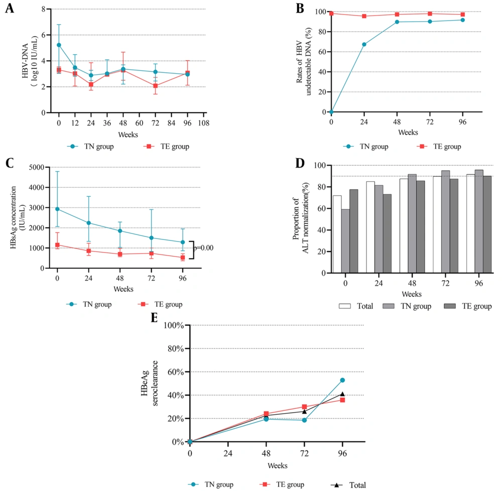
Figure 1.
The antiviral effectiveness and biochemical response from baseline to week 96 between the TN and TE groups. (A) median HBV DNA. Bars show median ± interquartile range. (B) the proportion of VR. (C) median HBsAg decline. Bars show median ± interquartile range. (D) proportion of patients with normal ALT (≤ 40 U/L). (E) rates of HBeAg seroclearance among the whole cohort, TN group, and TE group.
Among 110 TE patients whose HBV DNA was below 100 IU/mL, HBV DNA re-detectable occurred in six patients during 96 weeks of treatment. The exact HBsAg, HBV DNA, and ALT levels of patients who underwent virologic response are depicted in Figure 2D-F. One patient (No. 3) underwent a virologic breakthrough twice, and most of them (5/6) regained undetectable HBV DNA after 6 - 12 months of continuing TAF treatment.
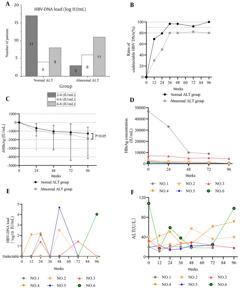
Figure 2.
Antiviral effectiveness of TN CHB patients divided into normal ALT group (ALT ≤ 40 U/L) and abnormal ALT group (ALT > 40 U/L). (A) number of patients with different levels of HBV DNA in the normal and abnormal ALT groups at baseline. (B) VR rates between the normal and abnormal ALT groups. (C) longitudinal changes of HBsAg levels between normal and abnormal ALT groups. (D) HBsAg levels of patients who underwent virologic breakthrough in the TE group. (E) HBV DNA of patients who underwent virologic breakthrough in the TE group. (F) ALT levels of patients who underwent virologic breakthrough in the TE group. Bars show median ± interquartile range.
4.3. Proportion of Normal ALT
The overall proportions of patients who achieved a normal ALT at baseline and weeks 48, 72, and 96 (ALT ≤ 40 U/L both for males and females) were 72.0% (116/161), 87.6% (141/161), 89.8% (123/137), and 91.6% (87/95), respectively (Figure 1D). In addition, for patients in the TN group, the proportions of normal ALT at baseline and weeks 48, 72, and 96 were 59.2% (29/49), 91.8% (45/49), 95.1% (39/41), and 95.8% (23/24), respectively (Figure 1D). For patients in the TE group, the proportions of normal ALT at baseline and weeks 48, 72, and 96 were 77.7% (87/112), 85.7% (96/112), 87.5% (84/96), and 90.1% (64/71), respectively (Figure 1D). Patients in the TN group had a similar trend but higher rates than patients in the TE group. The difference achieved statistical significance just at baseline (P = 0.02).
4.4. HBeAg Seroconversion and HBsAg Loss
The overall rate of HBeAg seroconversion was 22.5% (20/89), 26.0% (20/77), and 41.1 (23/56) at weeks 48, 72, and 96, respectively. Moreover, there was no significant difference between the TN and TE groups at week 48 (19.4% [6/31] vs. 24.1% [14/58], P = 0.17), week 72 (18.5% [5/27] vs. 30% [15/50], P = 0.27) and week 96 (52.9% [9/17] vs. 35.9% [14/39], P = 0.23) (Figure 1E).
Median qHBsAg levels decreased dramatically and consistently throughout the whole period, with 1763 (678.8, 3676) IU/mL at baseline versus 676 (144, 1442) IU/mL at week 96 (P = 0.00). In the TN group, the HBsAg level decreased from 2930 (1536.5, 6606) IU/mL to 1291.5 (770.5, 1991.2) IU/mL (P = 0.00). In the TE group, the HBsAg level decreased from 1158 (491.75, 2807) IU/mL to 533 (111, 1058) IU/mL (P = 0.00) (Figure 1C). There was a significant difference between the TN and TE groups at each time point. Only five out of 161 patients (3.72%) achieved HBsAg seroclearance at week 48. They were all HBeAg negative; most (4/5) were in the TE group and had low baseline HBsAg concentration.
4.5. TN patients Stratified by ALT Level
Patients in the TN group were divided into the normal ALT group (ALT ≤ 40U/L, n = 29) and the abnormal ALT group (ALT > 40U/L, n = 20) according to baseline ALT levels. The proportion of HBV DNA undetectable was numerically higher among patients in the normal ALT group than in the abnormal ALT group. In the normal ALT group, VR rates were 69.0% (20/29) at week 12, 79.3% (23/29) at week 24, 96.6% (28/29) at week 36, 96.6% (28/29) at week 48, 92.0% (23/25) at week 72, and 100.0% (14/14) at week 96 (Figure 2B). In the abnormal ALT group, VR rates were 30.0% (6/20) at week 12, 50.0% (10/20) at week 24, 80.0% (16/20) at week 36, 80.0% (16/20) at week 48, 82.4% (14/17) at week 72, and 80.0% (8/10) at week 96 (Figure 2B). There was a significant difference between the two groups at week 12 (P = 0.01) and week 24 (P = 0.03), but not thereafter. Regarding changes in qHBsAg levels from baseline, both groups showed a similar downward trend [normal ALT group: -1379.0 (-4325.5, -710.5) IU/mL, P = 0.01; abnormal ALT group: -2440.5 (-4601.2, -1146.0) IU/mL, P = 0.01] and no significant difference was observed between them at week 96 (P = 0.45) (Figure 2C).
4.6. Renal Function and Lipid Profile
Compared to baseline measurements, serum creatinine statistically increased (74.41 vs. 82.21 µmol/L, P = 0.00), and eGFR decreased (102.8 vs. 92.38 mL/min/1.73 m2, P = 0.00) in the TN group at week 96. In the TE group, serum creatinine increased from 74.21 µmol/L to 78.83 µmol/L (P = 0.00), and eGFR decreased from 103.75 mL/min/1.73 m2 to 94.85 mL/min/1.73 m2 (P = 0.00). None of them had eGFR below 60 mL/min/1.73 m2 at any time during the whole period.
Changes in eGFR (mL/min/1.73 m2) over time showed that the decreases were similar between the TN and TE groups at week 48 (-6.26 vs. -6.78, P = 0.71), week 72 (-6.97 vs. -9.66, P = 0.12), and week 96 (-11.46 vs. -10.90, P = 0.82) (Figure 3A). Changes in serum creatinine (µmol/L) were also similar between the two groups at week 48 (+4.22 vs. +4.51, P = 0.82), week 72 (+7.46 vs. +6.27, P = 0.39), and week 96 (+7.91 vs. +6.62, P = 0.52) (Figure 3B). The results showed that 25, 23, and 21 patients developed degradation in CKD stage (from CKD1 to CKD2) at weeks 48, 72, and 96, respectively, when compared with stages at baseline. There were three patients at week 48 and one patient at week 96 who showed CKD stage improvement (from CKD2 to CKD1) (Figure 3C). Importantly, only 1, 6, and 7 patients at weeks 42, 72, and 96 showed a percentage of eGFR decline > 25%. Only one patient showed a percentage of eGFR decline > 30%.
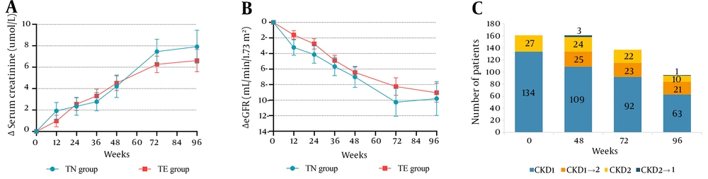
Figure 3.
Changes in Scr, eGFR, and CKD stages during 96-week follow-up. (A) changes from baseline in eGFR during 96-week follow-up of TAF treatment in TN and TE groups. (B) changes from baseline in Scr at weeks 12, 24, 48, 72, and 96 of TAF treatment in TN and TE groups. (C) changes of CKD stages for all patients compared to CKD stages at baseline. Bars show mean ± standard error of the mean.
For patients in the TN group, TG, TC, and LDL-C levels at baseline were 1.08 (0.82,1.48) mmol/L, 4.05 (3.56,4.66) mmol/L, and 3.72 (2.79,4.44) mmol/L, respectively. For patients in the TE group, TG, TC, and LDL-C levels at baseline were 0.92 (0.68,1.19) mmol/L, 3.96 (3.47,4.37) mmol/L, and 3.66 (2.79,4.00) mmol/L. They were comparable between the two groups at baseline, except for TG levels (P = 0.01) (Table 1). When comparing changes in lipids from baseline to week 96, serum TG, TC, and LDL-C levels increased gradually. In the TN group, lipidemia changes at week 96 were observed in TG [+0.39 (+0.18, +0.86) mmol/L], TC [+0.65 (+0.25, +1.10) mmol/L], and LDL-C [+0.25 (+0.24, +0.40) mmol/L]. In the TE group, changes at week 96 were observed in TG levels [+0.31 (+0.09, +0.63) mmol/L], TC levels [+0.96 (+0.52, +1.60) mmol/L], and LDL-C levels [+0.25 (+0.24, +0.35) mmol/L] (Figure 4A-C). The difference between the TN and TE groups was only significant in the TC increase (P = 0.02).
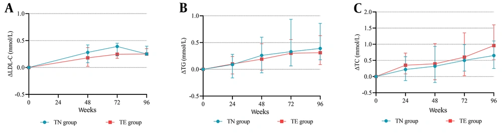
Figure 4.
Longitudinal changes of lipid profiles after TAF treatment during 96-week follow-up. (A) longitudinal changes in LDL-C levels. (B) longitudinal changes in TG levels. (C) longitudinal change in TC levels. Bars show median ± interquartile range.
5. Discussion
In this retrospective study, we investigated the effectiveness and safety of tenofovir alafenamide (TAF) monotherapy in patients with chronic hepatitis B (CHB) in both the treatment-naive (TN) and treatment-experienced (TE) groups over a 96-week follow-up period. We found that TAF is highly effective in controlling HBV replication. Moreover, there was no statistically significant difference between the TN and TE groups in VR and biochemical response. We also found that TAF seemed to have a “lipid-elevating" effect and a risk of persistently decreasing kidney function, primarily behaving as eGFR decline.
It has been demonstrated that persistently elevated HBV DNA was independently associated with the risk of hepatocellular carcinoma in CHB patients (12). Antiviral therapies may help to reduce the incidence rate of HCC and liver-related mortality by suppressing viral replication. In our study, TAF appeared to be highly effective in controlling HBV replication; the VR rates at week 96 were 91.7% in the TN group and 97.2% in the TE group. Similar studies also demonstrated the ability of TAF to suppress HBV replication. A multi-center, retrospective cohort study enrolled patients who were switched to TAF from other NAs and showed that TAF was superior in achieving HBV DNA suppression (HBV DNA < 20 IU/mL) from 88.19% at the time of the switch to 94.89% at week 96 post-switch (13), which was similar to other real-world studies (14, 15). Another real-world study among TN patients in Canada reported a 75% HBV undetectable (HBV DNA < 20 IU/mL) rate at week 48, which was lower than our study (90%) (15). The VR rates in TN patients of our study were also higher than in phase 3 clinical studies (8). The differences mentioned above may be attributed to varying test sensitivities in each study. This could be due to different detection lower limits and the inclusion of populations with varying levels of variability.
Also, ALT is a marker of liver necroinflammation, and achieving normal ALT levels during treatment may help reduce the risk of liver-related events (16). Our study demonstrated that TAF had a positive effect on reducing elevated ALT levels to within the normal range. Approximately 88% of patients (92% in the TN group and 86% in the TE group) achieved normal ALT at week 48, comparable with another study (17). Long-term treatment data of TAF in normalizing abnormal ALT are still to be confirmed by more studies. However, persistently normal ALT (PNALT) patients were also not “safe" during chronic HBV infection. Patients with PNALT and increased HBV DNA also underwent significant fibrosis (18). A Korean study enrolled 4,965 CHB patients and found that untreated HBeAg-positive patients with normal ALT levels and detectable HBV DNA had a higher incidence of HCC and death/transplantation when compared with treated patients with elevated ALT levels (19). When TN CHB patients in our study were divided into the normal and abnormal ALT groups, we found that the normal ALT group had a higher proportion of VR than the abnormal ALT group (100% vs. 80%), although no statistical significance was found.
Similarly, a study of CHB patients in China investigated the VR (HBV DNA < 100 IU/mL) of PNALT patients with elevated HBV DNA during 24 weeks of TAF monotherapy and reported a rate of 96.8% (20). One study enrolled 17 PNALT CHB patients and demonstrated that long-term lamivudine monotherapy could help improve liver histopathology, as proved by liver biopsy (21). The antiviral treatment seemed beneficial for PNALT CHB patients. However, we should correctly identify a special population in the true immune-tolerant phase who are recommended not to be aggressively treated. There are limited studies on treating patients with positive HBV-DNA and normal ALT levels. Further studies focusing on this problem are needed.
HBsAg is a significant marker of HBV infection derived from cccDNA and integrated HBV DNA (4). HBsAg seroclearance may further reduce the risk of HCC and liver-related death in CHB patients who have already achieved complete viral suppression (22). However, HBsAg loss rarely occurs in CHB patients treated with NAs. Our study showed that TAF decreased median qHBsAg during 96 weeks from 1763 to 676 IU/mL. However, only five patients achieved HBsAg loss; one was in the TN group, and the others were in the TE group. It was reported that HbsAg ≥ 3 log IU/mL and elevated ALT > 2 ULN at baseline might be a good predictor of HBsAg decline (23-25). We noted that qHBsAg decline in our study was more profound in the TN group than in the TE group, which may be explained by a higher proportion of higher HBsAg and ALT levels in the TN group at baseline.
Regarding the adverse effects of TAF on renal function, some differences were observed when contrasted to other real-world and RCT studies. Our study showed a more significant increase in serum creatinine and a greater decrease in eGFR. Our study's median decline in eGFR was -11.0 mL/min/1.73 m2, which is more profound than in other 96-week TAF studies (8, 14). Except for absolute levels of eGFR, there were other methods to evaluate renal dysfunction. Views that a decline in eGFR of more than 25% during one year or a decline of more than 30% over two years may indicate significant fluctuations (26). If we define eGFR declining more than 25% as deterioration in renal function, there were only 1, 6, and 7 patients at weeks 48, 72, and 96 that showed meaningful decline; if we define eGFR declining more than 30% as the endpoint of worsening renal function, only one patient was meeting the criterion during the entire period. Because of the unmeasurable variation of eGFR based on serum creatinine, more sensitive biomarkers reflecting renal dysfunction need to be included.
We found that TAF appeared to have a “lipid-increasing" effect in this study. Serum cholesterol and lipoproteins were associated with atherosclerotic cardiovascular disease (ASCVD) events, especially LDL-C. The recent ESC/EAS guideline (27) proposed that people with LDL-C levels greater than 4.9 mmol/L (190 mg/mL) were classified as a high-risk group for ASCVD, even without other risk factors, such as age, sex, and smoking. In our study, the median baseline LDL-C was 3.72 mmol/L, which was relatively high, with 14 patients exceeding 4.9 mmol/L. Approximately 12 additional patients met these criteria at the end of the follow-up. A randomized clinical trial found that people with LDL-C levels > 4.9 mmol/L without previous vascular disease may benefit from statins in the short and long term, mainly by reducing CHD incidence and cardiovascular disease-related mortality (28, 29). Although some studies investigating lipidemia changes in CHB patients treated with TAF showed an increase in LDL-C (17, 30), the exact clinical significance and mechanism of this change remain to be confirmed. However, based on the available evidence, we should pay attention to LDL-C levels, and timely drug intervention may be necessary when LDL-C levels are elevated to a higher level.
Our study is one of the clinical trials investigating TAF treatment under real-world conditions in China. We observed the potent antiviral effectiveness of TAF and related adverse effects, including renal and lipidemia changes. It may help clinics select antiviral therapies based on individual conditions. However, there were also limitations in our study. First, this was a single-center, retrospective study with a small sample size. It was a single-arm research study and lacked a control group, so its anti-HBV efficacy could not be compared with other antiviral drugs. Moreover, we could not conduct an in-depth statistical analysis due to the small sample size. Second, the enrollment time was different from January 2019 to October 2020. So, not every patient completed 96 weeks of follow-up. However, all patients included in our study completed clinical follow-up every three months until the end of the study. The decreasing numbers of patients with time were a reflection of the time since entry into the study. Third, adverse biomarkers of renal damage and serum lipidemia were not comprehensive. More laboratory tests, such as serum phosphate levels, urinary albumin to creatinine ratio, urinary retinol-binding protein to creatinine ratio, urinary beta-2-microglobulin to creatinine ratio, and lipoprotein (a) were more suitable to evaluate early renal tubular damage and further investigate the risk of ASCVD. However, due to these unconventional examinations and limitations of retrospective analysis, we were unable to obtain these data. Further studies with larger sample sizes and more extended follow-up periods are needed to verify the long-term use of TAF.
5.1. Conclusions
In summary, our study showed that TAF was proved to be highly effective no matter in TN and TE CHB patients. Meanwhile, a decrease in eGFR and an increase in lipidemia were observed during the 96-week follow-up. Further studies are necessary to investigate the long-term use of TAF in CHB patients.