
1. Background
Acute esophageal variceal bleeding (EVB) is a life-threatening complication of portal hypertension in cirrhosis and remains a major contributor to short-term mortality and health care burden (1-3).
Current international guidelines recommend upper gastrointestinal endoscopy within 12 hours of presentation, provided that adequate hemodynamic resuscitation has been achieved; however, the optimal time window within the first 24 hours remains debated (1, 2, 4). In observational cohorts, patients undergoing very early endoscopy (< 6 hours) often present with greater physiologic derangement, which may confound comparisons of outcomes across timing groups that are unadjusted (5-7).
2. Objectives
3. Methods
3.1. Study Design and Participants
This retrospective cohort study was conducted at The Affiliated Hospital of Guangdong Medical University, which has expertise in hepatology and gastroenterology, between January 2018 and December 2023. The study aimed to identify prognostic factors for 30-day rebleeding in patients with cirrhosis presenting with acute EVB.
The study was approved by the Ethics Committee of the Affiliated Hospital of Guangdong Medical University. Owing to the retrospective design and use of anonymized data, the Ethics Committee waived the requirement for written informed consent. All patient data were fully anonymized before analysis. Identifiable information was removed, and the deidentified dataset was stored in a secure, password-protected database accessible only to authorized personnel. Data management adhered to institutional governance policies and relevant privacy regulations, including the Health Insurance Portability and Accountability Act, to ensure the confidentiality and integrity of personal health information.
Eligible participants were adults with cirrhosis and endoscopically confirmed EVB who underwent emergency endoscopy within 24 hours of presentation. Patients were excluded if they had nonvariceal gastrointestinal bleeding, prior variceal interventions within 6 months, incomplete 30-day follow-up, or severe comorbidities likely to confound outcomes, such as advanced cardiac or renal failure. A total of 350 patients met the inclusion criteria. Baseline demographic, laboratory, and clinical data, including endoscopy timing, hemoglobin level, INR, and shock status, were extracted from the medical records. Patients were stratified by endoscopy timing into super-early (< 6 hours; n = 50), standard (6 - 12 hours; n = 180), and delayed (> 12 hours; n = 120) groups.
3.2. Patient Characteristics and Data Collection
Clinical and demographic data were extracted from electronic health records using a standardized protocol to ensure consistency and completeness. Baseline variables included age, sex, cirrhosis etiology, and comorbidities. Hemodynamic parameters, including blood pressure, heart rate, and signs of shock, and laboratory indices, including hemoglobin, INR, total bilirubin, albumin, serum creatinine, and estimated glomerular filtration rate, were recorded at admission. These variables served as surrogate markers of liver function and physiologic status.
Endoscopic variables included procedure timing, therapeutic modality (endoscopic variceal ligation [EVL] alone or combined with sclerotherapy), variceal grade and size, and the presence of active bleeding. Clinical outcomes included 30-day rebleeding, all-cause mortality, infection, and transfusion requirement. Rebleeding was defined as recurrent hematemesis, melena, or a decrease in hemoglobin level requiring intervention. Follow-up was conducted through outpatient visits or structured telephone interviews. Data were independently reviewed by 2 investigators, and discrepancies were resolved by consensus to ensure internal validity.
3.3. Variables and Predictors
The primary outcome was 30-day rebleeding, defined as recurrent variceal hemorrhage confirmed by clinical criteria. Secondary outcomes included all-cause mortality within 30 days, early rebleeding within 5 days, intensive care unit admission, hospital length of stay, and transfusion volume.
Predictor variables were selected based on prior literature and clinical plausibility. Endoscopy timing was categorized as super-early (< 6 hours), standard (6 - 12 hours), or delayed (> 12 hours). Hepatic dysfunction was assessed using the Child-Pugh classification. Shock was defined as hypotension requiring vasopressors or clinical evidence of hypoperfusion. Admission hemoglobin and INR were included to reflect bleeding severity and coagulation status, respectively. Total bilirubin was assessed as a marker of liver excretory function. Additional variables included endoscopic treatment modality, age, sex, cirrhosis etiology, and bleeding history. All candidate variables were entered into univariable and multivariable logistic regression models to identify independent predictors of 30-day rebleeding. The final multivariable model served as the basis for nomogram development.
3.4. Statistical Analysis
All analyses were conducted using SPSS version 26.0 (IBM Corp.) and R version 4.0.2 (R Foundation for Statistical Computing). Continuous variables were expressed as the mean ± standard deviation or median with interquartile range, depending on their distribution. Categorical variables were presented as frequencies and percentages. Between-group comparisons were performed using 1-way analysis of variance or Kruskal-Wallis tests for continuous variables and chi-square or Fisher exact tests for categorical variables. Univariable logistic regression was used to identify candidate predictors of 30-day rebleeding. Variables with P < 0.05 were entered into a multivariable logistic regression model to determine independent associations. Adjusted odds ratios (ORs) with 95% confidence intervals (CIs) were reported. Variables included in the final model were endoscopy timing, Child-Pugh class, shock at admission, hemoglobin, INR, total bilirubin, first bleeding episode, and treatment modality. Two-tailed tests were used throughout, with P < 0.05 considered statistically significant.
Model performance was assessed in terms of discrimination and calibration. Discriminative ability was evaluated using receiver operating characteristic analysis, with area under the curve (AUC) values ≥ 0.80 indicating good performance. Calibration was assessed through visual inspection of the calibration curve and the Hosmer-Lemeshow goodness-of-fit test, with P > 0.05 indicating acceptable fit.
4. Results
4.1. Baseline Characteristics of Included Patients
Among 583 assessed patients, 233 were excluded for various reasons, including nonvariceal bleeding, previous variceal interventions, loss to follow-up, and other reasons. The final cohort comprised 350 patients stratified into 3 groups according to endoscopy timing: super-early (< 6 hours), standard (6 - 12 hours), and delayed (> 12 hours) (Figure 1). Significant between-group differences were observed in baseline demographic and clinical characteristics. The mean age in the super-early group was 58.3 ± 8.2 years, significantly lower than that in the standard (60.2 ± 9.4 years) and delayed (62.5 ± 10.5 years) groups (P = 0.02). Male patients predominated across all groups, accounting for 60% of the super-early group, 58% of the standard group, and 60% of the delayed group. Liver function, assessed using the Child-Pugh classification, showed that 50% of patients in the super-early group were classified as Child-Pugh class B and 20% as class C. In comparison, the standard and delayed groups had a more balanced distribution; the standard group included 55.6% of patients with class B and 11.1% with class C disease. These findings are depicted in the Supplementary File.
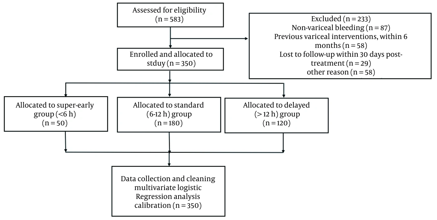
Figure 1.
Flowchart of patient selection and study design. Among 583 assessed patients, 233 were excluded for various reasons, including nonvariceal bleeding, prior variceal interventions, loss to follow-up, and other reasons. A total of 350 patients were enrolled and allocated to 3 groups based on endoscopic intervention timing: super-early (< 6 hours, n = 50), standard (6 - 12 hours, n = 180), and delayed (> 12 hours, n = 120). Statistical analysis included data cleaning, multivariable logistic regression, calibration, and external validation.
Regarding laboratory parameters, the super-early group had the highest Model for End-Stage Liver Disease (MELD) score (18.6 ± 3.4), which was significantly higher than that in the standard (16.8 ± 3.2) and delayed (17.4 ± 4.1) groups (P = 0.04). Mean hemoglobin levels were slightly higher in the super-early group (8.2 ± 1.1 g/dL) than in the standard (8.0 ± 1.0 g/dL) and delayed (7.8 ± 1.2 g/dL) groups; however, the difference was not statistically significant (P = 0.18). Initial INR values were highest in the super-early group (1.8 ± 0.3), suggesting more pronounced coagulopathy than in the standard (1.7 ± 0.2) and delayed (1.6 ± 0.3) groups. The prevalence of shock at admission was significantly higher in the super-early group (35%) than in the standard (30%) and delayed (25%) groups (P = 0.03). These findings are further illustrated in the Supplementary File. Baseline demographic and clinical characteristics are summarized in Table 1.
Table 1.
Baseline Characteristics of Patients and 2-Way Comparisons a
| Baseline Characteristics | Super-Early Group (< 6h, n = 50) | Standard Group (6 - 12h, n = 180) | Delayed Group (> 12h, n = 120) | Super-Early vs. Standard | Super-Early vs. Delayed | Standard vs. Delayed |
|---|---|---|---|---|---|---|
| Male | 30 (60) | 104 (58) | 72 (60) | χ² = 0.25, P = 0.45 | χ² = 0.05, P = 0.60 | χ² = 0.10, P = 0.77 |
| Age (y) | 58.3 ± 8.2 | 60.2 ± 9.4 | 62.5 ± 10.5 | t = 1.87, P = 0.07 | t = 2.30, P = 0.02 b | t = 1.12, P = 0.20 |
| Weight (kg) | 65.1 ± 10.5 | 68.4 ± 12.1 | 70.6 ± 13.3 | t = 1.75, P = 0.08 | t = 1.56, P = 0.12 | t = 2.20, P = 0.05 b |
| Blood pressure (mmHg) | 115.70 ± 15.1 | 120.75 ± 12.8 | 125.80 ± 14.9 | t = 1.63, P = 0.11 | t = 1.45, P = 0.15 | t = 0.93, P = 0.30 |
| Child-Pugh Class | χ² = 7.65, P = 0.03 b | χ² = 5.90, P = 0.06 | χ² = 2.25, P = 0.15 | |||
| A | 15 (30) | 60 (33.3) | 40 (33.3) | |||
| B | 25 (50) | 100 (55.6) | 60 (50) | |||
| C | 10 (20) | 20 (11.1) | 20 (16.7) | |||
| MELD score | 18.6 ± 3.4 | 16.8 ± 3.2 | 17.4 ± 4.1 | t = 2.22, P = 0.04 b | t = 1.79, P = 0.08 | t = 0.95, P = 0.30 |
| Hemoglobin (g/dL) | 8.2 ± 1.1 | 8.0 ± 1.0 | 7.8 ± 1.2 | t = 1.21, P = 0.18 | t = 1.35, P = 0.12 | t = 1.04, P = 0.27 |
| Initial INR | 1.8 ± 0.3 | 1.7 ± 0.2 | 1.6 ± 0.3 | t = 1.15, P = 0.22 | t = 1.25, P = 0.15 | t = 2.21, P = 0.05 b |
| Total Bilirubin (µmol/L) | 34.7 ± 6.5 | 32.5 ± 5.2 | 30.0 ± 4.5 | t = 1.78, P = 0.09 | t = 1.91, P = 0.07 | t = 1.60, P = 0.10 |
| First Bleeding | 30 (60) | 99 (55) | 60 (50) | χ² = 0.12, P = 0.45 | χ² = 1.48, P = 0.20 | χ² = 0.92, P = 0.35 |
| Shock | 17 (35) | 54 (30) | 30 (25) | χ² = 8.26, P = 0.03 b | χ² = 10.34, P = 0.01 c | χ² = 7.62, P = 0.04 b |
| Disease etiology | χ² = 5.10, P = 0.05 | χ² = 3.90, P = 0.10 | χ² = 2.30, P = 0.20 | |||
| HBV | 30 (60) | 99 (55) | 60 (50) | |||
| Alcoholic | 15 (30) | 45 (25) | 36 (30) |
a Values are expressed as mean ± SD or No. (%). Patients were stratified by endoscopy timing into super-early (< 6 hours), standard (6 - 12 hours), and delayed (> 12 hours) groups. Pairwise comparisons were performed using t tests or chi-square tests, as appropriate. Abbreviations: MELD, Model for End-Stage Liver Disease; INR, international normalized ratio; HBV, hepatitis B virus.
b P < 0.05 indicates statistical significance.
c P < 0.01 indicates statistical significance.
4.2. Clinical Outcomes According to Endoscopy Timing
Treatment outcomes varied significantly among the 3 endoscopy-timing groups. The 30-day rebleeding rate was lowest in the standard group at 5% (9/180), compared with 10% (5/50) in the super-early group and 12% (14/120) in the delayed group (P = 0.03). Pairwise comparisons indicated that the standard group had significantly lower rebleeding rates than both the super-early (P = 0.04) and delayed groups (P = 0.02). Kaplan-Meier analysis further demonstrated significant differences in 30-day rebleeding-free survival across the 3 groups, with the standard group showing the most favorable curve profile (log-rank P = 0.003) (Supplementary File).
The 30-day mortality rate ranged from 4% (7/180) in the standard group to 6% (3/50) and 8% (10/120) in the super-early and delayed groups, respectively; however, this difference was not statistically significant (P = 0.12). Kaplan-Meier survival analysis of 30-day mortality showed no statistically significant differences among the groups (log-rank P = 0.237) (Supplementary File). Early rebleeding within 5 days showed a similar pattern, with rates of 3% in the standard group, 5% in the super-early group, and 10% in the delayed group (P = 0.05). Intensive care unit admission was more frequent in the super-early group (25%) than in the standard (15%) and delayed (20%) groups (P = 0.11), with a significant difference between the super-early and standard groups (P = 0.04). Patients in the standard group had the shortest mean hospital stay (6.5 ± 1.8 days), followed by the super-early (7.5 ± 2.0 days) and delayed (9.0 ± 2.5 days) groups (P < 0.01). Transfusion requirements were also lowest in the standard group (2.0 ± 1.0 units) and highest in the delayed group (3.5 ± 1.6 units) (P = 0.02), with significant pairwise differences between the standard and delayed groups (P = 0.01) and between the super-early and standard groups (P = 0.02) (Supplementary File). Treatment outcome comparisons are summarized in Table 2.
Table 2.
Treatment Outcomes by Endoscopic Timing Group with Pairwise Comparisons a
| Outcomes | Super-Early (< 6 h) | Standard (6 - 12 h) | Delayed (> 12 h) | Overall P | Super-Early vs. Standard | Super-Early vs. Delayed | Standard vs. Delayed |
|---|---|---|---|---|---|---|---|
| 30-day Rebleeding Rate | 5/50 (10) | 9/180 (5) | 14/120 (12) | 0.03 | 0.04 | 0.72 | 0.02 |
| 30-day Mortality Rate | 3/50 (6) | 7/180 (4) | 10/120 (8) | 0.12 | 0.35 | 0.68 | 0.09 |
| Rebleeding within 5 days | 2/50 (5) | 6/180 (3) | 12/120 (10) | 0.05 | 0.18 | 0.03 | 0.02 |
| ICU Admission Rate | 12/50 (25) | 27/180 (15) | 24/120 (20) | 0.11 | 0.04 | 0.39 | 0.12 |
| Length of Hospital Stay (days) | 7.5 ± 2.0 | 6.5 ± 1.8 | 9.0 ± 2.5 | < 0.01 | 0.01 | 0.04 | < 0.01 |
| Transfusion Requirements (U) | 2.8 ± 1.3 | 2.0 ± 1.0 | 3.5 ± 1.6 | 0.02 | 0.02 | 0.09 | 0.01 |
a Values are expressed as mean ± SD or No. (%). The three groups were stratified based on endoscopic timing following admission: Super-Early (< 6 h), Standard (6 - 12 h), Delayed (> 12 - 24 h). Overall P-values represent comparisons across all three groups using the chi-square test for categorical outcomes and one-way ANOVA for continuous variables. Pairwise P-values were derived from post hoc pairwise comparisons, with Bonferroni correction applied to adjust for multiple testing. Statistical significance was set at a two-sided P < 0.05, and significant pairwise differences are indicated in bold. This table summarizes short-term clinical outcomes including 30-day rebleeding, mortality, early rebleeding, ICU admission, hospital stay, and transfusion requirements, providing comparative insights into the influence of endoscopic timing on prognosis in cirrhotic patients presenting with acute esophageal variceal hemorrhage. Abbreviations: ICU, intensive care unit; SD, standard deviation; ANOVA, analysis of variance; U, units.
4.3. Identification of Independent Predictors for 30-Day Rebleeding
To identify independent risk factors associated with 30-day rebleeding, a multivariable logistic regression model was constructed using clinically relevant covariates. Variables included endoscopy timing, Child-Pugh classification, shock at admission, baseline hemoglobin, INR, total bilirubin, history of previous variceal bleeding, and type of endoscopic intervention, as shown in Table 3.
Table 3.
Multivariable Logistic Regression Analysis for Predicting 30-Day Rebleeding a
| Variables | Odds Ratio (95% CI) | P Value |
|---|---|---|
| Endoscopic timing (reference: 6 - 12 h) | ||
| < 6 h (super-early) | 1.92 (0.83 - 4.48) | 0.13 |
| > 12 h (delayed) | 2.58 (1.14 - 5.86) | 0.02 |
| Child-Pugh class (reference: A) | ||
| B | 1.76 (0.89 - 3.48) | 0.10 |
| C | 3.42 (1.37 - 8.55) | 0.01 |
| Shock at admission | 2.11 (1.08 - 4.13) | 0.03 |
| Hemoglobin (per 1-g/dL decrease) | 1.26 (1.05 - 1.52) | 0.01 |
| INR (per 0.1-unit increase) | 1.19 (1.01 - 1.39) | 0.04 |
| Total bilirubin (per 10-μmol/L increase) | 1.08 (0.96 - 1.21) | 0.26 |
| First bleeding episode | 1.67 (0.88 - 3.16) | 0.12 |
| Treatment type (reference: EVL) | ||
| EVL + EIS | 1.42 (0.79 - 2.56) | 0.23 |
| Other | 1.89 (0.63 - 5.66) | 0.26 |
a Multivariate logistic regression was conducted to identify independent predictors of 30-day rebleeding. The model included clinically relevant covariates, regardless of univariate significance. Odds ratios (ORs) are presented with corresponding 95% confidence intervals (CIs). Endoscopic timing was referenced to the 6 - 12 hours group, Child-Pugh class to Class A, and endoscopic treatment modality to EVL (endoscopic variceal ligation) alone. Continuous variables (hemoglobin, INR, total bilirubin) were retained in their original form to preserve data granularity. Bolded categories represent reference groups for categorical variables. Statistical significance was defined as P < 0.05. Abbreviations: OR, odds ratio; CI, confidence interval; INR, international normalized ratio; EVL, endoscopic variceal ligation; EIS, endoscopic injection sclerotherapy.
Delayed endoscopic intervention (> 12 hours) was independently associated with an increased risk of rebleeding relative to the standard group (6 - 12 hours), with an OR of 2.58 (95% CI, 1.14 - 5.86; P = 0.02). Similarly, shock at presentation was significantly associated with an increased risk of rebleeding (OR, 2.11; 95% CI, 1.08 - 4.13; P = 0.03), as was advanced hepatic dysfunction, with patients classified as Child-Pugh class C having a higher risk than those with class A disease (OR, 3.42; 95% CI, 1.37 - 8.55; P = 0.01). Lower baseline hemoglobin levels were associated with an increased risk of rebleeding, with each 1-g/dL decrease conferring a 26% increase in the odds of rebleeding (OR, 1.26; 95% CI, 1.05 - 1.52; P = 0.01). Higher INR values were also associated with an increased risk, with each 0.1-unit increase associated with a 19% increase in the odds of rebleeding (OR, 1.19; 95% CI, 1.01 - 1.39; P = 0.04). Endoscopic intervention within 6 hours and Child-Pugh class B showed trends toward statistical significance; however, neither variable met the prespecified threshold for statistical significance.
These findings suggest that endoscopy timing beyond 12 hours, impaired hepatic reserve, coagulopathy, and initial circulatory instability are key predictors of early rebleeding. Supplementary File displays the adjusted ORs and corresponding 95% CIs for covariates identified as independent predictors in the multivariable logistic regression model.
4.4. Predictive Model Performance Evaluation
A multivariable logistic regression model was constructed to predict 30-day rebleeding in patients with cirrhosis and EVB. The final model incorporated 5 independent predictors: endoscopy timing, Child-Pugh class, shock at admission, baseline hemoglobin, and INR. Corresponding β coefficients and ORs are detailed in Table 4. Delayed endoscopy (> 12 hours) was associated with increased rebleeding risk (β = 1.20; OR = 3.3), as were Child-Pugh class C (β = 1.25; OR = 3.5) and shock at admission (β = 0.69; OR = 2.0). Lower hemoglobin and higher INR contributed incrementally, with each 1-g/dL decrease and 0.1-unit increase in INR corresponding to 6 - 7 and 5 - 6 points on the nomogram, respectively.
Table 4.
Predictors Included in the Nomogram for 30-Day Rebleeding a
| Predictors and Categories/Units | β Coefficient | Odds Ratio (95% CI) | Assigned Nomogram Points |
|---|---|---|---|
| Endoscopic timing | |||
| Standard (6 - 12 h) | Reference | – | 0 |
| Super-early (< 6 h) | 0.85 | 2.3 | 45 |
| Delayed (> 12 h) | 1.20 | 3.3 | 70 |
| Child-Pugh classification | |||
| Class A | Reference | – | 0 |
| Class B | 0.74 | 2.1 | 45 |
| Class C | 1.25 | 3.5 | 70 |
| Shock at admission | |||
| No | Reference | – | 0 |
| Yes | 0.69 | 2.0 | 45 |
| Hemoglobin | |||
| Per 1-g/dL decrease | 0.14 | 1.15 | 6 per unit |
| INR | |||
| Per 0.1-unit increase | 0.095 | 1.10 | 5 per 0.1 unit |
a β coefficients and odds ratios (ORs) were derived from a multivariable logistic regression model predicting 30-day rebleeding. Nomogram points were calculated based on the relative contribution of each predictor to the model. Reference categories indicate baseline risk and were assigned zero points. For continuous variables, points reflect the estimated increase in risk per unit change. ORs are approximate values derived from exponentiation of β coefficients and are provided for interpretative clarity. Abbreviations: OR, odds ratio; CI, confidence interval; INR, international normalized ratio.
A nomogram was developed to facilitate clinical application, with total scores mapped to the estimated 30-day rebleeding risk (Supplementary File). The model demonstrated good discriminatory performance, with an AUC of 0.82 (95% CI, 0.75 - 0.89) (Supplementary File). Calibration was acceptable, with a smoothed calibration curve showing alignment between predicted and observed rebleeding probabilities (Supplementary File). The Hosmer-Lemeshow test yielded a nonsignificant result (P = 0.09), indicating adequate model fit.
5. Discussion
Acute bleeding from esophageal varices is a critical decompensating event in the progression of cirrhosis. In this retrospective cohort, endoscopy performed within 6 - 12 hours was associated with the lowest 30-day rebleeding rate compared with endoscopy performed < 6 hours or > 12 hours; however, given the nonrandomized design, these findings should be interpreted as associations and may have been influenced by residual confounding.
Interestingly, the super-early (< 6 hours) group had a higher observed rebleeding rate; however, these patients also had markers of greater baseline severity, including a higher MELD score and more frequent shock on admission, suggesting important indication bias. Therefore, the present data do not support a causal inference that super-early endoscopy is harmful; rather, they highlight the need to prioritize adequate resuscitation and physiologic stabilization before endoscopic intervention.
Multivariable modeling identified endoscopy timing, Child-Pugh class C, hypotensive shock at presentation, lower hemoglobin concentration, and coagulopathy, as measured by an elevated INR, as independent correlates of early rebleeding. These findings are consistent with, and expand on, established prognostic frameworks. Notably, this study integrates these parameters into a cohesive risk-stratification algorithm, resulting in a well-calibrated predictive model with an AUC of 0.82 and externally validated discriminatory accuracy, underscoring its potential clinical applicability across diverse patient populations. Such a tool may serve as a point-of-care instrument for individualized clinical decision-making, particularly in settings in which resource triage and procedural prioritization are pressing concerns.
These results align with and further refine existing data and recommendations, such as those from the Baveno VII consensus, which endorse early intervention without defining a precise subwindow (10-12). By disaggregating the early-intervention time frame, our analysis adds empirical clarity to the therapeutic margin, illustrating that endoscopy performed too early or too late may both have clinical consequences. Including timing as an actionable variable in the predictive model enhances its translational value and distinguishes it from static prognostic indices that lack interventional modifiability.
From a practical perspective, these findings support considering 6 - 12 hours as a pragmatic target window for endoscopy, when feasible, after stabilization and according to resource availability. Nonetheless, because timing is influenced by clinical urgency and system factors, prospective studies or causal-inference designs with robust adjustment are needed to determine whether modifying timing alone improves outcomes.
5.1. Limitations
Several methodological constraints warrant acknowledgment. The retrospective nature of the analysis inherently increases susceptibility to selection bias and residual confounding, despite rigorous statistical adjustment and external validation efforts. The single-center design further limits generalizability across diverse health care contexts. Additionally, the exclusion of potentially contributory variables, such as active alcohol use, nutritional status, or direct portal-pressure measurements, may have limited the model’s comprehensiveness. Reliance on clinical and biochemical surrogates to define rebleeding, in the absence of confirmatory endoscopic reevaluation, also introduces the possibility of misclassification, particularly in borderline clinical scenarios. In particular, because endoscopy timing was not randomized and was closely linked to baseline severity and resuscitation status, causal inferences regarding timing and outcomes cannot be made.
Future investigations should prospectively evaluate the proposed model across geographically and demographically diverse populations. Incorporating real-time physiologic metrics, emerging biomarkers, and dynamic monitoring indices could improve predictive granularity. Moreover, constructing composite outcome models that integrate both rebleeding and mortality end points may better reflect the multidimensional nature of clinical deterioration in cirrhotic patients.
5.2. Conclusions
In this cohort, endoscopy performed within 6 - 12 hours was associated with lower 30-day rebleeding, and the nomogram provides an externally validated tool for individualized risk estimation. The less favorable outcomes observed in the < 6-hour and > 12-hour groups likely reflect baseline severity, resuscitation status, and system-related delays rather than a direct effect of timing itself. These findings support a patient-centered approach that integrates physiologic readiness with timely endoscopy.