
1. Background
Metabolic dysfunction-associated steatotic liver disease (MASLD) has emerged as a predominant cause of chronic liver disease worldwide, affecting approximately 30% of the global adult population. Characterized by hepatic steatosis in the absence of significant alcohol consumption, MASLD encompasses a spectrum ranging from simple steatosis to metabolic dysfunction-associated steatohepatitis (MASH), which may progress to cirrhosis and hepatocellular carcinoma (1). The rising prevalence of MASLD parallels the global epidemic of obesity and type 2 diabetes mellitus, positioning it as a major public health burden with hepatic and extrahepatic consequences, including cardiovascular disease, chronic kidney disease, and various malignancies (2-4).
The pathogenesis of MASLD is multifactorial, involving complex interactions among dietary factors, metabolic dysregulation, and inflammatory pathways. Overnutrition from high-carbohydrate and high-fat diets promotes abnormal fat storage, lipotoxicity, and increased de novo lipogenesis in hepatocytes, thereby driving disease initiation and progression (1, 5). Insulin resistance, a hallmark of MASLD, exacerbates hepatic lipid accumulation and is closely associated with elevated serum oxidized low-density lipoprotein (oxLDL) levels, which contribute to lipotoxicity and cellular injury (2, 3). The inflammatory milieu characteristic of MASLD is sustained by proinflammatory cytokines and adipose tissue-derived mediators, creating a state of chronic low-grade inflammation, termed metaflammation, that perpetuates hepatocellular damage and fibrosis (4, 6).
Among the inflammatory mediators implicated in MASLD pathophysiology, interleukin 6 (IL-6) plays a central role. Elevated IL-6 levels in patients with MASLD correlate with hepatic inflammation, fibrosis, and insulin resistance, suggesting its involvement in disease progression (7, 8). IL-6 orchestrates lipid homeostasis and systemic metabolic responses, fostering dynamic interactions that influence MASLD severity and clinical outcomes (8). Concurrently, oxidative and nitrosative stress, reflected by increased levels of nitric oxide derivatives (NOx), contribute to hepatocellular injury through protein nitration, mitochondrial dysfunction, and impaired metabolic homeostasis (9). Reactive nitrogen and oxygen species promote lipid peroxidation, generating oxidized LDL that binds to lectin-like oxidized low-density lipoprotein receptor 1 (LOX-1) (10).
LOX-1, a multiligand scavenger receptor primarily expressed in vascular endothelial cells, macrophages, and fibroblasts, is activated by diverse stimuli, including proinflammatory cytokines (IL-1β, IL-6, and TNF-α), oxidized LDL, angiotensin II, and shear stress (11, 12). Although LOX-1 expression remains low under physiological conditions, it is markedly upregulated during oxidative stress and inflammatory responses, conditions that are prevalent in MASLD (13, 14). Activation of LOX-1 triggers downstream signaling cascades that promote endothelial dysfunction, apoptosis, and additional inflammatory cytokine production, establishing a feed-forward loop that amplifies tissue injury (15). Emerging evidence from gene knockout studies implicates LOX-1 in the pathogenesis of MASH, although its precise role in exercise-mediated hepatic protection remains incompletely defined (13).
Given the limited pharmacological options for MASLD management, nonpharmacological interventions, particularly physical activity, have gained attention as cornerstone therapeutic strategies. Aerobic exercise exerts pleiotropic effects that counter metabolic dysfunction, including improved insulin sensitivity, enhanced mitochondrial biogenesis and function, reduced systemic inflammation, favorable modulation of lipid profiles, and increased antioxidant capacity (16, 17). Exercise-induced shear stress and associated hemodynamic changes may further enhance endothelial nitric oxide bioavailability and upregulate antioxidant enzyme systems, contributing to lipolytic and anti-inflammatory effects (17, 18). Moderate-intensity aerobic training has also been shown to reduce serum glucose, malondialdehyde, insulin resistance, and myocardial LOX-1 expression in diabetic rat models (10).
2. Objectives
Despite accumulating evidence linking exercise to improved metabolic homeostasis, the specific effects of chronic swimming exercise on hepatic LOX-1 expression and its relationship to MASLD pathophysiology remain incompletely characterized. This study was designed to investigate the association between moderate-intensity aerobic swimming exercise and hepatic LOX-1 expression in a high-fat diet-induced rat model of MASLD.
3. Methods
3.1. Animals
This experimental study included 32 male Wistar rats aged 6 to 8 weeks and weighing 200 ± 20 g, obtained from the Pasteur Institute Laboratory. Rats were housed in standard polyester cages (4 rats per cage) under controlled conditions (55 ± 5% humidity, 22 ± 2°C, 12-hour light/dark cycle) to ensure consistent environmental conditions. They had ad libitum access to high-fat pellets and/or a standard chow diet, according to the dietary interventions. All procedures complied with NIH regulations for animal care and were approved by the Ethics Committee of AJA University of Medical Sciences (IR.AJAUMS.REC.1399.256).
3.2. Experimental Design
After 1 week of acclimatization to the laboratory environment and swimming pool (110 × 100 × 80 cm, approximately 30°C), the rats were divided into the HFD (n = 16) and normal control (NC; n = 16) groups (phase I). During phase I (4 weeks), rats had free access to standard pellets and tap water (normal control group) or an HFD. The HFD pellets used for the HFD group were formulated by supplementing standard rodent chow with 2% cholesterol powder (Germany, Merck) and 60% animal fat. The fat composition comprised 71% monounsaturated, 23% trans-monounsaturated, and 6% polyunsaturated fat ingredients, as presented in Table 1 (13, 15). After 4 weeks, rats in the HFD group were randomly assigned to 2 equal groups for phase II (8 weeks): 1) HFD: rats received an HFD and remained sedentary; and 2) Exercise plus HFD (Exe + HFD): rats received an HFD and undertook an 8-week exercise program. The control rats were also divided into 2 equal groups: 3) Control: rats were maintained on a standard diet and kept sedentary; and 4) Exercise (Exe): rats received a standard diet and underwent an 8-week exercise regimen (Figure 1).
Table 1.
Nutritional Composition of the Diets a
| Components | Normal Diet | High-fat Diet | ||
|---|---|---|---|---|
| g | kcal | g | kcal | |
| Kcal/g | - | 3.71 | - | 5.05 |
| Carbohydrates | 60 | 64.4 | 35.7 | 28.2 |
| Protein | 23.1 | 24.8 | 20 | 16 |
| Total fat | 4.3 | 10.4 | 31.4 | 62.1 |
| Fat ingredients | ||||
| Saturated fatty acids | 37.3 | 62.69 b | ||
a Values are expressed as percentage.
b Structured fatty acids include: monounsaturated (71.62%), trans monounsaturated (23.12%) and polyunsaturated (5.21%).
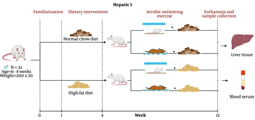
Figure 1.
Study design of the study
3.3. Aerobic Swimming Training Protocol and Load Measurements
Load-free swimming exercise was used as a model of moderate aerobic training. To ensure safe adaptation, rats initially completed a 10-minute swimming session to minimize stress and fatigue before the formal program. After this introductory period, rats swam freely at moderate intensity, with the daily duration increased by 10 minutes/day until it reached 60 minutes by the end of the second week. This protocol was then continued for 1 hour/day, 5 days/week, for 8 weeks.
For the inactive groups (Control and HFD), animals were placed in a low, shallow tank in which their paws touched the bottom while their heads remained above water. This procedure provided similar environmental exposure without actual swimming. To ensure animal safety and comfort, animals were thoroughly dried and placed in a warm chamber after each swim session to prevent hypothermia.
3.4. Tissue Extraction and Blood Collection
At the beginning and end of the experimental protocol, body weights were recorded using a digital scale. Forty-eight hours after the final exercise session and after a 12-hour fast, blood samples were collected via heart puncture and centrifuged at 2500 rpm for 15 minutes at 4°C to separate plasma, which was then stored at -20°C for biochemical analysis. Liver tissue was removed and embedded in formalin-fixed paraffin-embedded blocks for immunohistochemistry, and another portion was fixed for histological assessment.
3.5. Nitric Oxide Assay
Plasma levels of nitric oxide metabolites (NOx) were determined using the Griess colorimetric reaction. Plasma proteins were removed by deproteinizing 0.5 mL of plasma with 0.1 mL of zinc sulfate. The mixture was centrifuged for 20 minutes at 4°C and 4000 rpm, and the clear supernatant was collected for analysis. Nitrite was produced by reacting 0.1 mL of nitrate with 0.1 mL of vanadium (III) chloride, 0.05 mL of 0.01% sulfanilamide, and 0.05 mL of N-(1-naphthyl) ethylenediamine. Absorbance was measured spectrophotometrically, and NOx concentrations were calculated according to the standard curve.
3.6. Immunohistochemistry Assay
The expression of LOX-1 receptors and IL-6 proteins was evaluated using immunohistochemistry. For LOX-1, tissue sections were incubated with goat-derived anti-rabbit LOX-1 conjugated with FITC (ab5926; RRID: AB_920795) at a dilution of 1:1000. IL-6 protein levels were assessed using a specific anti-IL-6 antibody (ab9324; RRID: AB_307178). After antibody incubation, hematoxylin was used as a counterstain to delineate cellular structures. Protein expression was quantified by determining the percentage of the area exhibiting positive staining using ImageJ software (9, 10).
3.7. Histological Changes
After washing the liver in physiological saline, the tissue was fixed in 4% paraformaldehyde for approximately 48 hours. It was then dehydrated through a graded alcohol series, cleared with xylene solution, and embedded in liquid paraffin blocks. The tissue was cut into 5-µm sections using a microtome and placed on slides. To evaluate morphological changes, hematoxylin and eosin staining was performed, and the sections were examined under a light microscope.
3.8. Statistical Analysis
SPSS version 22 was used for statistical analyses. Intergroup differences were compared using 1-way ANOVA followed by the Tukey post hoc test, with significance set at P < 0.05.
4. Results
4.1. Effects of High-Fat Diet and Exercise on Body Weight
After 8 weeks of HFD consumption, the HFD group had a higher final body weight than the Control, Exercise, and Exercise plus HFD groups, and these differences were statistically significant (P < 0.05). These findings underscore the effect of diet on body weight management.
4.2. Effects of High-Fat Diet and Exercise on Serum Glucose and Lipid Profile
Eight weeks of HFD significantly altered the lipid profile. The HFD group exhibited elevated cholesterol levels compared with the Control group (P < 0.01). However, the exercise intervention did not significantly reduce serum cholesterol levels, as the Exercise plus HFD group maintained similarly elevated cholesterol levels compared with the HFD group (P > 0.05). Additionally, triglyceride levels were markedly higher in the HFD group than in the Control group (P < 0.01). Conversely, 8 weeks of exercise significantly reduced triglyceride levels in the Exercise plus HFD group compared with the HFD group (P < 0.05). Exercise did not significantly alter LDL-C levels, as no significant difference was observed between the HFD and Exercise plus HFD groups (P > 0.05). Glucose levels increased slightly with the high-fat diet, and exercise did not significantly affect this variable (Table 2).
Table 2.
Effects of High-Fat Diet and Exercise on Body Weight, Serum Lipid Profile, Glucose, and NOx Levels a
| Groups | Control | Exercise | High-Fat Diet | Exercise + High-Fat Diet | P-Value |
|---|---|---|---|---|---|
| Initial body weight (g) | 194 ± 7 | 205 ± 3 | 224 ± 38 | 228 ± 14 | 0.780 |
| Final body weight (g) | 324.4 ± 41 | 348.6 ± 28 | 394 ± 29 b, c, d | 352.4 ± 23 | 0.037 |
| Gonadosomatic index (%) | 1.49 ± 0.09 | 1.55 ± 0.10 | 1.20 ± 0.15 b, c, d | 1.40 ± 0.12 | 0.044 |
| Cholesterol (mg/dL) | 79.6 ± 20 | 97.6 ± 28 | 164.5 ± 50 b, c | 223 ± 64 ab | 0.001 |
| Triglyceride (mg/dL) | 90.8 ± 17 | 84.6 ± 20 | 169.6 ± 47 b, c, d | 154 ± 40 | 0.001 |
| LDL-C (mg/dL) | 65.73 ± 3 | 50.7 ± 7 | 157.2 ± 43 ab | 168.7 ± 56 ab | 0.029 |
| Glucose (mg/dL) | 124 ± 23 | 111.33 ± 22 | 141.5 ± 22 b, c | 144 ± 25 | 0.550 |
| NOx (µmol/L) | 3.7 ± 0.02 | 5.1 ± 1.3 | 6.1 ± 1.8 b, c, d | 4.6 ± 0.7 | 0.041 |
a Values are expressed as mean ± SD. Abbreviations: LDL-C, low-density lipoprotein cholesterol; NOx, nitrogen oxides.
b Versus the Control group.
c versus the Exercise group.
d versus the High-Fat Diet and Exercise plus High-Fat Diet group.
4.3. Effects of High-Fat Diet and Exercise on Serum Nitric Oxide
After 8 weeks on an HFD, the HFD group showed a significant elevation in serum NOx levels compared with the Control group (P < 0.05). The exercise intervention reduced NOx levels in the Exercise plus HFD group compared with the HFD group, supporting the potential of lifestyle modification to mitigate diet-related oxidative and nitrosative changes.
4.4. Immunohistochemical Analysis of Hepatic IL-6 and LOX-1 Protein Expression
Immunohistochemical analysis of hepatic expression of the proinflammatory cytokine IL-6 and the oxidative stress receptor LOX-1 revealed distinct group-specific patterns. Minimal immunoreactivity for both targets was observed in the Control and Exercise groups, indicating low basal expression under standard conditions and confirming that aerobic exercise alone did not induce inflammatory or oxidative stress signaling in hepatic tissue (Figures 2 and 3)
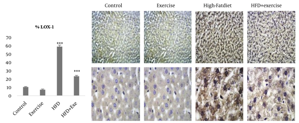
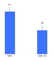
Figure 2.
Protein levels of LOX 1 in liver tissue. Brown color in the context is the sign of LOX 1 protein expression. The images are magnified at 200 X and 400 X with a scale bar set at 200μm. The data are presented as the mean ± standard deviation (SD). The groups are labeled as: Exe: Exercise, HFD: High fat diet group. Significant differences are denoted as *** P < 0.001 for the HFD group when compared to the Cont, Exe, and HFD + Exe groups, and *** P < 0.001 for the HFD + Exe group versus the Cont and Exe groups
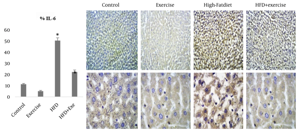
Figure 3.
Protein levels of IL-6 in liver tissue. The data are presented as the mean ± standard deviation (SD). Brown color in the tissue is the sign of IL 6 protein expression. Images were captured at magnifications of 200 X and 400 X, with a scale bar representing 200 μm. The groups are labeled as: HFD: High fat diet. Statistical significance is denoted as: *** P < 0.001 for comparisons between the HFD group and the Cont, Exe, and HFD + Exe groups. Also, *** P < 0.001 for the HFD + Exe group when compared to the Cont and Exe groups.
Concurrent implementation of the 8-week aerobic exercise intervention in the Exercise plus HFD group significantly attenuated this diet-induced upregulation, resulting in a marked reduction in immunostaining intensity for both IL-6 and LOX-1 relative to the sedentary HFD group (P < 0.01). These observations highlight the potential of exercise to attenuate hepatic inflammation and oxidative stress pathways associated with metabolic dysfunction. Nevertheless, despite this exercise-mediated reduction, IL-6 and LOX-1 expression levels in the Exercise plus HFD group remained higher than those in the Control group, suggesting partial rather than complete normalization of hepatic inflammatory and oxidative responses.
4.5. Histopathological Analyses
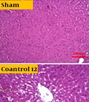
Histopathological evaluation revealed preserved hepatic architecture in the Control and Exercise groups, with normal hepatocyte morphology and no evidence of steatosis or inflammatory infiltration. In contrast, the HFD group exhibited marked pathological alterations, including severe hepatocellular ballooning, cytoplasmic lipid droplet accumulation, cellular swelling, and degeneration. Pyknotic nuclei with margination were observed in most hepatocytes, accompanied by cellular shrinkage and inflammatory infiltration. In the Exercise plus HFD group, these pathological changes were attenuated, and hepatic morphology was improved compared with the sedentary HFD group (Figure 4).
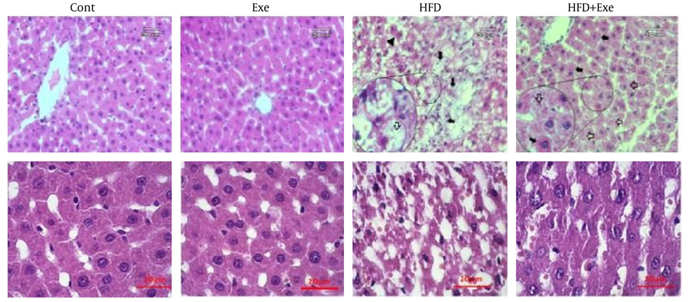
Figure 4.
Photomicrographs of liver with hematoxylin and eosin (H and E) staining. The images highlight different cellular conditions: the tip of the black arrow indicates shrinkage and necrotic hepatocytes, while the full black arrow points to ballooning and degenerated cells. The white arrow marks lipid droplets within the cytoplasm. Magnifications used are 20x and 40x, with scale bars measuring 20 μm and 60 μm respectively. The groups represented include Control (Cont), Exercise (Exe), High-Fat Diet (HFD) group, and High-Fat Diet combined with Exercise (HFD+Exe).
5. Discussion
The present study explored how moderate-intensity swimming exercise modulates high-fat diet-induced liver structural changes and metaflammation, with a focus on the role of the LOX-1 receptor. The findings indicate that regular aerobic exercise can reverse some manifestations of high-fat diet-induced MASLD, partly by improving histopathological appearance and metabolic profile and by attenuating the LOX-1/IL-6/NOx axis.
It is widely recognized that changes in dietary composition affect body mass and body weight by altering lipid profiles. Obesity, driven by a sedentary lifestyle and nutritional changes, is inversely associated with liver homeostasis, possibly through hepatocyte inflammation and nitrosative stress (13, 15, 16). The present findings indicate that high-fat diets lead to weight gain, whereas continuous aerobic activity prevents weight gain. Although the HFD increased cholesterol, LDL-C, and triglyceride levels, continuous swimming exercise specifically lowered triglycerides to levels comparable to those in the Control group. Notably, exercise did not significantly reduce serum cholesterol or LDL-C levels, suggesting a selective effect of exercise on triglyceride metabolism in this model.
In this study, chronic consumption of an HFD resulted in a significant increase in serum NOx levels. However, aerobic swimming exercise reduced NOx levels to those in the Control group, resulting in no pronounced difference in NOx levels between the Exercise plus HFD group and the Control group. Because measurement of total plasma NOx provides an integrated assessment of systemic nitric oxide production, elevated NOx levels in the context of MASLD have been attributed primarily to iNOS upregulation in inflamed hepatic tissue, contributing to nitrosative stress, protein nitration, and hepatocellular injury (8). The HFD produced a modest elevation in glucose levels that was not attenuated by the exercise intervention. Serum triglycerides were reduced following exercise, whereas cholesterol and LDL-C levels remained unchanged, suggesting a selective effect of exercise on triglyceride metabolism. This differential response may be attributed to the structural properties of cholesterol, because skeletal muscle cannot use cholesterol as an energy substrate in the same manner as free fatty acids. Notably, although total cholesterol levels may remain unaltered, exercise has been associated with shifts in the distribution of LDL and HDL subfractions, potentially reflecting improved lipoprotein quality rather than quantity (17).
Dyslipidemia-induced lipotoxicity increases IL-6 release, which is linked to MASLD progression (18). IL-6 levels in the liver and blood are also higher in patients with nonalcoholic steatohepatitis (NASH), and IL-6 may worsen hepatic inflammation and steatosis in MASLD (19). Kupffer cells may overproduce proinflammatory cytokines, a pathogenic characteristic of hepatic steatosis. Additionally, obesity recruits macrophages to the liver and changes their phenotype from M2 to M1 (20). Obesity-related hepatic inflammation leads to steatosis and liver lymphocyte infiltration by activating the IL-6 signaling pathway, which increases liver cell proliferation and hepatocyte degradation (21).
Immunohistochemical analysis revealed a substantial elevation in IL-6 and LOX-1 protein expression levels in the HFD group (P < 0.001). After 8 weeks of aerobic activity, both proteins showed lower expression levels relative to the HFD group but remained elevated compared with those in the Control group (P < 0.001). These correlative findings demonstrate a strong association between LOX-1 expression and markers of MASLD severity. The HFD increased liver parenchymal volume, mean liver cell volume, and lipid accumulation, whereas exercise attenuated these morphological changes.
Consistent with previous reports, HFD feeding induced significant weight gain and hypercholesterolemia, accompanied by cytoplasmic lipid vacuolation in hepatocytes (13, 15, 22). In this study, the HFD increased total cholesterol, triglycerides, and LDL-C, whereas aerobic exercise selectively reduced triglycerides without affecting cholesterol or LDL-C levels. This selective effect may be attributed to obesity-related dyslipidemia and nitrosative stress, which have been shown to alter apolipoprotein B/ApoB100 synthesis and secretion, thereby reducing very low-density lipoprotein (VLDL) export and promoting triglyceride accumulation in hepatocytes (23). Stereological analysis revealed a decreased hepatocyte number with increased mean cell volume and a higher prevalence of swelling, cytoplasmic disorganization, and clear space formation resulting from cytoplasmic dilution and organelle loss (24, 25). Such degenerative lesions, which typically precede apoptosis, reflect the progressive nature of diet-induced hepatocellular injury (25). Furthermore, HFD-induced hepatic inflammation contributed to lobular architectural disruption through the accumulation of fat, protein, and fluid within the parenchyma and perivascular spaces, leading to focal necrosis (26). Subsequent inflammatory cell infiltration stimulated perisinusoidal and perivenular collagen deposition, which extended through the hepatic lobule and culminated in early bridging fibrosis (27). Ultimately, progressive fibrosis results in the replacement of functional parenchyma with connective tissue (25-27).
MASLD is intricately associated with hepatic metaflammation, and weight reduction is strongly linked to improvements in histological features of NASH (1-5). The present results demonstrated that chronic aerobic swimming exercise mitigated the effects of HFD by preventing weight gain and significantly reducing serum triglyceride levels. Exercise also modulated HFD-induced histological alterations, decreasing hepatic lipid accumulation and inflammatory cytokine expression. In MASLD, circulating leukocytes infiltrate the liver and produce inflammatory mediators. Activated immune cells and damaged hepatocytes release proinflammatory cytokines, perpetuating disease progression (25-27). Chronic inflammation represents a critical risk factor in the progression from uncomplicated steatosis to advanced NASH and fibrosis. Exercise-mediated reductions in IL-6 and NOx levels in the present study suggest partial attenuation of this inflammatory cycle. These results support the concept that exercise improves MASLD-related histological changes and metabolic stress.
LOX-1 has been established as a downstream effector of endoplasmic reticulum stress signaling. Upregulation of LOX-1 contributes to MASLD progression by activating endoplasmic reticulum stress, and LOX-1 inhibition has been shown to alleviate endoplasmic reticulum stress (23, 28, 29). Moreover, proinflammatory cytokines, including IL-6, upregulate LOX-1 expression, thereby mediating inflammatory responses and apoptosis and underscoring the interplay between inflammation and endoplasmic reticulum stress (30). Supporting this, Deng et al. (31) demonstrated that chronic aerobic exercise attenuated HFD-induced metabolic dysregulation, insulin resistance, and hepatic fat accumulation in MASLD rats, concomitant with improved liver function markers.
The findings also demonstrate that exercise reduces serum triglycerides, nitric oxide, and hepatic IL-6 production. Consistent with these observations, Ho et al. associated LOX-1 with portal venous inflammation in NASH, supporting the link between nonalcoholic fatty liver disease (NAFLD), cholesterol accumulation, inflammation, and fibrosis (32). Exercise also reduces intrahepatic triglycerides and protects against lipotoxicity-induced obesity and steatohepatitis (33). A meta-analysis by Chen et al. confirmed that physical activity, including aerobic exercise, improves MASLD by reducing body fat percentage and waist-to-hip ratio (34). Mechanistically, regular exercise may attenuate chronic inflammation by promoting macrophage polarization from the proinflammatory M1 phenotype to the anti-inflammatory M2 phenotype, thereby modulating cytokine production in adipose tissue and visceral fat (35). Furthermore, aerobic exercise enhances lipid oxidation and suppresses hepatic lipogenesis through inhibition of stearoyl-CoA desaturase 1 (SCD-1) activity and activation of the AMP-activated protein kinase (AMPK) pathway (36).
Following exercise, sustained AMPK activation in muscle, liver, and adipose tissue occurs in response to decreased energy charge, as indicated by an increased AMP/ATP ratio. In the liver, AMPK suppresses lipid synthesis through acetyl-CoA carboxylase inactivation, malonyl-CoA decarboxylase activation, and downregulation of lipogenic enzymes (36). Aerobic exercise further reduces hepatic fat through sterol regulatory element-binding protein 1c (SREBP-1c) downregulation, PPARγ upregulation, and reduced nonesterified fatty acid delivery to the liver. These adaptations collectively reduce lipid accumulation and inflammation, as evidenced by reduced tumor necrosis factor alpha, while enhancing energy expenditure and fat oxidation (37). Additionally, exercise elevates hepatocyte growth factor, a key promoter of hepatic regeneration, angiogenesis, and vascular remodeling, and exercise training supports liver regeneration, with effects modulated by training intensity and modality (38). The observed co-occurrence of attenuation of the LOX-1/IL-6/NOx axis in the exercise group further implies restoration of nitrosative and inflammatory balance.
This study offers the advantage of concurrently evaluating serum levels and immunohistochemical markers of oxidative stress and inflammation, enabling stronger conclusions regarding the modulation of inflammatory conditions and the improvement of liver-related morphological and biochemical parameters. The findings establish significant correlations between exercise-mediated LOX-1 downregulation and improved MASLD-related parameters, providing a foundation for future mechanistic investigations.
5.1. Study Limitations
The current investigation did not evaluate other inflammatory pathways that concurrently interact with LOX-1, such as TNF-α, the NLRP3 inflammasome, and other scavenger receptors, which may have enhanced the applicability of the findings. The observed reduction in total NOx following exercise could reflect decreased nitrosative stress, but the contribution of altered endothelial nitric oxide synthase-mediated protective signaling cannot be determined from these data. Most importantly, the correlative nature of the study design must be acknowledged. Although strong associations were observed between LOX-1 downregulation and MASLD improvement following exercise intervention, causal mechanisms remain to be established through pathway-specific inhibition or genetic manipulation studies.
5.2. Conclusions
In conclusion, obesity-induced steatohepatitis was characterized by elevated inflammatory cytokine levels, LOX-1 protein upregulation, and increased nitrosative stress. Regular aerobic exercise was associated with a predictable decrease in serum nitrosative stress and inflammatory cytokines in liver cells, consistent with the known anti-inflammatory and antistress therapeutic effects of regular aerobic exercise, rather than with reductions in cholesterol or LDL-C. Importantly, conclusions regarding improved liver function and metabolic improvement are based on indirect markers, including the lipid profile, NOx levels, and histopathological analysis. The findings demonstrate that exercise is associated with improvements in morphological and biochemical parameters, but definitive conclusions regarding liver function improvement require direct enzymatic and functional assessment of hepatocellular injury markers, including ALT, AST, and insulin sensitivity indices. Given that oxidative stress and inflammation enhance LOX-1 expression and increased LOX-1 expression in turn exacerbates oxidative stress and inflammation, elevated LOX-1 expression appears to play a critical role in the progression of fatty liver and subsequent syndromes. Therefore, moderate-intensity swimming exercise appears to have a protective effect against hepatic steatosis and fatty liver, in part by reducing LOX-1 receptor expression, primarily through anti-inflammatory mechanisms.