1. Context
Hepatocellular carcinoma (HCC) is the most common primary liver malignancy and represents a substantial global health burden, particularly in regions with a high prevalence of hepatitis B and C virus infections (1-3). As the third leading cause of cancer-related death worldwide, HCC is often diagnosed at an advanced stage, when curative treatments such as resection or transplantation are no longer feasible (4, 5). In this context, medical imaging plays a central role in clinical decision-making, from initial diagnosis to treatment selection and follow-up (6-8). Among available modalities, magnetic resonance imaging (MRI) has emerged as a preferred tool because of its superior contrast resolution, functional imaging capabilities, and ability to assess both tumor characteristics and underlying liver function (9, 10).
In recent years, the development of cell-specific contrast agents for MRI has introduced a new dimension of biologically informed imaging that emphasizes molecular and cellular features rather than relying solely on structural information (11, 12). Unlike conventional extracellular agents, these compounds are designed to target specific hepatic cell populations, receptors, or microenvironmental features, thereby improving lesion characterization, functional assessment, and early detection (13-15). Hepatocyte-targeted agents, such as gadoxetate disodium, have already reshaped liver imaging by enabling combined anatomical and hepatobiliary-phase evaluation (16, 17). Experimental agents directed toward tumor cells, macrophages, and fibrotic tissue are also being investigated for their potential to support therapy selection and response monitoring (18, 19). These agents are intended to serve biomarker-like functions by stratifying patients, informing prognosis, and tracking therapeutic outcomes, particularly as HCC treatment shifts toward molecularly targeted and immune-based therapies (20, 21). These innovations represent a shift toward functional and molecular imaging, with the potential to improve diagnostic precision, guide biologically tailored interventions, and monitor treatment response in real time (22-24).
Despite this promise, the structure and direction of research on cell-specific MRI contrast agents in HCC remain poorly defined (25-28). There is limited understanding of how the literature is thematically organized, whether emerging approaches are gaining traction, and the extent to which collaborative networks shape the evolution of the field. This lack of clarity may contribute to fragmentation, duplication, or uneven clinical translation, particularly as new imaging agents face regulatory, technical, and implementation hurdles. Contrast-enhanced MRI is widely used to evaluate treatment response, disease progression, and prognosis in HCC; therefore, the literature on MRI contrast agents often overlaps with broader clinical studies assessing therapeutic outcomes.
To address this gap, we systematically mapped the global research landscape on cell-specific MRI contrast agents in HCC using bibliometric techniques to examine publication activity, collaboration patterns, and thematic structure over time. By assessing publication trends, citation impact, authorship networks, and conceptual clustering, this study aimed to identify where innovation is occurring and how well it is integrated into broader research and clinical contexts.
The primary aims of this study were to 1) quantify global publication, authorship, and citation patterns related to cell-specific MRI contrast agents in HCC; 2) identify the most influential contributors, institutions, journals, and countries shaping the field; 3) examine citation bursts to track shifting research impact over time; and 4) map the conceptual structure of the literature through co-citation analysis, keyword co-occurrence, cluster identification, and thematic categorization. Together, these objectives provide a comprehensive view of how the field is evolving, highlight areas of consolidation, identify gaps in translation, and clarify where future innovation is most needed.
2. Evidence Acquisition
2.1. Study Objective and Design
A bibliometric analysis was conducted to evaluate research trends in cell-specific contrast agents for MRI use in HCC, using data obtained from the Web of Science Core Collection for the years 2015 to 2025 (Figure 1). The 2015 to 2025 period was selected to assess recent research trends over a complete 10-year interval, enabling consistent bibliometric comparisons of publication activity, collaboration patterns, and thematic development in MRI-based HCC research. A total of 136465 records were initially retrieved. After a 2-step screening process, the first round excluded non-English articles (n = 1111), review articles (n = 20720), meeting abstracts (n = 13101), and unrelated publications (n = 78929), resulting in 22604 records. In the second screening, studies not directly focused on HCC were excluded (n = 18089), yielding a final dataset of 4515 articles for analysis.
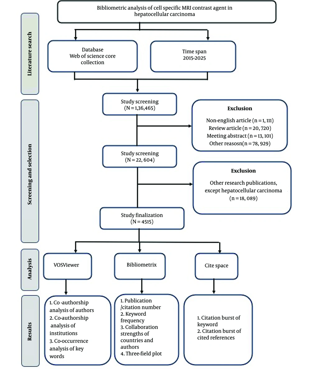
Figure 1.
Study design for the bibliometric analysis on cell-specific MRI contrast agents in hepatocellular carcinoma
Subsequent bibliometric evaluation used 3 analytical tools: VOSviewer, Bibliometrix, and CiteSpace. VOSviewer was used for co-authorship analysis among authors and co-occurrence analysis of keywords. Bibliometrix was applied to assess publication and citation counts, keyword frequency, and collaboration strengths between countries. CiteSpace was used to identify citation bursts in keywords and cited references and to generate keyword clustering and thematic mapping.
2.2. Data Source, Search Criteria, and Keywords
The Web of Science (WoS) was selected as the primary data source for this study because of its comprehensive coverage of peer-reviewed literature and robust citation tracking. To ensure retrieval of all relevant publications, the following search terms were used: "All Fields = Contrast agent" AND "All Fields = Magnetic resonance imaging" AND "All Fields = Hepatocellular carcinoma."
2.3. Data Analysis and Visualization Using Software Tools
The retrieved data were processed and analyzed using a combination of bibliometric software tools, including VOSviewer (version 1.6.20), CiteSpace (version 6.1.R3), Bibliometrix (R package), and built-in utilities from the Web of Science platform. These tools enabled evaluation of co-authorship patterns, keyword co-occurrence, citation dynamics, and other indicators of scholarly activity. VOSviewer (29), developed by van Eck and Waltman, is commonly used to construct and visualize bibliometric networks, in which node size reflects publication or citation volume and line thickness indicates the strength of associations. CiteSpace (30), developed by Chaomei Chen, is designed to detect citation bursts and map the temporal evolution of research topics. Bibliometrix (31), which includes the user-friendly Biblioshiny interface, provides an integrated environment for analyzing publication trends, author contributions, journal performance, and thematic structures across large datasets.
3. Results
3.1. Publication Trend
Between 2015 and 2025, 4515 publications on cell-specific MRI contrast agents in HCC were indexed across 131 sources (Figure 2). The annual publication rate declined by 1.75%, indicating a modest decrease in research activity. A total of 19086 authors contributed, with only 27 single-authored papers, highlighting a strong preference for collaboration. International co-authorship was present in 12.65% of publications, with an average of 10.6 coauthors per article. Authors used 6450 distinct keywords, and the dataset included 66067 references. The average document age was 4.26 years, and each publication received 16.66 citations on average, indicating moderate citation engagement.
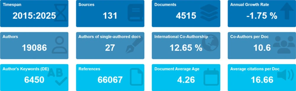
Figure 2.
Summary of descriptive bibliometric indicators for publications on cell-specific MRI contrast agents in hepatocellular carcinoma from 2015 to 2025
3.2. Author Contributions
Over the decade from 2015 to 2025, authorship in liver MRI research showed concentrated productivity among a select group of contributors. Zhang Y was the leading contributor (Figure 3), with 180 publications. Wang Y and Li J followed with 166 and 134 papers, respectively, forming a small cluster of dominant authors. A second group included Li X, with 130 publications; Wang J, with 126 publications; Chen J, with 125 publications; and Li Y, with 121 publications. Their outputs were closely aligned, indicating a competitive yet consistent presence in the field. A final tier of prolific contributors included Wang X, with 118 publications; Chen Y, with 116 publications; and Liu J, with 109 publications, all exceeding 100 publications.
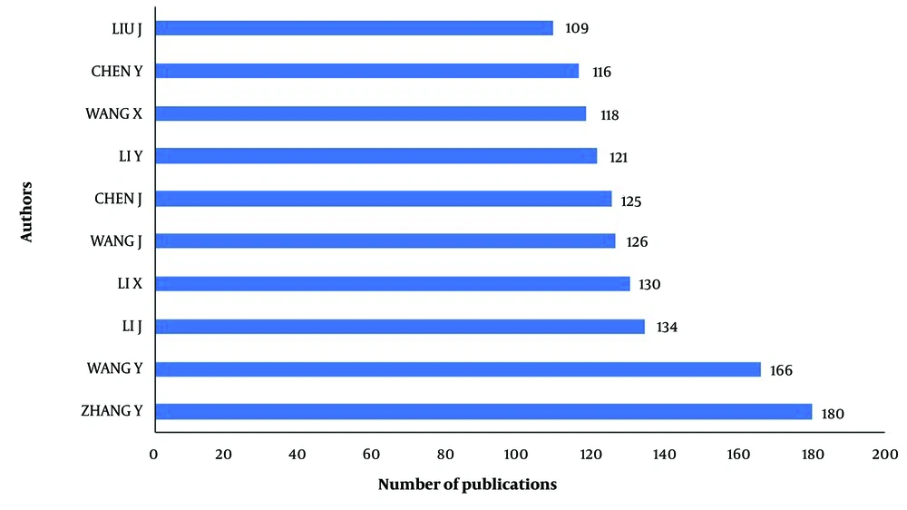
Figure 3.
Most relevant authors based on total publications
3.3. Collaborations and Affiliations
The co-authorship network was used to map collaborative relationships among participating researchers. Using a minimum publication threshold of 5 documents per author, 1233 authors met the inclusion criteria. Of these, 990 were connected to at least 1 other author, forming an integrated co-authorship network (Figure 4A). The remaining authors appeared as isolated nodes without collaborative links. Details regarding the top 5 performing authors within the network are provided in Table 1.
Table 1.
Publications and Citations of the Top 5 Authors in the Network
| Authors | Documents | Citations |
|---|---|---|
| Kudo, Masatoshi | 48 | 5181 |
| Shimose, Shiego | 39 | 735 |
| Niizeki, Takashi | 39 | 735 |
| Iwamoto, Hideki | 38 | 656 |
| Zhou Jian | 36 | 955 |
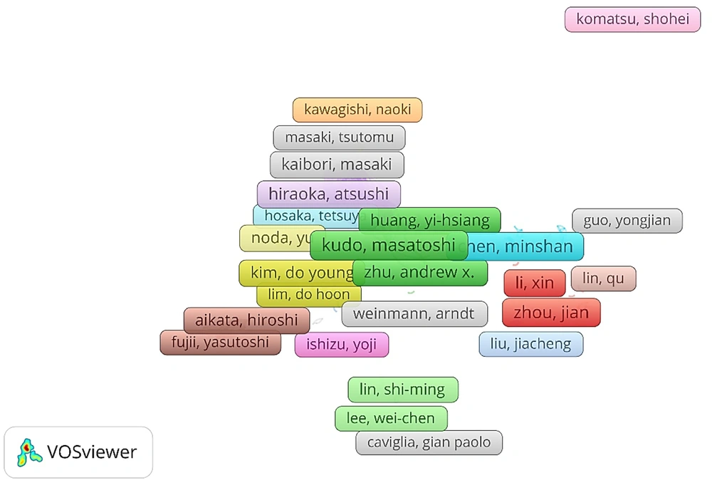
Figure 4.
Co-authorship analysis of authors
3.4. Geographic Distribution of Authors
Country-level participation in the dataset (Figure 5) showed that China contributed the most liver MRI studies (n = 13823), followed by the United States (n = 3176) and South Korea (n = 1374). Other notable contributors included the United Kingdom (n = 388), Spain (n = 271), and Turkey (n = 137). Countries with moderate output included Singapore (n = 112), Sweden (n = 78), Switzerland (n = 74), Thailand (n = 59), and Saudi Arabia (n = 56). These data indicate that both Western and Asian countries are active in this research area, with China and the United States serving as the primary publication hubs.
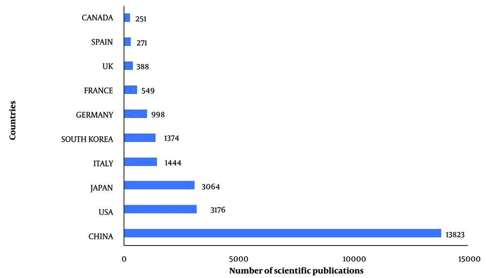
Figure 5.
Productive countries based on related publications
3.5. Prolific Journals and Most Impactful Documents
The most active sources for liver MRI research (Figure 6A) were predominantly fully open-access journals. Frontiers in Oncology (n = 608), Cancers (n = 455), Journal of Hepatocellular Carcinoma (n = 368), and BMC Cancer (n = 289) led the list, all of which offer immediate, unrestricted access. This finding suggests a clear preference for platforms that enhance visibility and dissemination. In contrast, hybrid or subscription-based journals, such as Anticancer Research and Journal of Cancer Research and Therapeutics, ranked lower, suggesting that researchers may prioritize accessibility and speed over conventional publishing models. These data reflect a broader shift toward open, translational publication venues in the field.
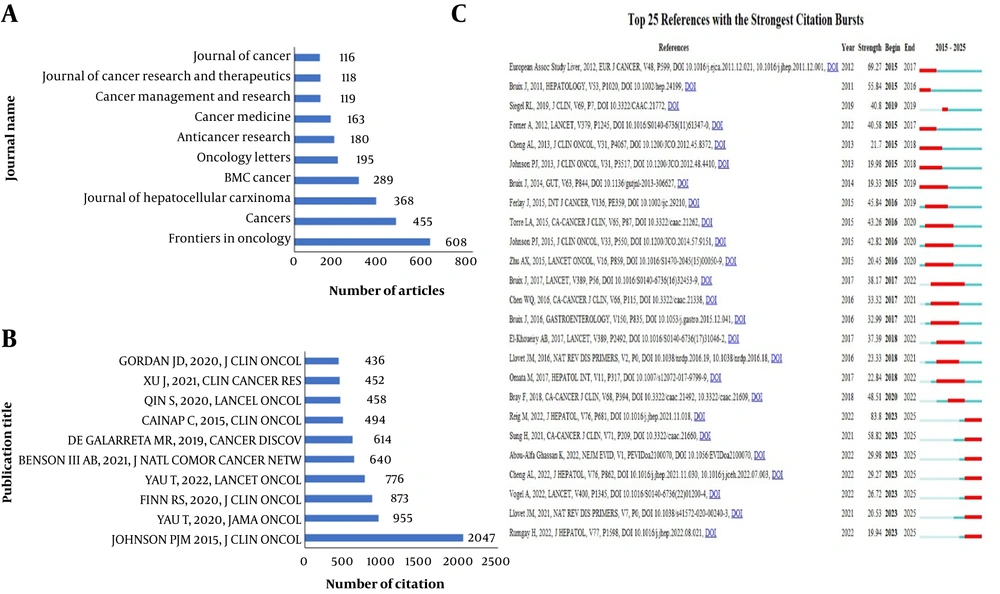
Figure 6.
A, Most active publication sources based on publication count; B, globally cited documents with the highest citation counts, and C, top 25 cited references with the strongest citation bursts over the analysis period, sorted by publication year
The most cited document (Figure 6B) was Johnson et al. (2015, J Clin Oncol) (32), with 2047 citations; this study introduced a prognostic model for liver function assessment. This was followed by Yau et al. (2020, JAMA Oncol) (33), cited 955 times, which focused on dual immunotherapy. Finn et al. (2020, J Clin Oncol) (34) ranked third, with 837 citations, and focused on combined targeted therapy and immunotherapy.
Among the top references with the strongest citation bursts (Figure 6C), Reig et al. (35) ranked first, with a burst strength of 83.80, reflecting the rapid uptake of the updated BCLC strategy in HCC treatment planning. The 2012 EASL-EORTC clinical guidelines, published in both Eur J Cancer and J Hepatol (36, 37), followed with a burst strength of 69.27, representing a foundational contribution to HCC management. Sung et al. (38) ranked third, with a burst strength of 58.82, providing widely cited cancer incidence and mortality estimates relevant to liver cancer epidemiology.
3.6. Most Used Keywords
A Co-occurrence Analysis (Figure 7A) Was Performed To Identify Prominent Research Themes And Terminology Within The Field. Using A Minimum Occurrence Threshold Of 5, 424 Author-provided Keywords Met The Inclusion Criteria. The 5 Most Frequently Used Keywords Are Listed In Table 2
Table 2.
Top 5 Most Used Keywords
| Keywords | Occurrences |
|---|---|
| Hepatocellular carcinoma | 2317 |
| Prognosis | 378 |
| Sorafenib | 303 |
| Transarterial chemoembolization | 288 |
| Lenvatinib | 216 |
Lenvatinib showed the strongest citation burst (Figure 7B) among therapy-related keywords, with a strength of 17.41 from 2023 to 2025, indicating rapidly increasing attention in the most recent period. Atezolizumab plus bevacizumab followed closely, with a burst strength of 15.74 from 2022 to 2025, reflecting sustained interest in combination immunotherapy. Bevacizumab alone also showed a strong burst of 15.54 over the same period. The near-simultaneous citation bursts of lenvatinib, bevacizumab, and atezolizumab plus bevacizumab indicate an ongoing emphasis on systemic therapies, particularly those involving targeted agents and immune checkpoint inhibition, in HCC treatment.
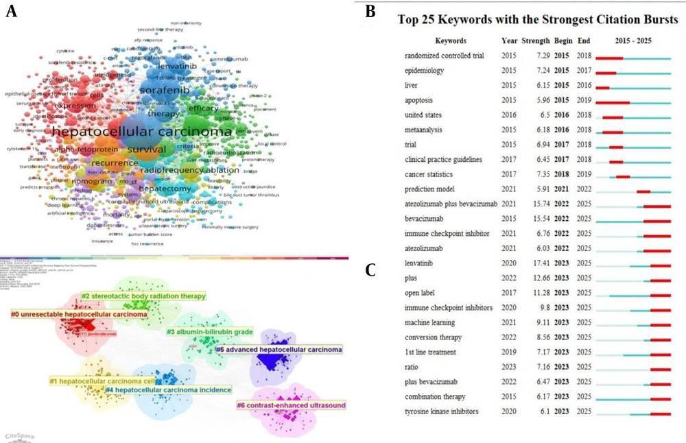
Figure 7.
A, Most frequently occurring keywords based on co-occurrence analysis; B, top 25 keywords with the strongest citation bursts, sorted by publication year; and C, thematic clusters based on keyword burst, indicating emerging trends and research hotspots
A clustering analysis highlighted research trends in the field based on keyword analysis. The clusters indicated a literature landscape centered on HCC, particularly advanced or unresectable forms, with additional focus on imaging and liver function assessment. Among the clusters observed (Figure 7C), Cluster #0 ("unresectable hepatocellular carcinoma") was the largest, indicating concentrated research on non-surgical disease management. Cluster #1 ("hepatocellular carcinoma cell") reflected cellular-level investigations, while Cluster #2 ("stereotactic body radiation therapy") captured interest in localized treatment strategies. Cluster #3 ("albumin-bilirubin grade") and Cluster #4 ("hepatocellular carcinoma incidence") indicated liver function scoring and epidemiological monitoring. Cluster #5 ("advanced hepatocellular carcinoma") emphasized disease progression, and Cluster #6 ("contrast-enhanced ultrasound") highlighted advances in diagnostic imaging.
3.7. Functional Categorization of Key Concepts
The thematic map organized keyword clusters (Figure 8) according to centrality, defined as relevance to the broader field, and density, defined as the degree of internal development. In the upper-right quadrant, terms such as prognosis, resection, and recurrence formed a motor theme; this theme was well developed and highly integrated within the field, reflecting mature areas related to clinical outcomes and disease management. The lower-right quadrant, containing hepatocellular carcinoma, cancer, and survival, represented basic themes: foundational topics that are central but comparatively less specialized. The upper-left quadrant included sorafenib, therapy, and efficacy, indicating niche themes with strong internal coherence but limited cross-field relevance, likely focused on targeted treatment studies. In contrast, expression, metastasis, and cells appeared in the lower-left quadrant as emerging or declining themes, characterized by low development and centrality and possibly reflecting nascent or waning interest in molecular and cellular aspects of disease.
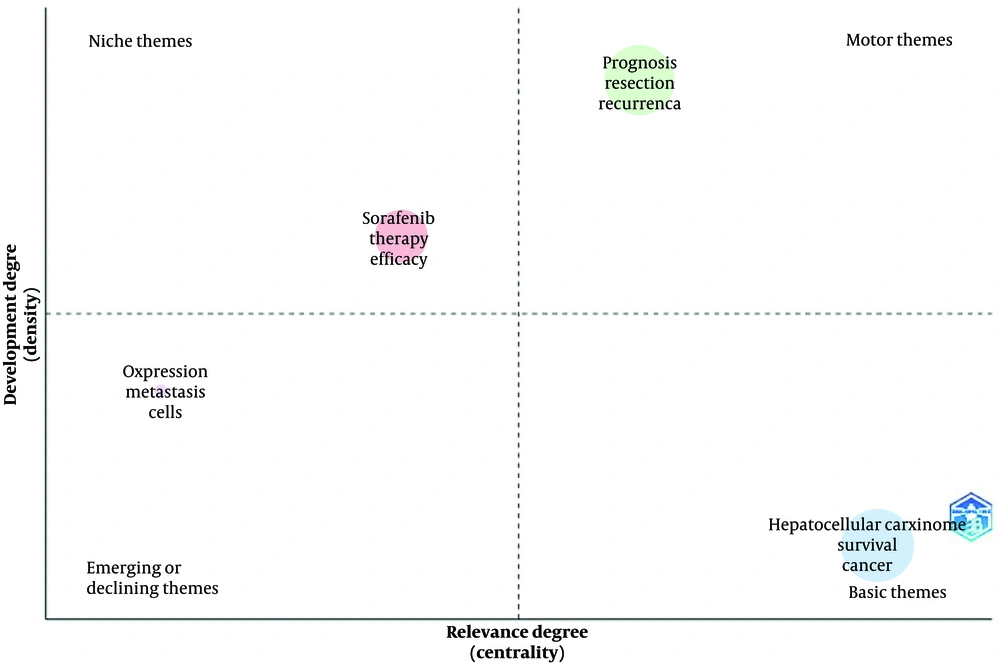
Figure 8.
Thematic map showing major and emerging research topics
4. Conclusions
4.1. Interpretation of Bibliometric Patterns
Research on cell-specific MRI contrast agents in HCC appears to be transitioning from a phase of rapid methodological development to one more strongly focused on clinical application. The bibliometric patterns observed in this study suggest that the field has reached a level of maturity in which existing imaging approaches are increasingly used to address specific clinical questions rather than to introduce entirely new imaging technologies. Although publication output showed a modest decline over the study period (Figure 2), citation activity remained relatively stable, indicating continued scientific interest and sustained clinical relevance. This pattern is frequently observed in medical imaging research once diagnostic technologies become integrated into clinical practice. At that stage, research tends to focus on improving interpretation, refining clinical indications, and expanding the use of imaging in treatment planning.
Authorship patterns further support the interpretation that the field is organized around established research groups. A relatively small number of investigators contributed a large share of the published literature. Authors such as Zhang Y, Wang Y, and Li J accounted for a substantial number of publications (Figure 3), suggesting the presence of stable research networks that continue to influence developments in the MRI-based evaluation of HCC. Although collaboration within research teams was extensive, with more than 10 authors per paper on average, collaboration between countries remained limited, with only 12.65% of publications involving international co-authorship. Geographic distribution patterns showed that research activity was concentrated in several major regions (Figure 5). China produced the largest number of publications, followed by the United States and South Korea, with additional contributions from the United Kingdom, Spain, and Turkey. These findings indicate that MRI research in HCC is globally distributed but still centered in a limited number of highly productive countries. The distribution of publication venues also reflects the applied orientation of the field. High publication volumes in open-access journals, such as Frontiers in Oncology and Cancers, facilitate rapid dissemination of imaging studies, whereas highly influential papers often appear in selective clinical journals, such as the Journal of Clinical Oncology (Figure 6A). Together, these patterns describe a research area characterized by concentrated expertise, strong within-institution collaboration, and sustained clinical interest in MRI-based investigation of HCC.
Over the past decade, certain landmark publications (Figure 6B) have shaped the trajectory of HCC research, influencing both the questions investigators ask and the strategies clinicians adopt. The most widely cited work, Johnson et al. (32), did not introduce a new therapy but instead transformed the way liver function is assessed in HCC. By establishing the albumin-bilirubin (ALBI) grade as an objective and reproducible measure, this study provided a prognostic tool that could be applied across treatment settings and offered a more nuanced alternative to the traditional Child-Pugh classification. Its rapid uptake is reflected not only in its citation count but also in its integration into clinical trial designs and guidelines, underscoring the value of methodological advances in driving the field forward. Therapeutic innovation was equally prominent among the most influential studies. Yau et al. (33) provided compelling evidence that combining checkpoint inhibitors could yield durable clinical benefit in advanced HCC. In the CheckMate 040 cohort evaluating nivolumab plus ipilimumab, the high-dose ipilimumab arm achieved a median overall survival approaching 2 years, an outcome that, at the time, markedly exceeded expectations for patients who had progressed on sorafenib. These results expanded the horizon of immunotherapy in liver cancer and catalyzed further trials exploring dual checkpoint blockade. Finn et al. (34) pursued treatment intensification by combining the antiangiogenic multikinase inhibitor lenvatinib with the programmed death 1 inhibitor pembrolizumab. This phase Ib study reported an objective response rate of 36%, with responses that often lasted beyond 1 year, suggesting a synergistic effect between angiogenesis inhibition and immune modulation. Although early phase, the study anticipated the combinatorial strategies that now dominate first- and second-line therapy trials.
The prominence of treatment-linked keywords (Figure 7A), such as "prognosis," "sorafenib," and "transarterial chemoembolization (TACE)," suggests that MRI-based research in HCC is primarily aligned with postdiagnostic decision-making. Rather than focusing on early detection or surveillance, the literature emphasizes how imaging informs therapeutic selection and response evaluation (39, 40). This pattern aligns with the broader clinical context in which HCC is often diagnosed at intermediate or advanced stages (41, 42), where management decisions depend on treatment eligibility assessment. The relatively high frequency of "prognosis" also indicates that imaging is routinely used to stratify patients based on survival expectations rather than solely for lesion detection or staging (43, 44). This finding reinforces the idea that contrast agent development and administration are shaped not only by technical novelty (26) but also by their perceived utility in guiding interventions such as sorafenib administration or TACE. This keyword profile underscores the alignment of MRI-based research with established treatment planning and prognostic assessment in HCC.
The CiteSpace clustering map (Figure 7C) showed that MRI research in HCC is organized around a small number of clinically centered keywords, reflecting a field more focused on integration than expansion. The prominence of clusters on unresectable (#0) and advanced HCC (#5) highlights the role of MRI in therapeutic decision-making, particularly in guiding transarterial chemoembolization, radiation therapy, or immunotherapy (45-47). The HCC cell (#1) cluster suggests efforts to correlate imaging features with tumor biology, offering noninvasive alternatives to biopsy (48). The stereotactic body radiation therapy (#2) cluster underscores the utility of MRI in treatment planning and posttherapy monitoring, including margin assessment and functional evaluation (49, 50). Functional imaging is also linked to biochemical stratification, as seen in the albumin-bilirubin grade (#3) cluster, which reflects efforts to align hepatobiliary contrast uptake with serum-based prognostic models (51). The HCC incidence (#4) cluster links MRI use with broader epidemiological trends, including risk from hepatitis viruses, alcohol, and nonalcoholic fatty liver disease, supporting its role in surveillance (52-54). Finally, the contrast-enhanced ultrasound (#6) cluster suggests a comparative focus across modalities, such as contrast-enhanced ultrasound and MRI (55, 56). The identified clusters capture both dominant clinical themes and specific diagnostic or prognostic applications of MRI reported in the literature.
The thematic map (Figure 8) showed that MRI research in HCC is concentrated around clinical topics, with limited discussion of experimental approaches. Core clinical themes, such as prognosis, resection, and recurrence, occupied the motor quadrant, highlighting the central role of MRI in surgical planning and outcome prediction. These areas were well developed and structurally important, supported by recent studies linking imaging features to recurrence risk after resection (57, 58). The basic themes quadrant, containing terms such as hepatocellular carcinoma, survival, and cancer, anchored the field's identity. These themes represent stable, general-purpose research areas that underpin much of the literature (59, 60). In contrast, niche themes, such as sorafenib, therapy, and efficacy, suggest ongoing efforts to use imaging for treatment monitoring, particularly for systemic therapies such as tyrosine kinase inhibitors (61). However, their peripheral position reflects limited integration with mainstream research. Emerging or declining themes, such as expression, metastasis, and cells, remain weakly connected. These themes point to preliminary efforts to link MRI with tumor biology or early spread, but current studies are fragmented and exploratory (62). The thematic structure highlights well-developed clinical topics alongside less frequently addressed molecular and cellular investigations.
Our bibliometric mapping, derived from a deliberately broad literature search, showed that even the most comprehensive dataset available on this topic still leaves important gaps.
Future studies should link MRI features to genomic profiles, immune cell infiltration, and tumor microenvironment characteristics, and incorporate radiogenomic and radiomic analyses into prospective, multi-institutional designs (46, 63-65). Although MRI research in HCC is well established in staging, treatment planning, and liver function assessment, it remains underrepresented in areas aligned with current molecular and immunologic treatment strategies. Standardized acquisition protocols for functional measures, such as apparent diffusion coefficients and dynamic contrast-enhanced kinetics, would improve reproducibility and enable pooled analyses. Integration with histopathology, circulating biomarkers, and complementary imaging modalities could facilitate composite biomarkers that capture disease biology and predict therapeutic response. Dynamic imaging approaches capable of monitoring biological change over the course of treatment should also be prioritized to align imaging endpoints with evolving systemic therapies (66-69).
4.2. Limitations
Even with a broad search strategy, this study has inherent limitations. The bibliometric results indicate a field that is thematically narrow and structurally conservative. High-centrality keywords and major clusters focus on prognosis, resection, recurrence, and liver function assessment, whereas molecular and immunologic themes appear only at the margins. Studies integrating MRI with genomic or immunologic data are rare, and biologically driven imaging approaches remain peripheral to the mainstream literature. Most studies are retrospective, single-center, and concentrated within small authorship networks, with limited international collaboration. Quantitative parameters are reported inconsistently, with no clear adoption of standardized acquisition or analysis methods. Multimodal integration is limited, and comparative work with computed tomography or contrast-enhanced ultrasound is often framed as isolated superiority testing rather than as part of coordinated diagnostic workflows. Addressing these gaps will require expanded thematic coverage, broader collaboration networks, and greater standardization of imaging protocols across studies.
4.3. Summary Conclusion
This analysis shows that MRI research in HCC continues to focus on well-established clinical roles, such as staging, treatment planning, and liver function assessment. However, it has not yet kept pace with the shift toward treatment strategies guided by molecular and immunologic profiling. To remain relevant in this changing landscape, future work should link imaging findings with biological markers of disease, apply standardized quantitative methods, and position MRI as part of integrated predictive workflows that can guide personalized care.