
1. Background
Hashimoto’s thyroiditis (HT), also known as chronic autoimmune thyroiditis, is the most common cause of hypothyroidism in developed countries (1-6). The disease is characterized by gradual destruction of thyroid follicular cells mediated by antibody- and T cell-dependent immune mechanisms and is commonly associated with lymphocytic infiltration of the thyroid parenchyma (7, 8). Initially described by Haruto Hashimoto in 1912 as “struma lymphomatosa,” HT may demonstrate histopathological changes such as follicular epithelial dysplasia (FED). FED is defined by architectural and cytological atypia within lymphocyte-rich regions of the thyroid and is increasingly regarded as a potential premalignant alteration within the spectrum of autoimmune thyroid disease (9, 10). Papillary thyroid carcinoma (PTC), the most frequent thyroid malignancy, accounts for approximately 80% of thyroid cancers (11). Originating from follicular cells, PTC is characterized by distinctive nuclear features and, although usually indolent, can metastasize via lymphatic pathways and, less frequently, to distant organs. Evidence from several studies suggests that chronic inflammation associated with HT may promote thyroid tumorigenesis, and a higher incidence of PTC has been reported among patients with HT (10, 12).
Cyclin D1 plays a fundamental role in regulating the transition from the G1 phase to the S phase of the cell cycle and has been shown to be overexpressed in various cancers, including breast, esophageal, and thyroid malignancies (13). In PTC, increased Cyclin D1 expression is commonly observed and may contribute to dysregulated cellular proliferation (13, 14). Nevertheless, the expression profile of Cyclin D1 in FED, particularly when arising in the background of HT, has not been fully elucidated.
Recent immunohistochemical evidence suggests that Cyclin D1 expression may be stronger in FED lesions associated with malignancy than in isolated FED lesions (12). However, comparative investigations of Cyclin D1 expression in FED and PTC in the background of HT remain limited, particularly in regional populations.
2. Objectives
This study aimed to evaluate and compare Cyclin D1 protein expression in patients with PTC and FED associated with Hashimoto’s thyroiditis. Identifying meaningful differences may support the role of Cyclin D1 as a predictive biomarker of malignancy in autoimmune-inflamed thyroid tissue.
3. Methods
3.1. Sample Selection and Procedure
This retrospective cohort study was conducted on thyroidectomy specimens with a histopathological diagnosis of Hashimoto’s thyroiditis archived in the pathology laboratory of Shahid Sadoughi Hospital, Yazd, Iran, from 2014 to 2023. A census sampling approach was used; therefore, all eligible cases referred during this period were included. After ethics committee approval, patients’ medical records were retrieved and reviewed. All thyroidectomy specimens with a confirmed histopathological diagnosis of Hashimoto’s thyroiditis during the study period were identified and considered for initial assessment.
Each case was independently re-evaluated on hematoxylin and eosin (H&E)-stained sections by 2 experienced pathologists to confirm the diagnosis of underlying Hashimoto’s thyroiditis and to determine the presence of either PTC or FED. Cases with discordant interpretations were reviewed jointly using a multihead microscope until diagnostic agreement was reached. Specimens showing concurrent PTC and FED, as well as those without evidence of either lesion, were excluded from the cohort.
The inclusion criteria comprised thyroidectomy specimens showing histopathological evidence of Hashimoto’s thyroiditis in association with either PTC or FED. FED was defined as foci smaller than 1 cm composed of atypical follicular epithelial cells with mild to moderate nuclear enlargement, irregular nuclear membranes, chromatin margination, nuclear grooves, nuclear crowding, and variable optical clearing, arranged in follicular, trabecular, nested, or solid patterns without papillary formation, infiltrative growth, or stromal desmoplasia. By contrast, PTC was diagnosed when unequivocal PTC-type nuclear features were present, including diffuse nuclear enlargement, irregular nuclear contours, chromatin clearing, nuclear grooves, and intranuclear pseudoinclusions, together with characteristic papillary architecture and/or infiltrative growth. Cases meeting the established histopathological criteria for PTC were classified as PTC rather than FED (15).
Exclusion criteria included incomplete clinical information, inability to obtain adequate follow-up data, poor-quality histological slides, insufficient tissue for Cyclin D1 immunohistochemical staining, synchronous malignancies, and coexistence of FED and PTC in the same thyroid specimen.
Initially, 450 thyroidectomy specimens with histopathologically confirmed Hashimoto’s thyroiditis were identified. After applying the inclusion and exclusion criteria, 340 cases were excluded: 302 cases without either PTC or FED, 30 cases with incomplete clinicopathological information, and 8 cases with concomitant PTC and FED. Finally, 110 eligible cases remained for analysis, including 81 PTC-associated cases and 29 FED cases. No diagnostically ambiguous, equivocal, or overlapping cases were identified during histopathological reassessment.
3.2. Cyclin D1 Immunohistochemical Staining
After selection of the histopathological slides, 4-µm sections were cut from the corresponding FFPE tissue blocks of each specimen and mounted on poly-L-lysine-coated adhesive slides. Immunohistochemical staining was then performed using the streptavidin-biotin horseradish peroxidase detection method with the Immunoperoxidase secondary detection system, according to the manufacturer’s protocol.
Tissue sections were deparaffinized by heating at 60°C, cleared with xylene, and rehydrated through graded ethanol concentrations. Antigen retrieval was performed in citrate buffer (0.01 mol/L, pH 6.8) using microwave heating, followed by rinsing in phosphate-buffered saline and inhibition of endogenous peroxidase activity with 3% hydrogen peroxide in methanol.
For immunohistochemical analysis, sections were incubated overnight at 4°C with Cyclin D1 primary antibody (clone FP12; Dako) diluted 1:50. After washing, the slides were incubated with biotinylated goat anti-mouse immunoglobulin and streptavidin-peroxidase conjugate. The reaction was visualized with 3,3'-diaminobenzidine and hydrogen peroxide, followed by hematoxylin counterstaining, dehydration, and mounting.
Positive and negative controls were included in every staining run, with mantle cell lymphoma serving as the positive control and follicular lymphoma as the negative control.
Cyclin D1-immunostained sections were examined using an Olympus CX23 microscope (Japan). Immunostained cells were initially identified throughout the PTC or FED area at ×40 or ×100 magnification and subsequently confirmed at ×200 or ×400 magnification. A clearly granular brown nuclear staining pattern was considered positive. Only nuclear staining in at least 5% of tumor cell nuclei was considered positive. Cyclin D1-stained slides were evaluated independently by 2 experienced pathologists using predefined criteria. During immunohistochemical assessment, the pathologists were blinded to the histopathological diagnosis and clinicopathological data. When discrepancies occurred, a joint review was performed, and final consensus was reached by mutual agreement.
3.3. Clinicopathological Variables and Follow-Up
Demographic and clinicopathological variables, including age, sex, and lymph node involvement, were obtained from the pathology records. Follow-up information was collected uniformly from included patients through telephone interviews conducted at the end of the study period. In patients with PTC, follow-up questions addressed survival status and any self-reported history of recurrence. In patients with FED, follow-up was limited to newly diagnosed thyroid-related clinical problems. Persistent disease, biochemical surveillance, imaging results, and disease-specific survival were not assessed systematically. Because admission and surgery dates were not consistently recorded in the archived files, the exact follow-up duration and range could not be calculated.
3.4. Ethical Statement
The study protocol complied with the ethical principles of the Declaration of Helsinki. Approval was granted by the Ethics Committee of Shahid Sadoughi University under approval number IR.SSU.MEDICINE.REC.1403.044. Owing to the retrospective design, informed consent was waived.
3.5. Statistical Analysis
Statistical analyses were performed using SPSS version 26 (IBM Corp., Armonk, NY, USA). Continuous variables were reported as mean ± standard deviation (SD). The normality of the age distribution was assessed using the Shapiro-Wilk test, and age was compared between groups using the independent-samples t-test. Categorical variables were summarized as frequencies and percentages and compared using the chi-square test or Fisher exact test when expected cell counts were low. Because 1 cell in the Cyclin D1 contingency table had a zero count, the Fisher exact test was used to compare Cyclin D1 expression between the PTC and FED groups. The effect size for the primary Cyclin D1 comparison was expressed as the absolute difference in positivity rates with 95% confidence intervals (CIs). Inferential statistical testing was not performed for follow-up outcomes because all patients had identical follow-up status and no between-group variability was present. A 2-sided P value < 0.05 was considered statistically significant.
4. Results
4.1. Cohort Characteristics
Of the 450 thyroidectomy specimens diagnosed with Hashimoto’s thyroiditis, 110 cases met the eligibility criteria and were included in the final analysis. Overall, 340 cases were excluded, including 302 cases without either PTC or FED, 30 cases with incomplete clinicopathological data, and 8 cases demonstrating concurrent PTC and FED. The final cohort comprised 81 patients (73.6%) in the PTC group and 29 patients (26.4%) in the FED group. No diagnostically equivocal or overlapping cases were identified during histopathological review.
The mean age was 37.64 ± 12.16 years in the PTC group and 41.86 ± 12.02 years in the FED group. The age distribution was approximately normal, and comparison using the independent-samples t-test showed no statistically significant difference between the groups (P = 0.111). Men comprised 13.6% of patients with PTC and 13.8% of patients with FED, whereas women accounted for 86.4% and 86.2%, respectively. Sex distribution did not differ significantly between the 2 groups (P = 0.977).
4.2. Cyclin D1 Expression
Table 1 shows the distribution of Cyclin D1 expression across the 2 groups.
Table 1.
Frequency Distribution of Cyclin D1 in the Two Mutually Exclusive Groups a
| Cyclin D1 | PTC | FED | Total | P-Value |
|---|---|---|---|---|
| Negative | 0 (0) | 4 (13.8) | 4 (3.6) | 0.001 |
| Positive | 81 (100) | 25 (86.2) | 106 (96.4) | 0.001 |
| Total | 81 (100) | 29 (100) | 110 (100) | 0.001 |
a Values are expressed as No. (%).
As presented in Table 1, Fisher exact test showed a significant difference in Cyclin D1 expression between the 2 mutually exclusive groups (P = 0.001). Cyclin D1 positivity was detected in 100% of PTC cases and 86.2% of FED cases, yielding an absolute difference of 13.8% (95% CI, 1.3% - 26.3%). Table 2 displays the frequency distribution of lymph node metastasis among patients with PTC.
Table 2.
Frequency Distribution of Metastasis in Patients with PTC a
| Metastasis | PTC |
|---|---|
| No | 58 (71.6) |
| Yes | 23 (28.4) |
| Total | 81 (100) |
a Values are expressed as No. (%).
4.3. Lymph Node Metastasis
As indicated in Table 2, metastasis was detected in 28.4% of patients with PTC.
4.4. Telephone Follow-Up and Histopathological Figures
Table 3 summarizes the findings obtained through telephone follow-up.
Table 3.
Telephone Follow-Up Findings in Patients with PTC and FED a
| Follow-up Finding | PTC | FED | Total |
|---|---|---|---|
| Alive at follow-up | 81 (100) | 29 (100) | 110 (100) |
| Reported recurrence/new thyroid-related disease | 0 (0) | 0 (0) | 0 (0) |
a Values are expressed as No. (%).
At the time of contact, all patients were reported to be alive. No patient with PTC reported recurrence, and no patient with FED reported a newly diagnosed thyroid-related clinical problem. However, persistent disease, biochemical follow-up, and imaging findings were not assessed systematically.
Positive Cyclin D1 staining in papillary thyroid carcinoma is illustrated in Figure 1. Classic papillary thyroid carcinoma (H&E stain, ×250) is shown in Figure 2. Follicular epithelial dysplasia in the background of Hashimoto thyroiditis (H&E stain, ×250) is demonstrated in Figure 3. Figure 4 depicts Cyclin D1 expression in FED with positive nuclear staining (IHC, ×400).
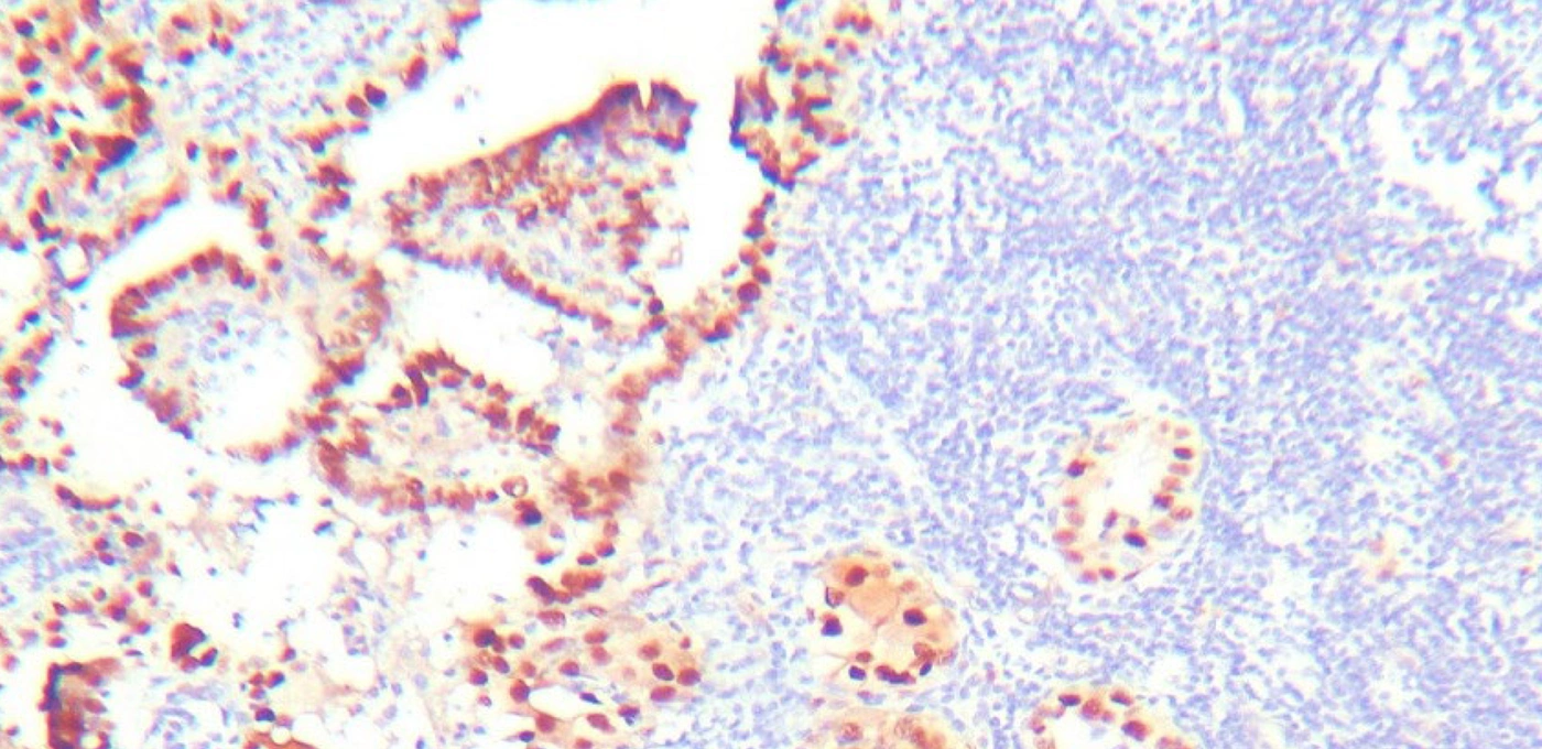
Figure 1.
Positive Cyclin D1 staining in papillary thyroid carcinoma.
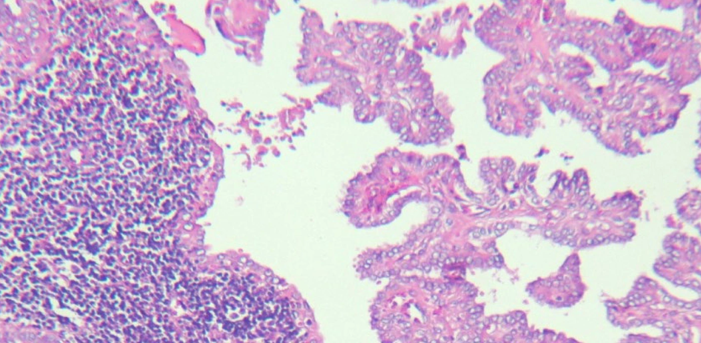
Figure 2.
Classic papillary thyroid carcinoma (H&E stain, ×250).
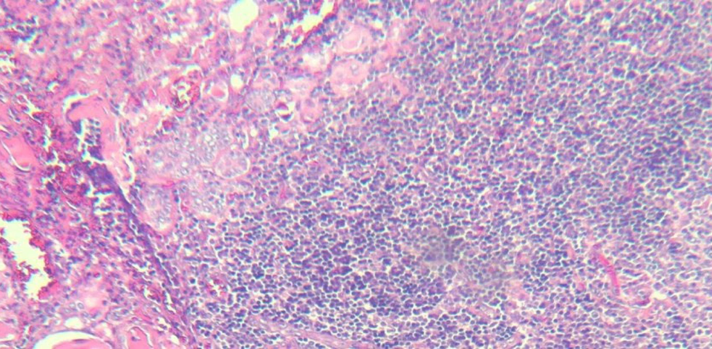
Figure 3.
Follicular epithelial dysplasia in the background of Hashimoto thyroiditis (H&E stain, ×250).
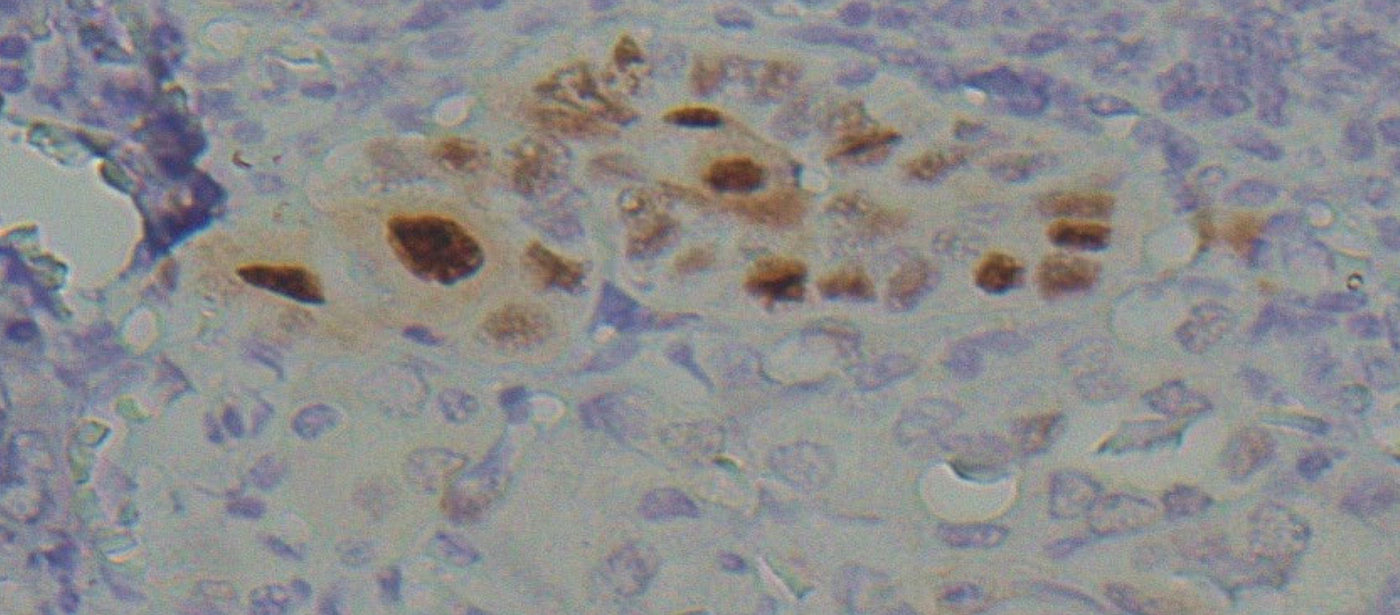
Figure 4.
Cyclin D1 expression in FED with positive nuclear staining (IHC, ×400).
5. Discussion
The present study evaluated 110 patients, including 81 diagnosed with PTC and 29 diagnosed with FED. Assessment of age and sex distributions revealed no statistically significant differences between the groups, indicating that the two groups were demographically comparable.
Cyclin D1 expression was examined in both groups. A statistically significant difference in Cyclin D1 expression was observed between patients with PTC and those with FED. Cyclin D1 positivity was observed in 100% of PTC cases compared with 86.2% of FED cases, suggesting that these two lesions exhibit distinct expression profiles. Nevertheless, because of the inherent constraints of the retrospective cohort design and the absence of standardized longitudinal follow-up, the present findings cannot establish whether FED functions as a precursor lesion that later develops into PTC.
These results are consistent with the study by Chui et al., who investigated Cyclin D1 expression in PTC and FED and reported higher positivity in PTC than in FED (15). Conversely, Seybt et al. found no significant association between Cyclin D1 expression and PTC; this discrepancy may be explained by differences in study methods or the demographic characteristics of the populations examined (16).
Although Gvianishvili et al., in a Georgian cohort, concluded that Cyclin D1 expression alone is not a dependable predictor of malignancy in patients with HT (17), most available studies have indicated a strong relationship between Cyclin D1 expression and PTC. Nevertheless, additional research is necessary to define its precise contribution to tumor progression and to determine whether it can be used as a reliable biomarker in the management of PTC.
Kholová et al. documented Cyclin D1 expression in 67.5% of FED cases, a frequency lower than that found in the current study. This variation may be related to differences in study populations, methodological approaches, or laboratory sensitivity. It may also indicate underlying clinical or genetic heterogeneity of FED across different patient cohorts (10).
Furthermore, the present findings showed lymph node metastasis in 28.4% of PTC cases. This observation indicates that metastasis is an important feature of PTC and may be linked to its specific biological and pathological mechanisms.
Toraih et al. performed a large-scale investigation involving 89,694 patients with PTC over a 21-year period and reported metastasis in nearly 42% of cases (18). Likewise, Tang et al. observed central lymph node metastasis in 67.8% of patients with PTC (19). These reports highlight variability in metastatic rates among patients with PTC. By comparison, the current study identified a metastasis rate of 28.4% among PTC cases, reflecting a relatively lower prevalence. Such differences may stem from variations in methodology, sample size, patient demographics, or study duration. The lower metastasis rate in the present cohort may also be associated with population-specific characteristics.
In a 2020 systematic review from China, Mao et al. reported several major predictors of lymph node metastasis in PTC, including age younger than 45 years, male sex, multifocality, tumor size greater than 1 cm, tumor location, capsular invasion, and extrathyroidal extension. Conversely, bilateral tumor involvement and Hashimoto’s thyroiditis were not significantly associated with lymph node metastasis (20).
In this study, follow-up information was collected through telephone interviews. For patients with PTC, the follow-up assessment addressed survival status and history of recurrence. For patients with FED, follow-up was restricted to survival status and questions regarding subsequent thyroid-related clinical problems. At follow-up, all patients in both groups were reported to be alive. However, because outcome assessment was not conducted according to a standardized protocol and information on persistent disease, biochemical markers, and imaging findings was not systematically obtained, the prognostic value of these observations remains limited. Therefore, further prospective studies are required to more clearly define the clinical significance and biological behavior of FED and its relationship with PTC.
Grogan et al. evaluated mortality during long-term follow-up of patients with PTC and reported that disease-specific deaths may occur many years after treatment, underscoring the need for lifelong surveillance (21). Although the short-term prognosis in the present study appears favorable, the possibility of recurrence or delayed complications may persist.
Dong et al. carried out a 10-year investigation including 466 patients with PTC and reported mortality rates of 2.1% during 10 - 20 years and 0.85% during 20 - 30 years (22). Compared with the present findings, in which all patients were alive and free of reported recurrence at follow-up, these results emphasize that long-term risks may remain despite favorable early outcomes. These observations further support the importance of continued follow-up even after apparent clinical resolution.
5.1. Conclusions
In this study, Cyclin D1 expression differed significantly between PTC and FED developing in the background of HT. Cyclin D1 positivity was identified in all PTC cases, whereas a lower frequency was observed among FED cases. These results suggest distinct Cyclin D1 expression patterns in PTC and FED. However, because of the retrospective cohort design and the absence of standardized longitudinal follow-up data, it cannot be determined whether FED represents a precursor lesion that progresses to PTC. Although some investigators have proposed that FED may be a potential premalignant lesion in the setting of HT, this possibility remains hypothetical and cannot be confirmed from the current findings. Further studies with larger sample sizes and extended long-term follow-up are needed to clarify the potential clinicopathological relevance of Cyclin D1 expression in these lesions.
5.2. Limitations
A major limitation of this retrospective cohort study is the lack of standardized long-term longitudinal follow-up. Therefore, although Cyclin D1 expression differed between PTC and FED, the present study cannot determine whether FED acts as a precursor lesion that progresses to PTC. Moreover, only short-term clinical status was assessed, whereas a comprehensive evaluation of disease outcomes requires long-term follow-up and analysis of long-term clinical endpoints. Another limitation is that Cyclin D1 was evaluated as the only biological marker, although other molecular and clinicopathological factors may also influence the biological behavior of these lesions. In addition, treatment-related adverse effects and their possible impact on patients’ quality of life were not investigated.
Finally, the relatively limited sample size, particularly in the FED subgroup, may have reduced statistical power and limited the generalizability of the findings. Larger multicenter studies incorporating standardized long-term follow-up and comprehensive molecular profiling are required to more accurately define the biological significance of FED and its potential association with PTC.