
1. Background
Triple-negative breast cancer (TNBC) is a distinct molecular subtype of breast cancer defined by the absence of estrogen receptor (ER), progesterone receptor (PR), and HER2 expression (1-3). TNBC accounts for nearly 10% - 20% of all breast cancer cases and is characterized by aggressive clinical behavior and a poorer prognosis than other breast cancer subtypes (1, 4). Patients with TNBC often present with high-grade tumors, rapid tumor growth, early recurrence, and a higher risk of distant metastasis (1, 4, 5). TNBC is also more common in younger women and is frequently diagnosed at advanced stages.
Despite major progress in breast cancer treatment, TNBC remains difficult to treat. Unlike other breast cancer subtypes, TNBC does not respond to hormonal therapy or HER2-targeted therapy because these receptors are absent (1, 3). Therefore, chemotherapy remains the main treatment option. However, patients with TNBC exhibit variable treatment responses: some respond well, whereas others experience early relapse and poor survival despite treatment (1, 6). This heterogeneity in clinical behavior highlights the need for reliable biomarkers to predict prognosis and guide patient management (2, 6).
Several biomarkers have been studied in TNBC to better characterize tumor behavior and predict five-year mortality. Markers such as Ki-67, EGFR, p53, androgen receptor, and basal cytokeratins have shown potential prognostic value in previous studies (7). However, the results remain inconsistent, and no single marker has demonstrated sufficient accuracy for routine clinical use. Although gene expression profiling has provided important insights into this heterogeneity, it is still rarely used in routine clinical workflows because of cost and accessibility limitations (8). Consequently, pathologists continue to rely heavily on immunohistochemistry-based markers for practical classification.
Cytokeratin 5/6 (CK5/6) is a basal-type intermediate filament protein expressed in epithelial cells and is commonly used as an immunohistochemical marker in breast cancer pathology (1, 3, 5). CK5/6 is frequently associated with the basal-like subtype of breast cancer, which shares several clinical and pathological features with TNBC. Previous studies have investigated the relationship between CK5/6 expression and clinicopathological characteristics in patients with TNBC. Some studies have reported associations between CK5/6 expression and tumor grade, stage, recurrence, and five-year mortality (1, 2, 6). Accordingly, CK5/6 has gained attention as a potential prognostic and classification marker in TNBC because of its possible role in tumor classification and prognosis. Despite growing interest in CK5/6, its relationship with mortality in patients with TNBC requires further evaluation. In addition, data on CK5/6 expression in patients with TNBC across different populations remain limited.
2. Objectives
The present study aimed to evaluate CK5/6 expression and examine its association with clinicopathological features and five-year mortality in patients with TNBC.
3. Methods
3.1. Study Design and Participants
This retrospective longitudinal study was conducted in 2021 among patients with TNBC diagnosed in the Department of Pathology at Shahid Sadoughi Hospital between 2011 and 2016. Sampling was performed using a census method, and all available TNBC cases during the study period were screened for eligibility. All eligible TNBC cases diagnosed during the study period were consecutively included using the census sampling method, and no additional sampling procedures were performed.
The inclusion criteria were histopathologically confirmed TNBC diagnosed between 2011 and 2016; availability of formalin-fixed, paraffin-embedded tissue blocks adequate for immunohistochemical evaluation of CK5/6; complete pathological records; and available five-year follow-up information obtained through medical records or telephone contact. The exclusion criteria were the absence of a TNBC diagnosis, incomplete pathological or clinical data, inadequate tissue samples for immunohistochemical analysis, and inability to determine five-year mortality status during follow-up.
3.2. Clinical Treatment
All patients underwent surgical treatment and received a similar chemotherapy regimen. The chemotherapy protocol included doxorubicin (60 mg/m2) and cyclophosphamide (600 mg/m2) every 21 days for four cycles, followed by taxane-based therapy for four additional cycles. Taxane therapy was administered as either weekly paclitaxel (85 mg/m2 for 12 cycles) or docetaxel plus carboplatin (AUC 6 every three weeks). Selection of paclitaxel versus docetaxel plus carboplatin was based on routine clinical decision-making and oncologist preference during the treatment period. Detailed comparative treatment data by CK5/6 expression or tumor stage were not consistently available for subgroup analysis.
3.3. Assessment of ER, PR, and HER2 Expression
ER, PR, and HER2 expression at initial diagnosis was assessed uniformly in all cases using immunohistochemistry (IHC). For ER and PR evaluation, moderate to strong nuclear staining in ≥ 1% of tumor cells was considered positive. HER2 positivity was defined as complete and intense membranous staining in more than 10% of tumor cells (3+ score), according to standard diagnostic criteria. All slides were reviewed by two experienced pathologists. Tumor stage was determined according to the American Joint Committee on Cancer (AJCC) staging system used at the time of diagnosis and was recorded based on pathology reports available in the medical records.
3.4. Immunohistochemical Evaluation of CK5/6
Formalin-fixed, paraffin-embedded tissue samples were retrieved from the pathology archives. Immunohistochemical staining was performed on 4-µm tissue sections using the streptavidin-biotin-peroxidase method. Tissue sections were deparaffinized in xylene and rehydrated with graded ethanol. Endogenous peroxidase activity was blocked using hydrogen peroxide for 10 minutes.
Antigen retrieval was performed in EDTA buffer (pH 9.0) using microwave heating for 20 minutes. After washing with phosphate-buffered saline (PBS), the slides were incubated for 60 minutes at room temperature with a ready-to-use monoclonal anti-cytokeratin 5/6 (EP24+EP67) antibody (Master Diagnostica, Spain). The sections were subsequently incubated with a polymer-based peroxidase detection system containing DAB chromogen (Master Polymer Plus Detection System; Master Diagnostica, Spain). The slides were then counterstained with hematoxylin.
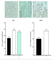
CK5/6 expression was identified by cytoplasmic and/or membranous brown staining. The CK5/6 staining index (range 0 - 9) was calculated by multiplying staining intensity by the percentage of stained tumor cells. Staining intensity was scored as negative (score 0), weak (score 1), moderate (score 2), or strong (score 3). Cases with no detectable staining were assigned a final score of 0. In cases showing any positive staining, the percentage of positive tumor cells was scored as < 10% (score 1), 10% - 50% (score 2), or ≥ 50% (score 3), and the staining index was calculated by multiplying the intensity and percentage scores. CK5/6 expression was considered negative when the final score was 0 and positive when the final score ranged from 1 to 9 (1). Slide evaluation was performed independently by two pathologists who were blinded to patient outcomes.
3.5. Mortality Rate Assessment
Follow-up was calculated from the date of pathological diagnosis to the date of death, the last clinical visit, or the last telephone contact. The primary outcome was five-year all-cause mortality status. Survival information was obtained from hospital medical records and telephone follow-up with patients or their relatives. For some deceased patients, exact dates of death were not available in the medical records, and approximate dates reported by relatives during telephone follow-up were recorded. Because the exact timing of death could not be reliably confirmed in all cases, detailed time-to-event analyses, such as Kaplan-Meier or Cox proportional hazards analyses, were not performed. Data regarding recurrence, disease-free survival, distant metastasis during follow-up, and breast cancer-specific mortality were not consistently available and therefore were not included in the analysis.
3.6. Statistical Analysis
Data were analyzed using IBM SPSS Statistics version 26. Continuous variables were presented as mean ± standard deviation (SD), and categorical variables were expressed as frequency and percentage. Comparisons between categorical variables were performed using the chi-square test or Fisher exact test, as appropriate. An independent-samples t-test was used to compare continuous variables between groups. Univariate logistic regression analysis was performed to evaluate the association between clinicopathological variables and five-year mortality. Variables analyzed included age, lymph node involvement, tumor grade, tumor stage, CK5/6 expression, and CK5/6 staining index. Lymph node involvement was analyzed as a binary variable (involved vs not involved) because detailed nodal burden and lymph node ratio data were not consistently available in the medical records. Odds ratios (ORs) and 95% confidence intervals (CIs) were calculated. Variables with clinical relevance and significant associations in univariate analysis were entered into the multivariate logistic regression model. Because only 10 mortality events were observed in the study population, the multivariable model was intentionally limited to three predictors: age, tumor stage, and CK5/6 expression. This restriction was intended to minimize the risk of model overfitting, in line with commonly recommended guidelines regarding the number of outcome events per predictor variable. However, the small number of events resulted in approximately 3.3 events per predictor, which is below conventional recommendations. Therefore, the adjusted model was considered exploratory and should be interpreted with caution.
A P value < 0.05 was considered statistically significant.
4. Results
4.1. Patient Characteristics and Clinicopathological Findings
A total of 67 medical records of patients with TNBC diagnosed between 2011 and 2016 were initially screened for eligibility. Of the 17 excluded patients, reasons for exclusion included inadequate tissue for immunohistochemical evaluation (n = 5), incomplete pathological or clinical records (n = 6), unavailable five-year follow-up information, or combinations of these factors (n = 6). Comparisons between included and excluded patients were performed using the available baseline data for each variable. No statistically significant differences were observed in the available demographic or clinicopathological characteristics, including age, tumor grade, tumor stage, and lymph node involvement (all P > 0.05). Because missing information varied across excluded patients, the number of observations differed by variable. Ultimately, 50 patients were included in the final analysis.
At the five-year follow-up, 10 (20%) patients had died and 40 (80%) were alive. The mean age of the study population was 48.16 ± 8.97 years. Patients who died had a mean age of 51.5 ± 12.3 years, whereas survivors had a mean age of 47.3 ± 7.8 years. This difference was not statistically significant (P = 0.33). The most common age group in the overall population was older than 50 years (32%), followed by 45 - 50 years (26%). Among patients who died, 60% were older than 50 years, whereas among survivors, the most frequent age group was 45 - 50 years (30%). No significant association was observed between age group and five-year mortality (P = 0.08).
Lymph node involvement was present in 13 (26%) patients, whereas 37 (74%) had no lymph node involvement. Lymph node involvement was observed in 10% of patients who died and 30% of survivors, with no significant difference between the groups (P = 0.19). Regarding tumor grade, 16 (32%) patients had grade 2 tumors and 34 (68%) had grade 3 tumors. Grade 3 tumors were found in 60% of patients who died and 70% of survivors. Tumor grade was not significantly associated with five-year mortality (P = 0.54).
Tumor stage was significantly associated with five-year mortality (P < 0.001). Stage 2 disease was the most common stage overall and was observed in 27 (54%) patients. Among patients who died, stage 3 disease was the most frequent stage (50%), whereas among survivors, stage 2 disease was the most common stage (65%). Stage 4 disease was present in 30% of patients who died compared with 5% of survivors.
4.2. CK5/6 Expression and Five-Year Mortality
CK5/6 expression was positive in 45 (90%) patients and negative in 5 (10%). Negative CK5/6 expression was observed in 30% of patients who died and 5% of survivors. CK5/6 expression was significantly associated with five-year mortality (P = 0.01). The distribution of the CK5/6 staining index did not differ significantly between patients who died and those who survived (P = 0.17). CK5/6 index score 2 was the most common score and was observed in 14 (28%) patients. Scores of 3, 4, and 6 were each observed in 8 (16%) patients.
4.3. Logistic Regression Analysis
Univariate logistic regression analysis showed that tumor stage (OR = 10.4, 95% CI, 2.40 - 45.08; P < 0.01) and CK5/6 expression (OR = 0.12, 95% CI, 0.01 - 0.87; P = 0.03) were significantly associated with five-year mortality. Age, lymph node involvement, tumor grade, and CK5/6 index were not significantly associated with mortality.
Variables with clinical relevance and significant findings in univariate analysis were included in the multivariate logistic regression model. In the adjusted analysis, age (OR = 1.12, 95% CI, 1.01 - 1.23; P = 0.02), tumor stage (OR = 6.36, 95% CI, 1.49 - 27.07; P = 0.01), and CK5/6 expression (OR = 0.04, 95% CI, 0.002 - 0.65; P = 0.02) remained significantly associated with five-year mortality. However, because the multivariable model was based on only 10 mortality events, the adjusted odds ratios should be interpreted cautiously, particularly for CK5/6 expression, which was represented by only five CK5/6-negative cases and yielded relatively wide confidence intervals.
5. Discussion
In the present study, the association between CK5/6 expression and five-year mortality was evaluated in patients with TNBC. CK5/6 positivity was observed in most patients and was significantly associated with lower five-year mortality. In the multivariate logistic regression analysis, CK5/6 expression remained significantly associated with five-year mortality after adjustment for age and tumor stage. In addition, advanced tumor stage was significantly associated with mortality, whereas the CK5/6 staining index, tumor grade, and lymph node involvement were not significantly associated with five-year mortality. These findings suggest that CK5/6 expression may have prognostic value in patients with TNBC and may contribute to risk assessment when considered alongside clinicopathological factors.
Although CK5/6 is widely used as a marker of basal-like differentiation, its biological role in TNBC remains incompletely understood. CK5/6 is a structural intermediate filament protein involved in maintaining epithelial cell integrity rather than a direct regulator of tumor proliferation or metastatic behavior (9). Therefore, its prognostic significance likely reflects the biological characteristics of the tumor subtype in which it is expressed rather than an independent effect on tumor progression (10). Experimental and molecular studies have demonstrated that basal-like TNBC comprises biologically heterogeneous tumors with distinct genomic alterations and treatment responses, suggesting that CK5/6-positive tumors may represent different biological entities across patient populations (11). These mechanisms remain hypothetical, and further studies integrating molecular profiling with immunohistochemical markers are required to clarify the biological significance of CK5/6 expression in TNBC.
Several previous studies have investigated the relationship between CK5/6 expression and prognosis in patients with TNBC; however, the findings have been inconsistent. Some studies have reported that CK5/6 positivity is associated with lower mortality and recurrence rates, suggesting a possible protective prognostic role in certain TNBC subgroups (2, 3). In contrast, other studies have associated CK5/6 expression with more aggressive tumor behavior, higher recurrence, and increased mortality, particularly in tumors with basal-like features (1, 12). Differences in patient characteristics, treatment protocols, sample size, follow-up duration, and immunohistochemical scoring methods may partly explain these variations across studies. The present study demonstrated a significant association between CK5/6 positivity and lower five-year mortality, supporting the potential prognostic value of CK5/6 expression in patients with TNBC. However, this association should be interpreted cautiously because only five patients were classified as CK5/6-negative, and the multivariable model was based on only 10 mortality events. Consequently, the adjusted odds ratios may be unstable and should be considered exploratory rather than definitive estimates of the independent prognostic effect of CK5/6.
In the present study, advanced tumor stage was significantly associated with higher five-year mortality in both univariate and multivariate analyses. This finding is consistent with previous studies showing that tumor stage remains one of the most important prognostic factors in patients with TNBC (13). Patients with advanced-stage TNBC generally have a higher risk of recurrence, distant metastasis, and mortality than patients diagnosed at earlier stages (14). In the current study, stage 3 and stage 4 disease were more frequent among patients who died during follow-up, whereas stage 2 disease was more common among survivors. Although age was not significantly associated with mortality in the univariate analysis, it became significant after adjustment for tumor stage and CK5/6 expression. This change may reflect confounding among these variables, allowing the independent association of age with mortality to become more apparent after adjustment. However, given the limited number of mortality events, this finding should be interpreted cautiously because the adjusted estimates may be unstable. Similar findings have been reported in previous studies indicating that increasing age and advanced disease stage may negatively affect mortality in patients with TNBC (15).
In the current study, lymph node involvement, tumor grade, and the CK5/6 staining index were not significantly associated with five-year mortality. Similar variability in the prognostic value of these clinicopathological factors has been reported in previous TNBC studies (16). Large cohort analyses have shown that although tumor stage remains strongly associated with mortality, the independent prognostic effect of histologic grade may become less evident after adjustment for other pathological variables (15). In addition, some studies have suggested that the extent of nodal burden and lymph node ratio may provide more prognostic information than nodal positivity alone in patients with TNBC (17). The absence of a significant association between the CK5/6 staining index and mortality indicates that staining intensity was not associated with five-year mortality in the present cohort. However, the study was not designed to compare the prognostic performance of binary CK5/6 expression and the staining index; therefore, no conclusions regarding the superiority of either approach can be drawn. The heterogeneous biological behavior of TNBC and differences in pathological characteristics across patient populations may also contribute to the variability in prognostic findings reported across studies (18).
The identification of accessible prognostic biomarkers remains an important challenge in TNBC because routine clinical management still depends largely on conventional clinicopathological parameters despite advances in molecular classification (19). Molecular profiling methods have improved the understanding of TNBC heterogeneity; however, their routine clinical use remains limited in many centers because of cost and technical limitations (20). As a result, immunohistochemical markers such as CK5/6 continue to receive attention as practical surrogate markers for identifying basal-like differentiation in TNBC (21). Several review studies have suggested that combining basal markers, including CK5/6, with clinicopathological variables may improve prognostic stratification and help identify TNBC subgroups with different clinical outcomes (22). In addition, biomarker-based classification models incorporating CK5/6 and other markers such as EGFR and Ki-67 have shown potential value for improving risk assessment and supporting more individualized management strategies in patients with TNBC (8). Because CK5/6 evaluation can be performed using standard immunohistochemistry techniques that are already available in most pathology laboratories, it may provide a practical and low-cost adjunct tool for pathological assessment, particularly in resource-limited settings where molecular profiling is not routinely available (23).
The present study had several strengths that should be considered when interpreting the findings. All patients were diagnosed and evaluated at a single pathology center using a uniform immunohistochemical protocol for ER, PR, HER2, and CK5/6 assessment, which reduced methodological variability between cases. In addition, all patients underwent surgical treatment and received similar chemotherapy regimens, providing a relatively homogeneous treatment setting for mortality evaluation. The study also included a five-year follow-up period, which allowed assessment of longer-term mortality in patients with TNBC. However, several limitations should also be acknowledged. The study was retrospective and conducted at a single center with a relatively small sample size, which may limit the generalizability of the findings. In addition, only 10 mortality events were observed during follow-up, which restricted the number of variables that could be included in the multivariate model and may have affected the stability of the regression estimates. The lack of molecular profiling and additional basal-like markers such as EGFR and Ki-67 was another limitation, as TNBC is a biologically heterogeneous disease and immunohistochemical markers alone may not fully reflect its molecular complexity. Furthermore, survival analysis was based on five-year mortality status and did not include detailed time-to-event analyses such as Kaplan-Meier or Cox proportional hazards modeling. Future multicenter studies with larger sample sizes, longer follow-up periods, and integrated molecular and immunohistochemical evaluation are recommended to further clarify the prognostic role of CK5/6 in patients with TNBC.
5.1. Conclusions
CK5/6 expression was frequently observed in patients with TNBC and was associated with five-year mortality in this cohort. Patients with positive CK5/6 expression had lower mortality during follow-up, whereas advanced tumor stage was associated with increased five-year mortality. In the adjusted analysis, age, tumor stage, and CK5/6 expression remained significantly associated with five-year mortality. However, the CK5/6 staining index, tumor grade, and lymph node involvement were not significantly associated with five-year mortality in this study. CK5/6 assessment may provide useful prognostic information, although the observed association should be regarded as hypothesis-generating until confirmed in larger, adequately powered prospective studies. The findings should be interpreted cautiously because of the retrospective design, small sample size, and limited number of mortality events. Larger prospective multicenter studies with more detailed survival analyses are needed to clarify the prognostic role of CK5/6 in TNBC.