
1. Background
Primary liver cancer remains a major global health concern, ranking as the sixth most commonly diagnosed cancer worldwide and the third leading cause of cancer-related mortality. Hepatocellular carcinoma (HCC) accounts for approximately 75% - 85% of all primary liver cancers (1). Its incidence varies substantially across geographic regions and is highest in East Asia and sub-Saharan Africa, where chronic hepatitis B and C virus infections are more prevalent (2).
Although the incidence of HCC in Iran remains relatively low compared with that in high-burden regions, national estimates indicate that its epidemiological pattern has changed over time. Between 2000 and 2016, incidence increased modestly overall despite a decline after 2010, and substantial provincial variation was observed, with higher rates in central, southern, and southwestern Iran (3). An independent analysis of national death-registry data also identified an upward trend in premature mortality from liver cancer (4). These studies provide valuable information on incidence and mortality but do not specifically describe survival among patients with HCC at the national level.
Evidence on HCC prognosis in Iran is even more limited. An institutional cohort of 227 patients reported 1-, 3-, and 5-year survival rates of 60.5%, 27.6%, and 8.37%, respectively (5). Although this study provided important clinical information, its single-center design limits the generalizability of its findings, particularly given that patient characteristics, referral patterns, diagnostic pathways, and access to treatment may differ across regions.
2. Objectives
To address this evidence gap, we analyzed national cancer-registry data linked to vital-status records to estimate overall survival among Iranian patients diagnosed with HCC between 2011 and 2017.
3. Methods
3.1. Study Design and Data Sources
This retrospective, registry-based cohort study was conducted in collaboration with the Cancer Research Center at Shahid Beheshti University of Medical Sciences. Data on cancer characteristics and diagnostic method for patients diagnosed with HCC in Iran between 2011 and 2017 were obtained from the national cancer registry, whereas vital status and dates of death were obtained via linkage with the national registration office.
3.2. Case Identification and Eligibility
Cases were identified using the registry topography, morphology, and diagnostic-method fields according to the International Classification of Diseases for Oncology, third edition (ICD-O-3). The initial dataset comprised raw hepatobiliary cancer registry records. Records were first restricted to primary liver cancer coded as C22.0 and then to HCC-compatible morphology codes. The eligible morphology codes were 8170/3 for conventional HCC, 8171/3 for fibrolamellar HCC, 8172/3 for scirrhous HCC, 8174/3 for clear-cell HCC, and 8175/3 for pleomorphic-type HCC. These topography and morphology criteria were applied to both clinically and pathologically diagnosed cases.
Non-HCC liver tumors were excluded using the combined topography and morphology fields. Excluded categories included intrahepatic cholangiocarcinoma, mixed cholangiocarcinoma-HCC, other non-HCC carcinomas, metastatic tumors, melanomas, mesenchymal tumors, lymphomas, non-solid malignancies, and records with inconsistent or inappropriate coding. Death-certificate-only cases were excluded because the timing and method of diagnosis could not be reliably verified.
Diagnostic method was classified according to the registry diagnostic-method field. Cases recorded as having histological or cytological confirmation were classified as pathological diagnoses. Cases recorded as clinical or clinical-only diagnoses without histological or cytological confirmation were classified as clinical diagnoses. Detailed information on imaging findings, serum biomarkers, or physician-reported criteria used to establish the clinical diagnosis was not available in the registry dataset. Clinically diagnosed cases were retained only when both the C22.0 topography code and an HCC-compatible morphology code were present; therefore, cases were not included solely on the basis of a clinical diagnosis or liver-site coding.
3.3. Duplicate Records and Vital-Status Ascertainment
Duplicate records were initially identified through repeated national identification numbers. Potential duplicates were then manually reviewed by comparing available identifying and registry variables, including first name, last name, father's name, sex, date of birth, date of diagnosis, diagnostic method, and cancer-registry coding. Records determined to represent the same patient were consolidated, and a single record with the most complete and internally consistent information was retained for analysis.
After exclusion of 210 death-certificate-only cases and removal of 34 duplicate records, 1,747 potentially eligible records entered the vital-status ascertainment process. Vital status and dates of death were determined primarily through linkage with the national registration office using national identification numbers. A total of 1,019 (58.3%) of the 1,747 records were successfully linked to the national registration office and had usable vital-status information. For the remaining 728 records without usable linkage to the national registration office, including records with absent, invalid, or unmatched national identification numbers, telephone follow-up was attempted when valid contact information was available. The available registry extract did not permit separate quantification of records with absent, invalid, or unmatched national identification numbers. Telephone follow-up was successfully completed for 60 additional patients. The remaining 668 records were excluded because vital status or calculable follow-up time could not be reliably established after linkage with the national registration office and telephone follow-up procedures. These records were not treated as censored observations. The final analytic cohort therefore comprised 1,079 patients with confirmed vital status and calculable survival time.
3.4. Ethical Considerations
The study protocol was approved by the Research Ethics Committees of the School of Medicine, Shahid Beheshti University of Medical Sciences, Tehran, Iran (approval ID: IR.SBMU.MSP.REC.1401.374; approval date: November 1, 2022).
3.5. Statistical Analysis
SPSS version 27 was used for statistical analysis and data visualization. A two-sided P value < 0.05 was considered statistically significant. The distribution of continuous variables was assessed using the Kolmogorov–Smirnov test. Categorical variables were compared using the chi-square test. Overall survival (OS) was estimated using the Kaplan–Meier method, and survival curves were compared using the log-rank test.
Overall survival was calculated from the date of diagnosis to the date of death or the last confirmed follow-up. Death was considered the event of interest. Patients who were alive at the last follow-up were censored at their last confirmed follow-up date in October 2024. For descriptive and Kaplan–Meier analyses, patients were categorized as younger than 65 years or 65 years or older.
A multivariable Cox proportional hazards regression model was used to evaluate factors associated with OS. Sex was coded as male versus female, with female as the reference category. Tumor pathology was coded as variant HCC morphology versus conventional HCC, with conventional HCC as the reference category. Variant HCC included fibrolamellar, scirrhous, clear-cell, and pleomorphic-type morphologies. Age was entered as a continuous variable, and the hazard ratio (HR) was interpreted as the change in the hazard of death per 1-year increase in age. The categorical age-group variable (< 65 vs ≥ 65 years) was used only for the Kaplan–Meier and log-rank analyses. Diagnostic method was initially coded as clinical diagnosis versus pathological diagnosis, with pathological diagnosis as the reference category.
Complete-case analysis was used for the Cox model. No patients were excluded because there were no missing values for sex, age, diagnostic method, tumor pathology, survival time, or vital status in the final analytic cohort. The proportional hazards assumption was assessed using log-minus-log survival plots and time-dependent covariate terms based on interactions with ln(time + 1). Diagnostic method showed evidence of non-proportional hazards and was therefore used as a stratification variable in the final Cox model, allowing separate baseline hazard functions for clinically and pathologically diagnosed patients. Consequently, a single adjusted HR for diagnostic method was not estimated. Sex, tumor pathology, and age were entered simultaneously in the stratified model, and the proportional hazards assumption was reassessed for these covariates.
4. Results
4.1. Cohort Selection and Follow-Up
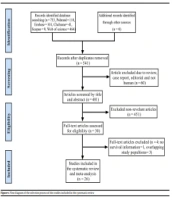
A total of 18,177 hepatobiliary cancer records for patients diagnosed between 2011 and 2017 were initially screened. After restricting the dataset to primary liver cancers coded as C22.0, 12,001 records remained. Application of the predefined HCC-compatible morphology criteria identified 1,991 potentially eligible records. We then excluded 210 death-certificate-only records because the timing and method of diagnosis could not be reliably established. After removing 34 duplicate records, 1,747 records entered the vital-status ascertainment process. Of these, 1,019 were successfully linked to the national registration office, and telephone follow-up was successfully completed for an additional 60 patients. The remaining 668 records were excluded because vital status or calculable follow-up time could not be reliably established. The final analytic cohort included 1,079 patients with HCC. The cohort-selection process is presented in Figure 1.
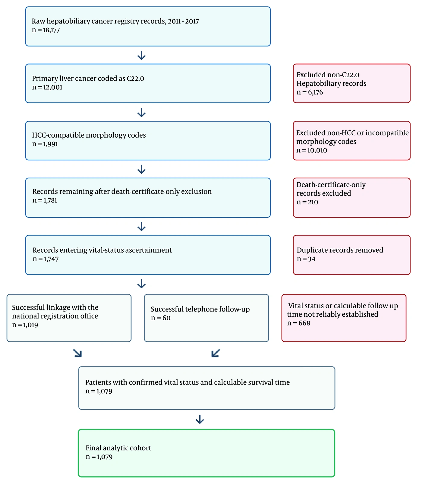
Figure 1.
Flow diagram of cohort selection.
The median observed follow-up time from diagnosis to death or censoring was 5 months in the overall cohort. Among the 141 censored patients, the median follow-up time was 119 months (interquartile range, 107 - 131 months).
4.2. Patient Characteristics and Survival Estimates
Descriptive characteristics of the included patients are summarized in Table 1. The mean age was 61.4 ± 14.8 years (range, 18 - 106 years). Most patients were male (n = 779, 72.2%). Overall, 604 patients (56.0%) were younger than 65 years, and 475 (44.0%) were 65 years or older. Diagnosis was pathological in 842 patients (78.0%) and clinical in 237 (22.0%). Conventional HCC was the most common morphology type (n = 1,048, 97.1%), followed by fibrolamellar HCC (n = 18, 1.7%), clear-cell HCC (n = 11, 1.0%), scirrhous HCC (n = 1, 0.1%), and pleomorphic-type HCC (n = 1, 0.1%).
Table 1.
Descriptive Characteristics and Survival Estimates a
| Variables | Value | Mean Survival (mo) | P-Value |
|---|---|---|---|
| Gender | 0.460 | ||
| Female | 300 (27.8) | 31.00 (25.11 - 36.89) | |
| Male | 779 (72.2) | 29.63 (27.05 - 33.22) | |
| Age group (y) | < 0.001 | ||
| < 65 | 604 (56.0) | 41.34 (36.63 - 46.06) | |
| ≥ 65 | 475 (44.0) | 15.56 (12.50 - 18.63) | |
| Diagnostic method | < 0.001 | ||
| Clinical | 237 (22.0) | 24.04 (18.70 - 31.38) | |
| Pathological | 842 (78.0) | 31.93 (27.90 - 34.88) | |
| Status at follow-up | |||
| Alive | 141 (13.1) | ||
| Deceased | 938 (86.9) | ||
| Tumor morphology | 0.300 | ||
| Conventional HCC | 1,048 (97.1) | 29.93 (26.82 - 33.04) | |
| Fibrolamellar HCC | 18 (1.7) | 38.66 (16.85 - 60.48) | |
| Scirrhous HCC | 1 (0.1) | 1.00 (1.00 - 1.00) | |
| Clear-cell HCC | 11 (1.0) | 15.06 (0.00 - 34.64) | |
| Pleomorphic-type HCC | 1 (0.1) | 13.00 (13.00 - 13.00) |
a Values are expressed as No. (%) or (95% CI). Survival estimates are reported in months. P values for survival comparisons were obtained using the log-rank test. Abbreviations: CI, confidence interval; HCC, hepatocellular carcinoma.
Diagnostic method was classified according to the registry diagnostic-method field. Pathological diagnosis included cases with histological or cytological confirmation. Clinical diagnosis included registry-coded clinical or clinical-only cases without pathological confirmation and was retained only when both C22.0 topography and an HCC-compatible morphology code were present.
At the last follow-up in October 2024, 141 patients were alive, corresponding to 13.1% of the final analytic cohort, whereas 938 patients had died. The mean and median survival times were 30.0 ± 1.6 months and 5.0 ± 0.5 months, respectively. The 1-, 3-, and 5-year overall survival rates were 37.5%, 21.0%, and 16.2%, respectively.
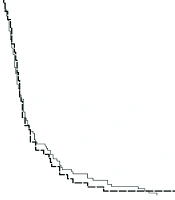
The provincial distribution of HCC cases is shown in Figure 2. The log-rank test showed no statistically significant difference in overall survival between males and females. Survival was significantly better among patients younger than 65 years than among those 65 years or older (Figure 3). Patients with a pathological diagnosis also had significantly longer survival than those with a clinical diagnosis (Figure 4). No statistically significant difference in survival was observed across tumor pathology subtypes.
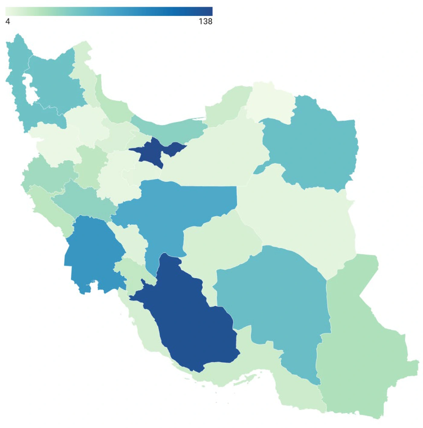
Figure 2.
Provincial distribution of HCC cases diagnosed between 2011 and 2017.
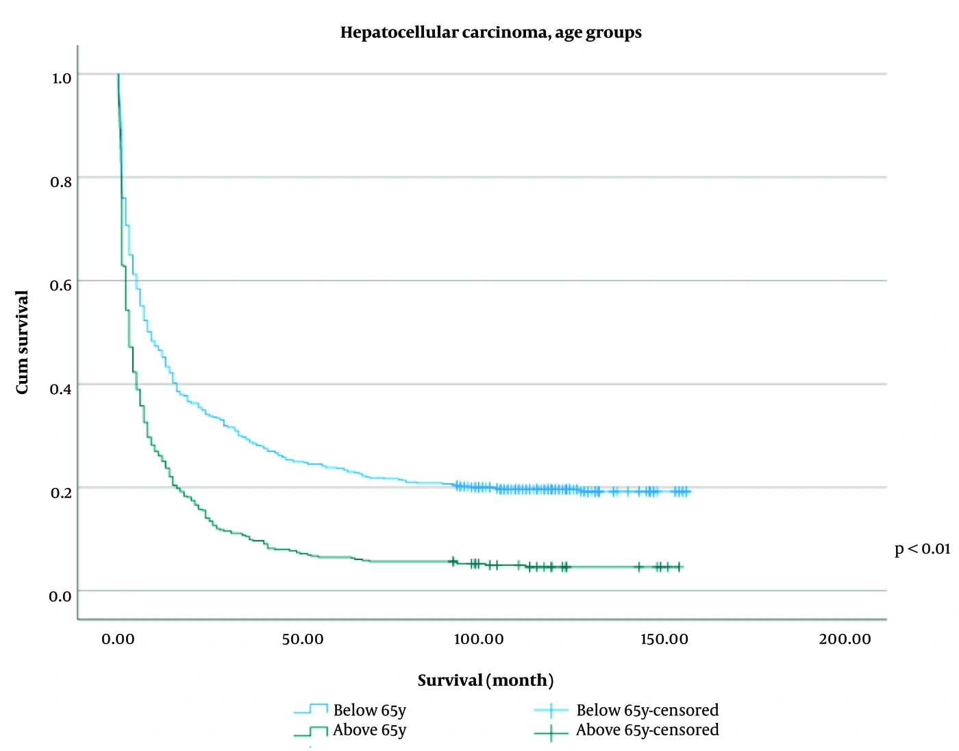
Figure 3.
Overall survival curves according to age group: < 65 years versus ≥ 65 years.
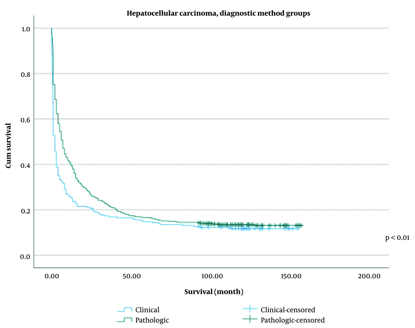
Figure 4.
Overall survival curves according to diagnostic method: Clinical diagnosis versus pathological diagnosis.
4.3. Multivariable Cox Regression
The initial proportional hazards assessment indicated non-proportionality for diagnostic method (time-dependent interaction, P < 0.001). The final Cox model was therefore stratified by diagnostic method and included all 1,079 patients, comprising 938 deaths and 141 censored observations. No patients were excluded because there were no missing data for the variables included in the model. Age was entered as a continuous variable, whereas the < 65 versus ≥ 65-year classification was used only for the Kaplan–Meier and log-rank analyses. No evidence of non-proportional hazards was observed for the covariates retained in the stratified model (global P = 0.893).
Older age was independently associated with poorer OS, with an approximately 2% increase in the hazard of death per 1-year increase in age (HR, 1.02; 95% CI, 1.02 - 1.03; P < 0.001). Sex was not significantly associated with OS (HR, 0.98; 95% CI, 0.85 - 1.14; P = 0.822), and tumor pathology was also not significantly associated with OS (HR, 1.25; 95% CI, 0.85 - 1.86; P = 0.260). Because diagnostic method was used as the stratification variable, a single adjusted HR for clinical versus pathological diagnosis was not estimated (Table 2).
Table 2.
Multivariable Cox Regression Analysis of Overall Survival Stratified by Diagnostic Method a
| Potential predictor | Comparison/Reference | HR (95% CI) | Wald Statistic | P-Value |
|---|---|---|---|---|
| Gender | Male vs female | 0.98 (0.85 - 1.14) | 0.05 | 0.822 |
| Diagnostic method | Clinical vs pathological | Stratification variable; HR not estimated | — | — |
| Tumor pathology | Variant HCC vs conventional HCC | 1.25 (0.85 - 1.86) | 1.27 | 0.260 |
| Age | Per 1-year increase | 1.02 (1.02 - 1.03) | 90.42 | < 0.001 |
a The final Cox model was stratified by diagnostic method because of non-proportional hazards; therefore, no adjusted HR was estimated for diagnostic method. Female and conventional HCC were used as the reference categories for sex and tumor pathology, respectively. Age was modeled as a continuous variable, with the HR representing the change in hazard per 1-year increase. Variant HCC included fibrolamellar, scirrhous, clear-cell, and pleomorphic-type morphologies. Abbreviations: CI, confidence interval; HCC, hepatocellular carcinoma; HR, hazard ratio.
5. Discussion
5.1. Principal Findings
In this national registry-based cohort, the 1-, 3-, and 5-year OS rates were 37.5%, 21.0%, and 16.2%, respectively. Patients aged 65 years or older had significantly poorer survival than those younger than 65 years. Kaplan–Meier analysis showed longer survival among pathologically diagnosed patients than among clinically diagnosed patients; however, the association with diagnostic method varied over time. There was no statistically significant difference in survival between males and females.
5.2. Comparison with Previous Studies
The survival estimates observed in this cohort fall within the broad ranges reported in several Asian registry-based studies, although these comparisons are descriptive rather than indicative of equivalent outcomes. A national study from China (6) reported 1-, 3-, and 5-year survival rates of 27.2%, 12.7%, and 8.9%, respectively. A nationwide registry-based report from Japan covering 1978 - 2005 documented a 5-year survival rate of 37.9% (7). In Taiwan, a study conducted from 2002 to 2012 reported a 5-year crude relative survival rate of 28.9% (8). A systematic review and meta-analysis of national and institutional or regional reports from Asian countries reported pooled 1-, 3-, and 5-year survival rates of 34.8%, 19.0%, and 18.1%, respectively (9). A 2024 Korean report indicated a 5-year survival rate of 39.3% (10).
European registry studies have also reported variable survival estimates. A Swedish registry study reported a median survival of 16.8 months among patients diagnosed between 2013 and 2016 (11). In the United Kingdom, 1-, 2-, and 5-year survival rates were 46.7%, 33.8%, and 18.3%, respectively, among patients diagnosed between 2013 and 2017 (12). A registry study from the Netherlands reported 1-year survival rates of 40% - 50%, 3-year rates of 20% - 27%, and 5-year rates of 14% - 21% among patients diagnosed between 2009 and 2016 (13), whereas a United States registry analysis also documented temporal improvements in survival (14). Direct cross-country comparisons should be interpreted with caution because study periods, registry completeness, population coverage, case ascertainment, stage distribution, treatment availability, and analytic methods vary across settings (15).
5.3. Age and Overall Survival
Older age was associated with poorer overall survival in this registry cohort. However, the reasons for this association cannot be determined from the available data. Contemporary HCC prognostic and treatment frameworks incorporate tumor burden, liver function, and performance status, and treatment selection is also influenced by clinical eligibility and competing health conditions (16, 17). Because disease stage, liver function, performance status, treatment, comorbidities, and liver-disease etiology were not available in our dataset, the observed age association may reflect residual confounding or treatment-selection effects. It should not be interpreted as evidence of an age-specific causal effect.
5.4. Sex and Overall Survival
In our study, the male-to-female ratio was 2.6:1, consistent with the internationally observed male predominance in HCC incidence (18, 19). There was no statistically significant difference in OS between males and females. Findings regarding sex-based survival differences in HCC remain inconsistent; some studies have reported better survival among female patients (20, 21), whereas others have found no significant or independent sex-based difference in overall survival (22, 23).
5.5. Diagnostic Method and Overall Survival
Patients with a pathological diagnosis had better overall survival in the registry than those with a clinical diagnosis, as assessed by Kaplan–Meier analysis. Diagnostic method did not satisfy the proportional hazards assumption, indicating that its association with mortality varied over follow-up. It was therefore used as a stratification variable in the final Cox model, and a single constant adjusted hazard ratio was not reported. The observed survival difference should not be interpreted as evidence that pathological confirmation itself improves survival. In at-risk populations, HCC may be diagnosed noninvasively using characteristic multiphase imaging findings, whereas biopsy is performed selectively based on clinical context (17). Consequently, the registry diagnostic method may serve as a marker of different diagnostic pathways, disease stage, performance status, treatment eligibility, treatment selection, or access to care. Because detailed imaging, laboratory, stage, treatment, and comorbidity data were unavailable, residual confounding and diagnostic misclassification cannot be excluded.
5.6. Study Limitations
The primary limitations of this study are its retrospective design and reliance on registry data. Determination of vital status mainly depended on national identification numbers and linkage to the national registration office. Some potentially eligible records lacked usable identifiers, and telephone follow-up could be completed only when valid contact information was available. Records with unverified vital status or follow-up time were excluded rather than censored, potentially introducing selection bias. Cases identified solely through death certificates were also excluded because the method and timing of diagnosis could not be confirmed. Data on disease stage, liver function, treatment, performance status, comorbidities, imaging findings, laboratory criteria, and liver-disease etiology were unavailable, limiting the ability to adjust for clinically important prognostic factors. Telephone follow-up may also have introduced recall error. Cases with survival of less than 1 month were retained because they represented observed outcomes in the registry population.
5.7. Implications for Future Research
Despite these limitations, this study provides national survival estimates for Iranian patients with HCC and may serve as a benchmark for future research. Further studies using more comprehensive registry linkages and detailed clinical, staging, and treatment data are required.
5.8. Conclusions
This retrospective national registry-based cohort study provides descriptive OS estimates for Iranian patients diagnosed with HCC between 2011 and 2017. Older age was independently associated with poorer OS, whereas sex and tumor pathology were not. Patients with a clinical diagnosis had poorer survival in the unadjusted Kaplan–Meier analysis. However, the association with diagnostic method varied over time and was not summarized by a single adjusted hazard ratio. These findings should not be interpreted causally because stage, liver function, treatment, performance status, comorbidities, and liver-disease etiology were unavailable. The results may serve as a national benchmark for future population-based studies with more comprehensive clinical and treatment data.