
1. Context
Premature ventricular complexes (PVCs) are prevalent arrhythmias in clinical practice (1). However, approaches to their evaluation and management remain inconsistent. Frequent PVCs are known to independently predict serious cardiovascular complications, yet current clinical practice typically lacks standardized protocols and often relies on individual clinical judgment (2). As new diagnostic and therapeutic modalities emerge, the traditional approach to managing these patients is evolving rapidly.
This narrative review synthesizes current evidence on PVC management, including PVC diagnosis, risk stratification, and treatment options, and explores a future paradigm that incorporates advances in PVC clinical practice. This future vision emphasizes diagnostic precision, refined patient selection criteria, and optimized treatment strategies. In this article, we aim to identify gaps in current practice and propose a more consistent, personalized, and effective approach to the future management of PVCs.
2. Evidence Acquisition
We conducted a narrative review of PVC management by searching PubMed and major cardiology journals for publications from 2015 to 2025 using search terms including "premature ventricular complex," "PVC management," "catheter ablation," and "ventricular ectopy." We prioritized recent systematic reviews, meta-analyses, randomized controlled trials, and major clinical practice guidelines, including the 2017 AHA/ACC/HRS and 2022 ESC guidelines. In addition, we searched ClinicalTrials.gov for ongoing trials relevant to the diagnosis and treatment of PVC.
3. Results
3.1. Epidemiology and Clinical Significance
Frequent PVCs are increasingly recognized as an independent risk factor for adverse cardiovascular events (2). This recognition underscores the importance of thorough diagnosis and an appropriate therapeutic approach in contemporary electrophysiology. PVCs are common, and their observed prevalence increases with longer ECG monitoring. Studies indicate that standard 2-minute resting ECGs detect PVCs in only a small fraction of the general population (approximately 1% to 4%), whereas extended ambulatory monitoring reveals a much higher frequency, with up to three-quarters of individuals exhibiting PVCs over a 24- to 48-hour period (1).
The clinical significance of frequent PVCs (≥ 500/day or ≥ 1% of total beats) is substantial (2). Recent meta-analyses indicate that frequent PVCs strongly predict long-term cardiovascular complications. Even in structurally normal hearts, a high PVC burden nearly doubles the risk of developing atrial fibrillation or heart failure (2). It is also statistically significantly associated with stroke and increased all-cause mortality (2). In addition, a high PVC burden can cause PVC-induced cardiomyopathy (PVC-CM), a potentially reversible form of dilated cardiomyopathy (3).
3.2. Diagnosis and Burden Assessment
Accurate diagnosis and treatment require precise assessment of PVC frequency. PVC burden, expressed as a percentage of total heartbeats, is an independent predictor of adverse cardiovascular outcomes and guides treatment decisions (2). Although the 12-lead ECG remains the most effective method for identifying PVC origin sites, it provides only a brief snapshot. Therefore, ambulatory monitoring is essential for determining the true burden and characterizing ectopy complexity (4, 5).
Because PVC frequency varies over time, extending monitoring to 6 - 7 days is recommended to capture an individual's maximum PVC burden (6). Short-term recordings (24 - 48 hours) can substantially misclassify burden because of day-to-day variability, making standardized monitoring duration essential, because treatment decisions depend on accurate burden thresholds.
In addition to electrocardiographic monitoring, transthoracic echocardiography is an important tool for initial evaluation and serial risk stratification. Echocardiography assesses left ventricular ejection fraction (LVEF), chamber dimensions, and wall-motion abnormalities to detect early PVC-induced cardiomyopathy and identify underlying structural heart disease (5, 7). Current guidelines recommend baseline and periodic echocardiographic follow-up in patients with frequent PVCs to monitor for LVEF decline before it becomes irreversible.
3.3. Current Guideline-Based Management
Current clinical practice guidelines, specifically the 2017 AHA/ACC/HRS guidelines and the 2022 ESC guidelines, structure PVC management around 3 fundamental considerations: symptom severity, PVC burden, and the presence of structural heart disease (SHD) (7, 8). The guidelines suggest intervention thresholds at PVC burdens ranging from 10% to 15% (7, 8). In addition, patients with frequent PVCs should undergo serial echocardiographic assessment and rhythm monitoring to detect early progression of structural complications (8).
Although these guideline recommendations provide a structured framework, therapeutic options remain limited in both efficacy and safety.
3.3.1. Medical Therapy: Efficacy and Limitations
For symptomatic patients with structurally normal hearts, initial pharmacological management typically includes beta-blockers or non-dihydropyridine calcium channel blockers (5, 7). These agents provide only modest efficacy for PVC suppression, with clinically meaningful PVC burden reduction, typically defined as ≥ 80% reduction or resolution of symptoms, achieved in only 12% to 24% of patients overall. Beta-blockers are particularly effective for sympathetically mediated or outflow-tract PVCs, whereas non-dihydropyridine calcium channel blockers are useful in fascicular PVCs; however, most patients experience < 50% burden reduction (5, 9).
When first-line agents are insufficient, Class Ic antiarrhythmic drugs such as flecainide may be considered (5, 7). Flecainide achieves substantially greater PVC suppression than first-line therapy, with recent studies reporting ≥ 80% burden reduction in approximately 64% of patients and complete or near-complete suppression (> 99% reduction) in 55% to 56% of patients with idiopathic PVCs (10, 11). Class Ic agents are recommended only in patients with structurally normal hearts and preserved ejection fraction. Flecainide is contraindicated in patients with ischemic heart disease, significant left ventricular hypertrophy, or reduced LVEF because of the proarrhythmic risk demonstrated in the CAST trial (5, 7, 12).
For high-risk patients with structural heart disease, Class III agents such as amiodarone and sotalol represent safer alternatives (7). Amiodarone is particularly effective in suppressing PVCs and improving LVEF in patients with heart failure, although its long-term use is limited by significant adverse effects, including thyroid dysfunction, pulmonary toxicity, and hepatotoxicity (7). Recent data from the CHF-STAT trial showed that amiodarone achieved successful PVC suppression (≥ 80% burden reduction) in 72% of patients with heart failure and frequent PVCs, with corresponding improvement in LVEF in approximately 39% of cases (13). Sotalol, another Class III agent, can suppress PVCs in selected patients with coronary artery disease, preserved renal function, and a normal baseline QT interval. Sotalol efficacy is lower than that of flecainide (≥ 80% burden reduction in approximately 33% of patients) and requires inpatient initiation with continuous QT monitoring because of the risk of torsades de pointes (11).
Current pharmacological options have important limitations. Beta-blockers and non-dihydropyridine calcium channel blockers achieve meaningful PVC reduction in only a minority of patients (5, 9). There remains a substantial unmet need for safer and more effective pharmacological agents. Class I and III antiarrhythmic drugs offer greater suppression but carry risks of proarrhythmia and extracardiac toxicity (5, 7, 14).
Given the modest efficacy and toxicity concerns of pharmacological therapy, catheter ablation has emerged as an increasingly important therapeutic option.
3.3.2. PVC Mapping and Ablation Techniques in Routine Practice
Current guidelines recommend ablation for patients experiencing LVEF decline secondary to frequent PVCs when antiarrhythmic drugs are ineffective or declined (5, 15). In addition, catheter ablation has emerged as the preferred treatment for PVC-induced cardiomyopathy, demonstrating superiority in eliminating PVCs compared with medical management alone (3, 15).
In contemporary catheter ablation, localization of the PVC origin typically begins with activation mapping, which is considered the gold standard technique when sufficient spontaneous ectopy is present (15). When PVCs are infrequent or noninducible, pace mapping serves as an alternative by matching the paced QRS complexes to the clinical arrhythmia. These approaches are usually combined with 3-dimensional electroanatomic mapping systems. However, pace mapping has limited spatial resolution (1 - 2 cm2), which is inferior to activation mapping (1.2 cm2), potentially leading to suboptimal ablation targets (15).
Radiofrequency ablation (RFA) is the standard energy source used in PVC ablation (16). It delivers thermal energy to create lesions at the PVC origin site, but it carries risks of collateral damage and may require long application times (16). The RFA achieves overall acute success rates of 80% to 95%, with the highest efficacy (> 90%) for right ventricular outflow tract (RVOT) origins (3, 5). RFA for epicardial origins has lower success rates, typically 60% to 80% acute procedural success with higher recurrence, and papillary muscle origins achieve acute success of 70% to 88% but remain technically challenging because of catheter instability and multiple morphologies (5, 17). Long-term clinical success for papillary muscle PVCs is approximately 69% (17).
Cryoablation, which uses tissue freezing, is an established alternative energy modality. It offers improved catheter stability, which is crucial for papillary muscles (15, 18). Cryoablation demonstrates comparable overall success rates, with studies showing acute success up to 100% and lower recurrence (0% at 6 months in direct comparison), specifically for papillary muscle origins of the left ventricle, where catheter stability is critical (15, 18). Despite these acute success rates, procedural limitations remain, including the requirement for spontaneous PVC activity during mapping, the possible need for repeat procedures, and the risk of complications.
3.4. Emerging Approaches Studied in Recent Research
3.4.1. Reconsidering Intervention Thresholds
Recent studies suggest that the accepted safety threshold for PVC burden may be substantially lower than current guideline recommendations (19). Studies show that LV dysfunction can emerge at PVC burdens as low as 5% to 10%, directly challenging the established 10% to 15% intervention threshold (20). These findings suggest that current guideline-recommended watchful waiting strategies may be inadequate to prevent early structural changes.
Beyond electrocardiographic assessment, cardiac magnetic resonance imaging (CMR) is emerging as a valuable tool for risk stratification. CMR demonstrates that myocardial fibrosis can develop in asymptomatic high-burden PVC patients (> 15% to 20%) before any detectable LVEF decline (21). The ongoing IRMESV trial (NCT06801743) is prospectively assessing when CMR should be performed in patients with frequent PVCs to define evidence-based indications and improve detection of subclinical structural abnormalities that could influence treatment decisions (22).
3.4.2. Paradigm Shift: Early Ablation as First-Line Therapy
Emerging research from 2023 to 2025 is challenging the traditional conservative approach (15). Recent systematic reviews indicate that early catheter ablation demonstrates significantly superior efficacy to antiarrhythmic drug therapy in reducing PVC recurrence and promoting ventricular function restoration (3, 15). This evidence is driving a paradigm shift toward considering ablation as first-line therapy, particularly for RVOT-originating PVCs, for which success rates consistently exceed 90% with favorable safety profiles (15, 18).
Nevertheless, the optimal timing of ablation in patients with modest LV dysfunction or intermediate PVC burden has not been clearly defined, and randomized data in this population remain limited. Further prospective studies are required to determine which subgroups benefit most from early ablation versus initial pharmacological therapy.
3.4.3. Advances in Mapping and Ablation Technologies
Although activation and pace mapping with RFA remain the routine approach, recent studies have explored strategies to overcome their limitations. A novel preprocedural tool, noninvasive electrocardiographic imaging (ECGI), has recently emerged. The ECGI workflow consists of 4 main steps: 1) application of a 252-electrode vest to the patient's torso to record body-surface potentials during PVCs; 2) acquisition of a thoracic CT scan to define patient-specific heart-torso geometry; 3) reconstruction of epicardial potentials and activation sequences by solving the inverse problem of electrocardiography using mathematical algorithms; and 4) generation of 3-dimensional epicardial activation maps that localize the PVC origin with errors typically < 10 mm (23). This noninvasive approach enables precise preprocedural planning and has demonstrated superior accuracy compared with conventional 12-lead ECG algorithms (23).
Most recently, pulsed field ablation (PFA) has been introduced as a novel nonthermal technology that uses high-voltage electrical fields to induce irreversible electroporation (24). Recent prospective data from 2024 specifically evaluating focal PFA for PVCs demonstrated high acute success rates with a favorable safety profile, particularly highlighting preservation of adjacent coronary arteries and nerve structures because of tissue selectivity (25). Although PFA significantly improves procedural efficiency, with shorter procedure times compared with thermal modalities in atrial fibrillation (AF), large-scale comparative data for ventricular applications are still evolving (26). The ongoing FOCUS-PFA trial (NCT06747013) is evaluating focal PFA for PVCs and focal ventricular tachycardia (27).
3.5. Future Paradigm: Personalized PVC Management With Machine Learning and Artificial Intelligence
3.5.1. Personalized Risk Prediction
To personalize PVC management, validated risk prediction models must be developed. These models must integrate PVC burden with clinical variables, including age, sex, comorbidities, and severity of left ventricular dysfunction, along with morphological features such as QRS duration and PVC origin site. Imaging biomarkers, particularly myocardial fibrosis on cardiac MRI, also provide critical prognostic information. These integrated models could identify patients at the highest risk of cardiomyopathy development and enable individualized burden thresholds rather than universal cutoffs.
3.5.2. Machine Learning for PVC Localization
Artificial intelligence and machine learning represent promising tools for improving PVC localization accuracy. Deep learning algorithms trained on 12-lead ECG data can predict the PVC site of origin with accuracies ranging from 78% to 98%, depending on classification complexity (28, 29, 30). More sophisticated systems have demonstrated accuracies exceeding 98% when classifying origins among 21 anatomical sites, surpassing human expert performance (30). Deep learning models achieve localization errors of 3 - 11 mm, whereas traditional activation mapping and pace mapping typically achieve spatial resolution of 12 - 20 mm (15, 29).
No large randomized controlled trials have yet directly compared machine learning-guided ablation with traditional electroanatomical mapping (31).
3.5.3. Cardiac Digital Twins and Noninvasive Mapping
A digital twin in medical research is a virtual model of a patient or biological system. It integrates personal clinical information, medical images, and physiological measurements to help researchers predict disease progression, treatment response, and the safety of new therapies before they are applied in real life.
The cardiac digital twin is another emerging technology in PVC management. This approach integrates body-surface potential mapping with patient-specific computational models of cardiac electrophysiology. Digital twins reconstruct epicardial activation patterns and predict arrhythmia origins from noninvasive ECG data (32). Early studies combining digital twins with ECGI demonstrate localization accuracy with a mean error of 7.8 mm, substantially superior to conventional ECGI alone (30 - 36 mm error) (32).
These artificial intelligence-based methods remain under investigation and are not yet implemented in routine clinical practice. Prospective multicenter validation studies are needed before widespread clinical adoption.
3.6. Future Directions and Knowledge Gaps
First, the optimal ambulatory ECG monitoring duration must be established as a clinical standard. As noted above, recent data suggest that 6 - 7 days of continuous monitoring provides substantially more accurate estimates than conventional 24- to 48-hour recordings (6). Standardizing monitoring duration is essential because treatment decisions depend on burden thresholds.
Second, pharmacological options remain limited. Beta-blockers and non-dihydropyridine calcium channel blockers have modest efficacy (5, 9). Class I and III antiarrhythmic drugs offer greater suppression but carry significant toxicity (5, 7, 14). The development of safer and more effective pharmacological agents represents a critical unmet need. As catheter ablation technology advances, future frameworks may progressively de-emphasize chronic medical therapy. However, current evidence does not support completely eliminating medical therapy from PVC management algorithms, and this hypothesis requires rigorous evaluation in well-designed comparative studies.
Third, the choice between catheter ablation and medical therapy remains an area of active investigation, particularly in patients with cardiomyopathy and reduced LVEF. Although recent guidelines favor ablation over antiarrhythmic drugs in patients with suspected PVC-induced cardiomyopathy and frequent monomorphic PVCs (7, 8), the optimal timing of ablation in patients with modest LV dysfunction or intermediate PVC burden requires further investigation through randomized controlled trials.
Finally, emerging technologies, including machine learning algorithms and cardiac digital twins, demonstrate superior spatial resolution compared with conventional mapping techniques (29, 32). However, prospective multicenter validation studies comparing these AI-guided approaches with traditional electroanatomical mapping are essential before routine clinical implementation (31).
4. Conclusions
In contemporary electrophysiology, PVCs are frequently observed in a substantial proportion of the population during cardiac monitoring. Moreover, emerging studies demonstrate an independent association between PVCs and adverse cardiovascular events. In view of this recognition, updating current PVC management protocols is essential. Multiple ongoing trials are evaluating improved diagnostic tools, localization and ablation techniques, and more efficient treatment options. Throughout this article, we have discussed the future of PVC management by using results from ongoing trials and highlighting the need for further well-designed studies. Although substantial work remains to define the truly optimal approach to PVC management, there is promising evidence that this goal is achievable. Innovations such as machine learning algorithms and cardiac digital twins offer new avenues that could fundamentally transform how we diagnose and treat these arrhythmias in the future. Figure 1 summarizes this evolving paradigm, contrasting established practices with emerging tools and future directions.
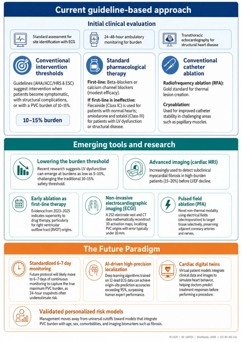
Figure 1.
Visual summary of the evolving paradigm in PVC management