
1. Introduction
Atrial fibrillation (AF) is a common arrhythmia with an increasing prevalence among adults (1). Individuals with cardiovascular or respiratory diseases who develop new-onset AF have a higher likelihood of hospitalization and mortality (2). In addition, AF is associated with an increased risk of thromboembolic events, such as stroke (3). AF is also associated with pulmonary hypertension (4).
Pulmonary hypertension can cause right-sided heart failure. Restricted blood flow from peripheral organs to the heart is characteristic of right-sided heart failure, leading to congestion that may affect other organs, such as the liver and gallbladder. In this case report, we describe atrial masses in a patient who was admitted with pruritus. We also discuss the implications of this finding and consider appropriate management strategies.
2. Case Presentation
A 73-year-old woman from the Middle East presented with mild generalized pruritus without associated cutaneous lesions. The symptoms did not improve or worsen with daily activities such as showering or exposure to sunlight. Over the previous 2 months, the patient had noticed weight loss and difficulty swallowing solid foods. She reported no constitutional symptoms, such as fever, night sweats, jaundice, or changes in urine or stool color. She had right upper quadrant (RUQ) abdominal pain and dyspnea.
On physical examination, the patient’s vital signs were within normal limits. No cutaneous lesions, splenomegaly, signs of hepatic disease, or jaundice were observed. Her medical history included hypertension, chronic kidney disease, and peptic ulcer disease. Her family history was unremarkable, and no other family members had pruritus. The patient had no history of smoking, alcohol consumption, or substance abuse.
Diagnostic evaluation included urinalysis, complete blood count, liver function tests, serum creatinine and urea measurements, alkaline phosphatase (AlkP), alanine aminotransferase (ALT), and gamma-glutamyl transferase (GGT). The patient had elevated serum urea, creatinine, ALT, AlkP, and GGT levels and a reduced hematocrit (Table 1). All other blood test results were within the normal range. All urinalysis findings were within the normal range except for proteinuria.
Table 1.
Results of Blood Tests Before Hospitalization of the Patient
| Test | Results (Reference) |
|---|---|
| Urea | 54 mg/dL (17 - 43) |
| Creatinine | 1.42 mg/dL (0.6 - 1.3 in females) |
| ALT | 45 IU/L (up to 31 in females) |
| Alkaline phosphatase (AlkP) | 447 IU/L (64 - 360) |
| Gamma-glutamyl transferase (GGT) | 288 U/L (up to 32 in females) |
| WBC | 7.7 mil/cumm (4 - 10) |
| RBC | 3.9 mil/cumm (3.7 - 5.2 in females) |
| Hemoglobin | 11.7 g/dL (11.5 - 15 in females) |
| Hematocrit | 34% (35 - 47 in females) |
Although elevated blood urea and creatinine levels were considered possible causes of pruritus, the presence of RUQ pain, elevated ALT, and high creatinine levels prompted comprehensive abdominopelvic sonography. Sludge and stones were found in the gallbladder. Because of dysphagia and weight loss, the physician recommended upper endoscopy. Given the increased vulnerability of older patients to complications during certain medical procedures, the physician requested a cardiology evaluation before endoscopy.
The cardiologist performed echocardiography and electrocardiography (ECG). The ECG showed AF rhythm (Figure 1). Atrial fibrillation was also detected during echocardiography. The estimated ejection fraction was 45%. During diastole, 3 large echogenic atrial masses measuring 36 × 25 mm were observed protruding into the left ventricle and left ventricular outflow tract (Video 1). Additional findings included severe right ventricular enlargement, moderate right ventricular systolic dysfunction, mild mitral regurgitation (MR), and mild-to-moderate mitral annular calcification. Abnormal septal motion was also detected. The tricuspid valve leaflets were thickened and retracted, and severe tricuspid regurgitation (TRVC = 13 mm) was observed. The systolic pulmonary artery pressure (SPAP) was 47 mm Hg.
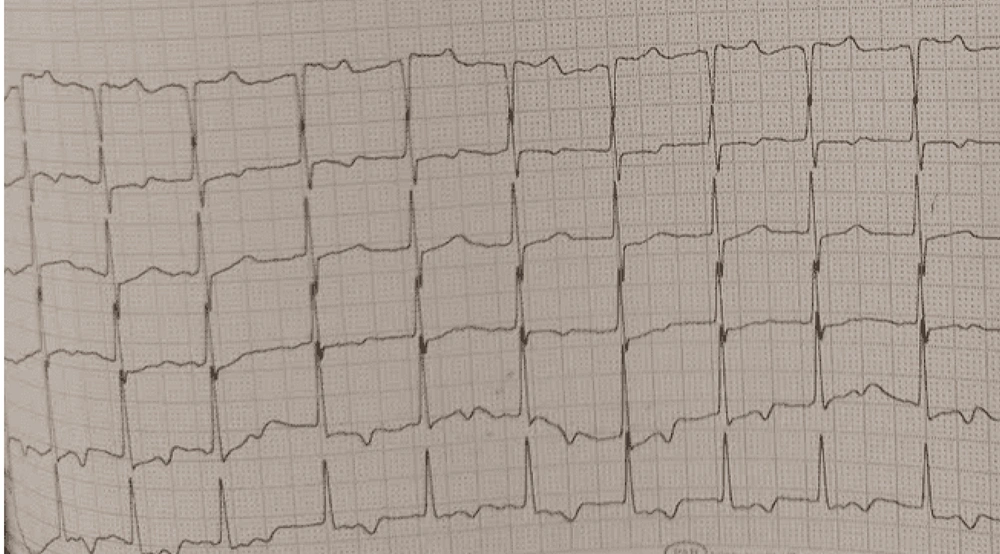
Figure 1.
ECG without P waves because of atrial fibrillation (AF).
After the masses and AF rhythm were detected on echocardiography, coronary angiography was promptly performed to rule out potential vascular disease. Coronary angiography showed no abnormalities in the coronary arteries. Therefore, the cardiologist recommended consultation with a cardiac surgeon. The cardiac surgeon advised surgical intervention to remove the masses and perform valvoplasty. During surgery, 4 masses were discovered, although echocardiography had shown 3 masses. After surgery, the masses were sent to a pathologist for further examination (Video 2) (Figure 2). The pathologist reported fibrin material admixed with leukocytes and clot formation with ingrowing vascular channels (Figure 3), suggesting a thrombus.
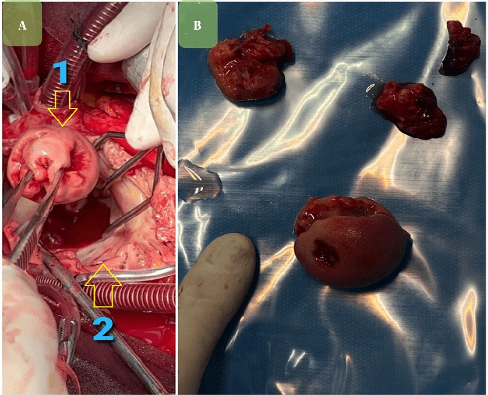
Figure 2.
This figure shows atrial masses. A, this part of the figure shows the largest atrial mass (1) and atrium (2); B, atrial masses after surgery.
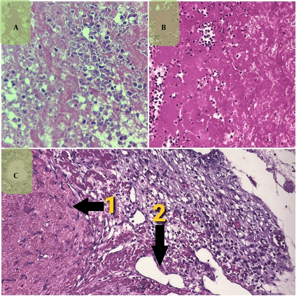
Figure 3.
A, histopathological morphology of the clot showing fibrin material admixed with some leukocytes, hematoxylin and eosin staining, high-power field (400×). B, histopathological morphology of the clot showing fibrin material admixed with some leukocytes, hematoxylin and eosin staining, low-power field (100×). C, Histopathology of organizing thrombi with ingrowing proliferating vascular channels, hematoxylin and eosin staining, low-power field (100×). The first arrow (1) shows the organizing thrombi, and the second arrow (2) shows the vascular channel.
The patient was transferred to the intensive care unit after cardiac surgery. Regrettably, she did not survive in the intensive care unit because of right-sided heart failure and hepatic failure.
3. Discussion
We present a case report of a patient whose chief complaint on admission was generalized pruritus. She also had RUQ abdominal pain and difficulty swallowing solid foods, and her condition warranted upper endoscopy. A cardiologist evaluated her cardiovascular risk before the endoscopic procedure. ECG showed an AF rhythm. Echocardiography also showed an AF rhythm and large atrial masses protruding into the left ventricle, with dilation of the right atrium and ventricle. Thus, generalized pruritus appeared to be the presenting symptom of heart failure complicated by an AF rhythm and multiple thrombotic masses.
Before cardiac surgery, the physician considered two possible differential diagnoses. Carcinoid masses were suspected because of the size of the masses and their possible association with dysphagia to solids. Many cardiac tumors result from the spread of primary tumors from other parts of the body, including the gastrointestinal tract (5). However, the pathologist confirmed thrombus and ruled out cancer in the cardiac masses. Therefore, heart failure and an AF rhythm were the most likely causes of these masses.
Hypertension is a significant risk factor for an AF rhythm and heart failure (6). Heart failure can cause pruritus (7). One plausible explanation in this case is that hypertension contributed to AF. Atrial fibrillation then led to the formation of 4 thrombotic masses because of blood stasis in the left atrium. However, chronic left-heart abnormalities related to the patient's long-standing hypertension and AF were the primary drivers of pulmonary hypertension, rather than the masses themselves. Pulmonary hypertension can cause right-sided heart failure. Heart failure affected the liver and gallbladder, leading to generalized pruritus. Notably, heart failure can also cause pruritus independently of gallbladder involvement.
Pruritus, a common symptom in patients with heart failure, should be addressed (7). Moreover, AF is associated with MR and mitral annular calcification, as observed in our patient (8). However, no research is currently available regarding the frequency of heart failure among individuals with pruritus. This case serves as a reminder to consider the cardiovascular system when evaluating pruritus, particularly when it is accompanied by hepatobiliary signs and symptoms.
Cholecystalgia is an unusual manifestation of heart failure (9). To our knowledge, this report highlights a unique case in which pruritus presented as a symptom of right-sided heart failure and AF with thrombotic masses. A similar case involved a 67-year-old woman with a previous diagnosis of pulmonary hypertension who was admitted with cholecystitis. In that patient, cholecystitis masked underlying right-sided heart failure (10).
4. Conclusions
Pruritus is a common cutaneous symptom that may be associated with serious underlying conditions. It is a known but often overlooked symptom in patients with heart failure and may be related to hepatic congestion. However, heart failure is frequently overlooked in patients who present with pruritus but have no prior history of heart failure. The potential association between pruritus and heart failure should be considered, particularly in patients with risk factors such as hypertension. Heart failure may also present with hepatobiliary manifestations. Furthermore, pruritus accompanied by hepatobiliary symptoms may indicate right-sided heart failure. When a patient presents with pruritus as the primary symptom, a thorough medical history and physical examination can help determine whether heart failure should be considered.