
1. Introduction
Sarcoidosis is a chronic multisystemic disease of unknown etiology characterized by granulomatous inflammation in lungs, liver, eyes, lymph nodes, skin, heart, nervous and musculoskeletal systems and gut (1). Diagnosis is typically confirmed in adults when three key features are present; positive radiological imaging (chest X-Ray), a suggestive clinical picture and the histological presence of non-caseating granulomas (2). However, there are no universal criteria especially in children and sarcoidosis is often a diagnosis of exclusion (3). The clinical value of using serum angiotensin converting enzyme (ACE) in diagnosis is debated as it can be raised non-specifically at moderately high levels in childhood. However, ACE is said to be elevated in 60% of patients with sarcoidosis and can be used to monitor disease activity (4).
The incidence of sarcoidosis varies worldwide as race, genetics and environment are implicated in the pathophysiology (5). The highest incidence is reported in Northern European and African-American individuals while Japanese populations are the least affected (6). The peak age of onset is between 25 - 45 years; paediatric presentations are very rare and often present a diagnostic challenge due to the nonspecific symptoms and the lack of specific biomarkers for diagnosis (7). The precise cause of sarcoidosis is still unknown. Current opinion suggests that exposure to antigens in genetically susceptible individuals may contribute to CD4+ T helper cell activation, triggering an abnormal immune response and leading to granuloma formation (8). Treatment with immunomodulatory drugs is often required.
The association of sarcoidosis with autoimmune disorders is well recognized and probably indicates a complex interplay of immunological and genetic mechanisms (8, 9). In general, autoimmune conditions are characterized by an immune response to self-antigens such as thyroperoxidase in Hashimoto’s thyroiditis (HT) or islet cell components in Type 1 diabetes (T1D). There is a polyclonal activation of B lymphocytes (delayed hypersensitivity, secondary autoimmunity) in response to one or several exogenous antigens in sarcoidosis (1, 8). Sarcoidosis may co-exist with several endocrine and non-endocrine autoimmune conditions (9). However, the reported cases with co-existing endocrine manifestations are rare in children.
We report a child who developed hypercalcemic pancreatitis after supplementation with vitamin D, subsequently leading to a diagnosis of sarcoidosis. Additionally, she was diagnosed with HT, T1D and secondary adrenal insufficiency. To our knowledge, such an unusual association of endocrinopathies has not been described previously in childhood sarcoidosis.
2. Case Presentation
A 9-year-old girl of mixed ethnic origin was referred to the endocrine team in view of hypercalcaemia, lethargy and weight loss. At 8 year of age, she was investigated elsewhere for lethargy, tiredness and reduced appetite and was diagnosed with autoimmune hypothyroidism and vitamin D deficiency (25-hydroxyvitamin D [25(OH)D] 11 nmol/L). At that point, she was also noted to have benign joint hypermobility and a functional cardiac murmur. There was no history of pain or swelling of joints. She did not complain of any significant ailments in the past and there was no history to suggest an endocrine or autoimmune disease in the family.
She was born at 42 weeks gestation with no neonatal concerns. Her symptoms had persisted despite satisfactory control of the hypothyroid state and intermittent vitamin D supplementation. She was treated with cholecalciferol (20,000 IU/d for 1 week and 800 IU/d later) for persistent vitamin D deficiency. A week later, she began to complain of low-grade fever and intermittent bouts of vomiting and abdominal pain. She was treated at the local hospital for a presumed abdominal infection in view of high C-reactive protein (CRP) levels but the screen for sepsis was negative. Her symptoms persisted and a weight loss of 4kg was noted over the preceding month. Additionally, she was detected to have hypercalcemia and was hospitalized.
On physical examination, her anthropometric parameters were normal for age. She was noted to have dark discoloration under the eyes and pallor. There was no goitre. The systemic examination was unremarkable. Investigations revealed hemoglobin of 96 g/L (normal, 115 - 155 g/L), erythrocyte sedimentation rate (ESR) 35 mm/h (normal, 3 - 13 mm/h) and platelet count was elevated at 685 × 109/L (normal, 140 – 400 × 109/L). Serum ferritin, CRP and albumin were 25 ng/mL (normal, 29 - 371 ng/mL), 44 mg/L and 25 g/L (normal, 35 - 55 g/L) respectively. Serial serum concentrations of calcium, phosphorus, alkaline phosphatase (ALP), parathyroid hormone (PTH), 25(OH)D and 1, 25 dihydroxy vitamin D [1, 25(OH)2D] are shown in Table 1. Serum magnesium was 0.58 mmol/L (normal, 0.7 - 1 mmol/L) and serum amylase was 395 U/L (normal, 23 - 85 U/L).
Table 1.
Biochemical Parameters Before, During and After the Episode of Hypercalcemic Pancreatitisa
| Biochemical Parameter | Before Episode | During Hypercalcemic Episode | After Episode | ||||||
|---|---|---|---|---|---|---|---|---|---|
| 1 y | 2 mo | 1 mo | D1 | D3 | D4 | D5 | 1 mo | 6 mo | |
| Serum calcium, mmol/L | 2.28 | 2.33 | 3.70 | 3.78 | 3.62 | 2.55 | 2.35 | 2.36 | |
| Serum phosphorus, mmol/L | 1.33 | 1.46 | 0.82 | 1.36 | 1.7 | ||||
| Serum Alkaline phosphatase, IU/L | 133 | 289 | 393 | 810 | |||||
| Serum parathyroid hormone, pmol/L | 0.1 | 0.7 | 3.2 | 3.6 | |||||
| Serum 25(OH)D, nmol/L | 44 | 29 | 11 | 65 | 63 | 69 | 81 | ||
| Serum 1,25(OH)2D, pmol/L | 161 | 102 | |||||||
| Urinary calcium/creatinine ratio, mmol/mmol | 3.03 | 0.9 | |||||||
aNormal levels: calcium, 2.15 - 2.74 mmol/L; phosphorus, 0.97 - 1.94 mmol/L; alkaline phosphatase, 203 - 1151 IU/L; 25(OH)D, 50 - 125 nmol/L; 1,25(OH)2D, 43 - 143 pmol/L; parathyroid hormone, 1.1 - 6.9 pmol/L; urine calcium creatinine ratio, 0.08 - 0.79 mmol/mmol.
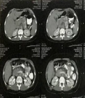
Abdominal ultrasonogram (USG) showed a diffusely enlarged hypoechoic pancreas surrounded by free fluid, and minimal ascites but no cholelithiasis. A lower gut endoscopy and abdominal magnetic resonance imaging (MRI) showed no evidence of inflammatory bowel disease. Anti-tissue transglutaminase antibodies were negative. A clinical diagnosis of acute pancreatitis probably caused by hypercalcemia was considered.
A rheumatological evaluation undertaken in view of her chronic non-specific symptoms revealed high ACE levels on 2 occasions (106 and 135 nmol/mL/min, normal range 10 - 43). Serum lactate dehydrogenase was 573 U/L (normal, 143 - 290 U/L). Tests for antinuclear antibody, anti-double-stranded DNA, perinuclear anti-neutrophil cytoplasmic antibodies and cytoplasmic anti-neutrophil cytoplasmic antibodies were negative. Ophthalmology examination showed no evidence of uveitis and conjunctival biopsy showed no granulomas.
Detailed investigations were conducted to exclude infectious and malignant diseases that may mimic sarcoidosis. Radiograph and CT scan of the chest, and echocardiography were normal. Brain MRI including pituitary gland and pelvic USG showed no abnormality. Bone marrow examination was normal. Total human chorionic gonadotropin and α-fetoprotein levels were < 1 mIU/mL (normal, 0 - 5 mIU/mL) and 2.2 ng/mL (normal, < 5 ng/mL) respectively. Urinary vanillylmandelic acid level was normal. Mantoux test showed no induration. HIV serology and PCR for cytomegalovirus, adenovirus and Epstein-Barr virus were negative. Urine microscopy was made and culture was negative. Creatine kinase was 9U/L (normal, 22 - 198 U/L). Immunoglobulin and lipid profiles were normal. Serum complement C3 and C4 levels were 88 mg/dL (normal, 90 - 180 mg/dL) and 20 mg/dL (normal, 20 - 50 mg/dL) respectively. Plasma concentrations of ceruloplasmin, copper, mercury and lead were 0.33 g/L (normal, 0.32 - 0.57 g/L), 15 µmol/L (normal, 13.2 - 21.4 µmol/L), 3 nmol/L (normal, 0 - 20 nmol/L) and 1 µg/dL (normal, < 5 µg/dL) respectively.
A short standard adrenocorticotropic hormone (ACTH) stimulation test showed serum cortisol concentrations of < 50, 320 and 378 nmol/L at baseline, 30 and 60 minutes respectively (normal stimulated peak concentration, > 500 nmol/L). Plasma ACTH level was < 1.1 pmol/L (normal, 2 - 11 pmol/L) suggestive of secondary adrenal insufficiency. Insulin-like growth factor-1 (IGF-1) level was < 3.3 nmol/L (normal, 4 - 20 nmol/L). The blood glucose concentrations during the 2nd week of hospital stay were ≥ 11.1 mmol/L on multiple occasions leading to a diagnosis of diabetes mellitus. Glycosylated hemoglobin (HbA1c) level was 31 mmol/mol (normal, < 42 mmol/mol). Titers of anti-thyroid peroxidase, anti-glutamic acid decarboxylase (GAD) and anti-adrenal antibodies were 253 IU/mL (normal, 0 - 60 IU/mL), 419 U/mL (normal, 0 - 5 U/mL) and negative (< 1 U/mL) respectively.
The vitamin D supplementation was stopped and intravenous hyperhydration was initiated along with intravenous furosemide for the management of hypercalcemia. On day 3 of hospitalization, a single subcutaneous dose of calcitonin (4 IU/kg) was administered in view of persistent hypercalcemia following which the hypercalcaemia gradually improved (Table 1) paralleling an improvement in abdominal symptoms. Hyperhydration was continued for another 2 days. Repeat USG of abdomen a week later showed resolving pancreatitis. There was no recurrence of hypercalcaemia over the next several months. Levothyroxine was continued. She was started on replacement oral hydrocortisone (10 mg/m2/day).
Subsequently methotrexate (15 mg once a week) and oral prednisolone (40 mg once daily) was added for the management of sarcoidosis (hydrocortisone was discontinued whilst on prednisolone). A basal-bolus insulin regimen was commenced with good effect on glycaemic control. She has shown consistent clinical improvement in the year following diagnosis and her symptoms have significantly improved. Prednisolone was discontinued after 6 months and she was restarted on replacement hydrocortisone. Her current growth is optimal (weight 75th centile, height 50th centile) and she continues on hydrocortisone, levothyroxine, insulin (HbA1c 42 mmol/mol) and methotrexate.
3. Discussion
In our patient, the diagnosis of sarcoidosis was based on the constellation of clinical features, with raised ESR and platelets and elevated ACE concentrations following exclusion of other conditions that mimic sarcoidosis (1, 3). Additionally, the low complement concentrations (10) and occurrence of hypercalcemia after routine doses of vitamin D (11) indicated the presence of granulomatous inflammation in the body. The serum levels of immunoglobulin and complement are usually low during disease activation due to their deposition in active sarcoid lesions and in circulating immune complexes (10).
An extensive radiological and laboratory workup and non-recurrence of symptoms over a fairly long-term follow-up helped exclude the usual chronic infectious and occult malignant disorders associated with hypercalcemia in children. However, the diagnosis of sarcoidosis remains probable, as the demonstration of granulomatous inflammation in tissues was not possible. Significant clinical improvement was noted on treatment with methotrexate and prednisolone.
Hypercalcemia occurs in 10% - 13% of patients with sarcoidosis and is due to dysregulated (non-PTH mediated) overproduction of 1,25(OH)2D by the granulomatous tissue and activated macrophages, that increases intestinal absorption of calcium (12). Hypercalcemia may also manifest after starting vitamin D supplements as the availability of more substrate 25(OH)D leads to its accelerated conversion to 1,25(OH)2D by autonomously produced 1 α-hydroxylases (11). Vitamin D levels are often low in sarcoidosis due to accelerated conversion of available 25(OH)D to 1,25(OH)2D (11, 13). In our patient, low vitamin D levels persisted despite routine cholecalciferol supplementation probably due to the enhanced utilization of 25(OH)D by 1 α-hydroxylases. Provision of higher doses of cholecalciferol had subsequently led to symptomatic hypercalcemia, similar to previous reports (11, 13). It may be mentioned that a diagnosis of sarcoidosis had not been entertained at the time of starting higher doses of cholecalciferol, similar to previous observations (11, 13).
Acute pancreatitis that occurred in our patient could either be related to sarcoidosis or hypercalcaemia. The sarcoid infiltration of the pancreas, noted in about 1% of autopsy series, may manifest as acute or chronic pancreatitis (14, 15). Hypercalcaemic pancreatitis often manifests acutely and resolves after treatment of hypercalcemia, as seen in our patient, whereas sarcoidosis-induced pancreatitis requires steroids (14). The proposed mechanisms of hypercalcaemic pancreatitis include increased output and activation of pancreatic enzymes, intracellular activation of pancreatic proteases through cholecystokinin release, and increased permeability of pancreatic ducts causing leakage of enzymes (14). A notable feature of hypercalcaemic pancreatitis is that the short-term hypercalcaemia is more likely to be associated with acute pancreatitis than chronic hypercalcemia (11, 13, 14). Acute pancreatitis may also be associated with a number of rheumatic disorders such as systemic lupus erythematosus, rheumatoid arthritis, polyarteritis nodosas and Wegener’s granulomatosis (14) that were excluded in our patient by appropriate investigations. Drug-induced pancreatitis was not considered as the child had not received any drugs known to cause acute pancreatitis (16). Furthermore, a diagnosis of drug-induced pancreatitis is usually made if no other cause can be detected (16). Thus, the temporal association with administration of cholecalciferol resulting in acute hypercalcaemia and prompt resolution after lowering of serum calcium levels makes hypercalcemia as the most likely etiology of acute pancreatitis in our patient.
Hypothyroidism is a common co-existing endocrinopathy in sarcoidosis (1, 3). This may result from sarcoidotic infiltration of the thyroid gland that occurs in < 1% of cases (12), but is more often due to associated thyroid autoimmunity (3), as seen in our patient. Furthermore, high titers of anti-GAD 65 antibodies indicate autoimmune diabetes in our patient similar to previous reports (17), although involvement of pancreas in sarcoidosis is occasionally noted to result in diabetes mellitus (18).
We found secondary adrenal insufficiency in our patient that occurs rarely in sarcoidosis (19). This usually follows sarcoid infiltration of pituitary gland visible as an enlarged gland or mass lesion on MRI (19). Low IGF-1 levels also indicated anterior pituitary dysfunction. However the MRI pituitary was normal and it is difficult to attribute the pituitary dysfunction in our patient to either sarcoidosis or autoimmune destruction. It may be mentioned here that the IGF-1 levels were measured during the phase of hyperglycemia and could well be due to the uncontrolled diabetes at that time (20).
In conclusion, we report a rare association of multiple autoimmune and non-autoimmune endocrinopathies in a girl with probable sarcoidosis. The disease is currently controlled on a complex medical regime and the patient has returned to normal activities but the long-term outcome remains uncertain. To the best of our knowledge, ours is the youngest ever patient with sarcoidosis who developed such diverse endocrine abnormalities.