1. Background
Pediatric lower urinary tract dysfunction (LUTD) includes a heterogeneous spectrum of storage and voiding abnormalities that impair quality of life and, if unrecognized, increase the risks of recurrent urinary tract infection, vesicoureteral reflux progression, and renal scarring. Noninvasive tests, particularly transabdominal bladder ultrasound and uroflowmetry, now anchor routine evaluation because they characterize complementary domains of LUTD. Ultrasound assesses bladder morphology, wall characteristics, and postvoid residual (PVR). Uroflowmetry quantifies voiding dynamics and flow curve morphology. Although both modalities are well established, it remains uncertain how closely structural sonographic parameters map onto specific uroflowmetry patterns in individual children, and how best to integrate these data for diagnosis and management.
Normative work has clarified expectations for uroflow parameters and typical bell-shaped curves in healthy school age children. Sonographic protocols increasingly emphasize standardized filling and posterior wall measurements to improve reproducibility. However, robust age and sex stratified reference ranges for bladder wall thickness in children remain limited, and prior studies have reported mixed results regarding the strength of structure-function associations. Clinically, this uncertainty can lead to overreliance on a single modality or to inconsistent phenotyping of LUTD subtypes.
To address these gaps, we conducted a two center, age and sex matched, retrospective case–control study of children aged 5 to 14 years. We compared bladder wall thickness, bladder volume, and PVR with uroflowmetry patterns and quantitative flow measures. We hypothesized that, while cases would differ markedly from controls on both structural and functional indices, specific uroflowmetry flow patterns would show at most weak associations with ultrasound parameters after accounting for age and sex.
2. Objectives
Our objectives were threefold: (1) To quantify between group differences in sonographic and uroflowmetry measures; (2) to describe the distribution of flow curve phenotypes in symptomatic versus asymptomatic children; and (3) to examine the adjusted relationships between flow patterns and bladder volume and wall thickness. These data aim to inform a multimodal, evidence based diagnostic approach that interprets ultrasound and uroflowmetry together, rather than in isolation, for optimized assessment of pediatric LUTD.
3. Methods
3.1. Study Design and Setting
This retrospective, two center case–control study was conducted at Shohadaye Kargar and Shahid Sadoughi hospitals (Yazd, Iran) from January 2022 to December 2023. The protocol was approved by the Ethics Committee of Shahid Sadoughi University of Medical Sciences (IR.IAU.KHUISF.REC.1404.408) and adhered to the Declaration of Helsinki. We enrolled 240 children aged 5 - 14 years and allocated them equally to case and control groups using individual 1:1 matching.
3.2. Participants and Sample Size Calculation
Cases were children presenting with urinary dysfunction symptoms, including daytime incontinence, urgency, frequency > 8 voids/day, dysuria, nocturnal enuresis (≥ 2 episodes/week in children > 5 years), or subjective voiding difficulty. Controls were recruited from routine health visits or non-urological evaluations and were confirmed asymptomatic via standardized questionnaires completed by parents and children. Individual 1:1 matching was performed on sex (identical) and age (within ± 6 months) using computer generated random selection from eligible controls.
Sample size was calculated a priori with G*Power v3.1 assuming a between group mean difference of 1.0 mm in bladder wall thickness (SD = 1.2 mm), α = 0.05, and power = 0.80, yielding 118 participants per group. To accommodate potential missingness, we included 120 per group (total n = 240). Exclusion criteria were incomplete diagnostic data, neurological disease affecting bladder function, congenital urinary tract abnormalities, active urinary tract infection at assessment, prior urological surgery, or lack of parental consent.
3.3. Sonographic Assessment
Transabdominal bladder ultrasound was performed on a Voluson™ E8 system (GE Healthcare, Chicago, IL) with a 2 - 8 MHz convex transducer by radiologists with ≥ 5 years’ pediatric experience, following standardized protocols. Children were scanned with moderate bladder filling {~50 - 75% of expected capacity; [age (years) + 1] × 30 mL if < 12 years and 350 - 400 mL if ≥ 12 years}.
Bladder volume was calculated with the ellipsoid formula (length × width × height × 0.52) using sagittal and transverse measurements. Bladder wall thickness was measured at the posterior wall in the midsagittal plane during moderate filling, avoiding trabeculation and artifacts; the average of three measurements was recorded. The PVR was measured within 5 minutes of complete voiding.
For reliability, a subset of 30 examinations underwent duplicate measurements by two independent operators, yielding intraclass correlation coefficients (ICC) > 0.90 for all parameters.
3.4. Uroflowmetry Assessment
Uroflowmetry was performed with the MMS UD 2000 system (Medical Measurement Systems, Enschede, Netherlands) according to International Children’s Continence Society guidelines. Children received brief verbal instructions and voided in a private setting in their natural position (standing for boys, sitting for girls) after achieving comfortable bladder fullness. Only voids ≥ 50 mL were analyzed; testing was repeated when initial volume was insufficient.
Recorded parameters included maximum flow rate (Qmax), average flow rate (Qavg), voided volume, flow time, and time to maximum flow (TQmax). Flow curves were classified as bell shaped (normal), plateau, tower shaped, staccato, or intermittent by two independent observers, with discrepancies resolved by consensus.
3.5. Data Collection and Statistical Analysis
Data extracted from medical records included age, sex, weight, height, BMI, presenting symptoms, family history of urinary problems, prior urinary tract infections, comorbid conditions (particularly constipation), and all sonographic and uroflowmetry measurements. Data were entered on standardized forms with double data entry by two independent researchers; discrepancies were resolved by record review. De identified datasets were stored in password protected databases.
Analyses were conducted in SPSS v26 (IBM, Armonk, NY). Distributions were assessed with the Shapiro-Wilk test and visual inspection. Continuous variables are reported as mean ± SD or median (IQR); categorical variables as No. (%). Between group comparisons used independent t-tests or Mann-Whitney U tests (continuous) and chi square or Fisher’s exact tests (categorical). Analysis of covariance (ANCOVA) adjusted for age and sex. Correlations were assessed with Spearman’s rank correlation (continuous) and contingency analysis (categorical). All tests were two tailed, with statistical significance at P < 0.05. Effect sizes are reported as Cohen’s d (pooled SD) for continuous outcomes and Cramer’s V for categorical associations.
4. Results
4.1. Participant Characteristics
A total of 240 children were enrolled (120 cases; 120 controls). Sex distribution did not differ between groups [147 males (61.3%), 93 females (38.8%); χ² = 0.235, P = 0.629]. Mean age was comparable (controls: 10.05 ± 2.77 years; cases: 9.92 ± 2.79 years; t = 0.348, P = 0.728). Anthropometric measures were similar (controls vs. cases):
- Weight: 32.8 ± 11.2 vs. 31.9 ± 10.8 kg.
- Height: 135.4 ± 16.3 vs. 134.2 ± 15.8 cm.
- BMI: 17.6 ± 3.2 vs. 17.3 ± 3.1 kg/m2 (all P > 0.05).
4.2. Clinical Features
In the case group, the most frequent presenting symptoms were incontinence (27/120, 22.5%), urinary frequency (26/120, 21.7%), dysuria (22/120, 18.3%), and nocturnal enuresis (21/120, 17.5%). Controls were confirmed asymptomatic via standardized parent- and child-reported questionnaires. Family history of urinary problems was more prevalent in cases than controls (38.3% vs. 10.8%; χ2 = 23.1, P < 0.001). Prior urinary tract infection was also more common in cases (42.5% vs. 9.2%; χ2 = 34.2, P < 0.001). Constipation occurred in 35.0% of cases vs. 10.0% of controls (χ2 = 20.8, P < 0.001). Comorbidities beyond constipation were not systematically assessed.
4.3. Sonographic Parameters
Bladder volume did not differ significantly between groups (controls: 217.92 ± 88.12 mL; cases: 205.67 ± 81.90 mL; t = 1.098, P = 0.625). In contrast, bladder wall thickness was greater in cases (3.65 ± 1.02 mm) than controls (2.48 ± 0.27 mm; t = 11.2, P < 0.001; Cohen’s d = 1.45). The PVR volume was markedly higher in cases (41.19 ± 19.25 mL) vs. controls (9.80 ± 4.77 mL; Mann-Whitney U = 1,847, P < 0.001; Cohen’s d = 2.31). Abnormal bladder morphology was more prevalent in cases (28.3% vs. 4.2%; χ2 = 24.1, P < 0.001; Cramer’s V = 0.32), as was trabeculation (30.8% vs. 5.0%; χ2 = 26.3, P < 0.001; Cramer’s V = 0.33). In controls only, males had larger mean bladder volumes than females (238.5 ± 91.2 vs. 201.3 ± 82.4 mL; t = 2.07, P = 0.042); no sex difference was observed in cases (P = 0.187). Sonographic parameters are summarized in Table 1.
Table 1.
Sonographic Parameters in Cases and Controls a
| Parameters | Controls (N = 120) | Cases (N = 120) | P-Value | Effect Size b |
|---|---|---|---|---|
| Bladder volume (mL) | 217.92 ± 88.12 | 205.67 ± 81.90 | 0.625 | 0.15 |
| Wall thickness (mm) | 2.48 ± 0.27 | 3.65 ± 1.02 | < 0.001 | 1.45 |
| Post-void residual (mL) | 9.80 ± 4.77 | 41.19 ± 19.25 | < 0.001 | 2.31 |
| Normal morphology | 115 (95.8) | 86 (71.7) | < 0.001 | 0.32 |
| Trabeculation present | 6 (5.0) | 37 (30.8) | < 0.001 | 0.33 |
a Values are expressed as mean ± SD or No. (%).
b Effect size denotes Cohen’s d for continuous variables and Cramer’s V for categorical variables.
4.4. Uroflowmetry Patterns and Parameters
Normal bell-shaped curves predominated in controls (105/120, 87.5%) but were uncommon in cases (17/120, 14.2%; χ2 = 128.7, P < 0.001; Cramer’s V = 0.73). Among cases, abnormal patterns included intermittent (30/120, 25.0%), staccato (30/120, 25.0%), plateau (25/120, 20.8%), and tower-shaped (18/120, 15.0%); tower-shaped and staccato were observed exclusively in cases.
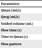
Maximum flow rate (Qmax) was lower in cases (8.88 ± 3.34 mL/s) than controls (13.82 ± 3.45 mL/s; t = 11.1, P < 0.001; Cohen’s d = 1.45). Average flow rate (Qavg) was likewise reduced (5.76 ± 2.23 vs. 8.82 ± 2.38 mL/s; t = 10.0, P < 0.001; Cohen’s d = 1.32). Voided volume (215.43 ± 92.79 vs. 209.54 ± 87.86 mL; P = 0.614), flow time (19.63 ± 4.81 vs. 19.86 ± 4.64 s; P = 0.703), and time to maximum flow (TQmax: 8.42 ± 2.89 vs. 8.15 ± 2.74 s; P = 0.482) did not differ significantly. Detailed uroflowmetry parameters and flow pattern distributions are presented in Table 2.
Table 2.
Uroflowmetry Parameters and Flow Patterns a
| Parameters | Controls (N = 120) | Cases (N = 120) | P-Value | Effect Size |
|---|---|---|---|---|
| Qmax (mL/s) | 13.82 ± 3.45 | 8.88 ± 3.34 | < 0.001 | 1.45 |
| Qavg (mL/s) | 8.82 ± 2.38 | 5.76 ± 2.23 | < 0.001 | 1.32 |
| Voided volume (mL) | 209.54 ± 87.86 | 215.43 ± 92.79 | 0.614 | 0.07 |
| Flow time (s) | 19.86 ± 4.64 | 19.63 ± 4.81 | 0.703 | 0.05 |
| Time to Qmax (s) | 8.15 ± 2.74 | 8.42 ± 2.89 | 0.482 | 0.10 |
| Flow pattern | < 0.001 | 0.73 b | ||
| Bell-shaped (normal) | 105 (87.5) | 17 (14.2) | ||
| Plateau | 10 (8.3) | 25 (20.8) | ||
| Tower-shaped | 0 (0) | 18 (15.0) | ||
| Staccato | 0 (0) | 30 (25.0) | ||
| Intermittent | 5 (4.2) | 30 (25.0) |
a Values are expressed as mean ± SD or No. (%).
b Effect size for flow pattern is Cramer’s V.
4.5. Structure-Function Correlations and Covariate Effects
No significant associations were detected between flow pattern categories and bladder volume or wall thickness (controls: Volume: F = 1.10, P = 0.335; wall thickness: F = 0.18, P = 0.835; cases: Volume: F = 1.17, P = 0.326; wall thickness: F = 0.75, P = 0.558). In ANCOVA models adjusting for age and sex, these findings remained non-significant. Age correlated positively with bladder volume in both groups (controls: R = 0.73, P < 0.001; cases: R = 0.69, P < 0.001). A sex effect on bladder volume was observed only among controls (F = 4.2, P = 0.042). No significant interactions between flow pattern and sex were identified (all P > 0.10). Comparisons of bladder sonographic parameters by flow pattern are shown in Table 3.
| Variables | Bladder Volume (mL) | Wall Thickness (mm) |
|---|---|---|
| Controls | ||
| Bell-shaped | 222.30 ± 87.98 | 2.49 ± 0.28 |
| Plateau | 184.90 ± 93.45 | 2.46 ± 0.26 |
| Intermittent | 192.20 ± 77.63 | 2.42 ± 0.08 |
| Cases | ||
| Bell-shaped | 189.88 ± 79.71 | 3.67 ± 1.13 |
| Plateau | 193.88 ± 80.07 | 3.42 ± 0.92 |
| Tower-shaped | 214.06 ± 77.38 | 3.56 ± 1.07 |
| Staccato | 230.57 ± 84.97 | 3.88 ± 1.01 |
| Intermittent | 194.50 ± 83.10 | 3.66 ± 1.01 |
a Values are expressed as mean ± SD.
b One-way analysis of covariance (ANCOVA, adjusted for age and sex) showed no significant association between flow pattern and bladder volume or wall thickness in either group (controls: F = 1.10, P = 0.335; F = 0.18, P = 0.835; cases: F = 1.17, P = 0.326; F = 0.75, P = 0.558).
5. Discussion
In these two centers, age and sex matched case-control study, children with urinary dysfunction exhibited significantly thicker bladder walls, markedly higher PVR volumes, and substantially more abnormal uroflowmetry patterns than asymptomatic peers, reinforcing the clinical value of a multimodal, noninvasive evaluation pathway advocated by the International Children’s Continence Society and related guidance (1-3). The magnitude of the between group differences for bladder wall thickness and PVR, together with the predominance of non–bell shaped curves in the case group, mirrors prior pediatric reports linking structural bladder changes and inefficient emptying with dysfunctional voiding phenotypes (4-11). At the same time, our analyses confirmed that, after adjustment for age and sex, specific uroflowmetry flow patterns did not correlate strongly with ultrasound parameters at the individual level, a finding that is consistent with the emerging view that these modalities provide complementary rather than redundant information (1, 12).
The absence of strong pattern specific correlations likely reflects the multifactorial pathophysiology of pediatric LUTD. Similar flow morphologies can arise from different underlying mechanisms; staccato curves, for example, may reflect pelvic floor discoordination, behavioral withholding, or pain, while increases in bladder wall thickness may result from chronic detrusor overactivity, reduced compliance, or compensatory responses to outlet resistance (6-8, 13-17). Sonographic measures also remain state dependent: Despite our standardization to moderate filling, residual variability in filling level and regional wall thickness can attenuate linear associations with concurrent flow morphology (5, 8, 18). Moreover, both ultrasound and uroflowmetry in routine practice are often obtained as single occasion tests; within child variability in hydration, stool burden, anxiety, and cooperation can affect volumes, PVR, and flow curves, blurring structure–function coupling on any one day (1, 11, 19, 20). Finally, relationships between parameters may be non-linear or threshold based, e.g., risk increases above certain PVR levels, so average differences across broad pattern categories may not capture clinically meaningful inflection points.
Clinically, these observations argue against inferring pathophysiology from either modality in isolation. Instead, ultrasound and uroflowmetry should be interpreted together and in clinical context, including symptom profiles, voiding diaries, and constipation assessment. In practice, clearly increased bladder wall thickness and consistently elevated PVR, particularly when accompanied by staccato or intermittent flow patterns and reduced flow rates, should prompt structured urotherapy (timed voiding, fluid optimization, constipation treatment), consideration of pelvic floor biofeedback for suspected dysfunctional voiding, and escalation to urodynamic testing when abnormalities persist or when findings are discordant. Where local protocols apply numeric triggers, thresholds such as bladder wall thickness above approximately 3.0 mm for this age range or repeated PVR values above 20 mL may justify earlier referral, especially in the setting of recurrent urinary tract infections or refractory symptoms.
Our results fit well within the literature. The predominance of bell-shaped curves among controls and the lower Qmax and Qavg in cases align with normative uroflow data for school age children (5) and with studies reporting reduced flow in dysfunctional voiding syndromes (21-25). The larger bladder wall thickness in symptomatic children and the markedly higher PVR echo prior work that associates structural bladder changes with impaired emptying and greater risk for infectious or reflux related sequelae (5-11, 26, 27). At the same time, the weak individual level structure-function coupling we observed has been noted in multimodal pediatric series, supporting a diagnostic strategy that synthesizes rather than substitutes across modalities (1, 12).
This study has several strengths. We used individual 1:1 matching on age and sex, standardized acquisition protocols for both ultrasound and uroflowmetry, and demonstrated excellent inter operator reliability for sonographic measurements (ICC > 0.90). We captured both curve morphology and quantitative flow parameters, including time to maximum flow, and we adjusted analyses for age and sex using ANCOVA, acknowledging known developmental effects on bladder capacity and voiding function. The sample size afforded robust power for the primary between group comparisons.
Limitations must also be acknowledged. As a retrospective, hospital based, two center study, our design limits causal inference and may not fully represent community populations. Although ultrasounds were performed at moderate filling, bladder wall thickness remains filling dependent, and single occasion imaging and uroflowmetry cannot account for day-to-day variability. Flow pattern classification, while based on established criteria, is susceptible to residual inter observer differences; similarly, we concentrated comorbidity ascertainment on constipation and did not systematically quantify other modifiers such as medication use, psychological factors, or stool burden by imaging. Finally, we did not incorporate invasive urodynamics or surface electromyography, which could have validated noninvasive phenotypes in discordant cases and refined mechanistic interpretation.
Future research should move beyond cross sectional snapshots to prospective, longitudinal, multicenter designs with repeated same day ultrasound and uroflowmetry under standardized filling targets, permitting within child modeling of variability and response to urotherapy. Parallel efforts should generate age and sex stratified normative nomograms for pediatric bladder wall thickness and PVR, ideally reported as Z scores to facilitate interpretation across the 5 - 14-year range (2, 3, 5, 19, 20, 28, 29). Analytically, pre specified multivariable models and modern methods such as penalized regression or machine learning classifiers could integrate quantitative flow features, curve shape descriptors, PVR, and sonographic metrics to improve discrimination and calibration for clinically relevant outcomes. Mechanistic substudies incorporating selective urodynamics and, where feasible, surface EMG would help link noninvasive signatures to detrusor pressure and sphincter activity. Comprehensive comorbidity assessment, including standardized constipation scoring, stool burden quantification, hydration tracking, and patient reported outcomes, should be embedded to contextualize structure–function relationships and tailor therapy. Finally, implementation studies should test the cost effectiveness and clinical impact of “ultrasound plus uroflowmetry for all” versus risk stratified pathways in real world pediatric urology clinics.
5.1. Conclusions
Children with urinary dysfunction have greater bladder wall thickness, higher PVR, and abnormal uroflowmetry compared with asymptomatic peers, yet individual level correlations between ultrasound parameters and specific flow morphologies are weak. These findings support a multimodal diagnostic approach in which ultrasound and uroflowmetry are interpreted together and against the clinical backdrop, rather than relied upon singly. Prospective, age and sex stratified, and analytically advanced studies are needed to establish normative sonographic values, validate noninvasive phenotypes against mechanistic measures, and determine the pathway level impact of integrated testing on outcomes and resource use.