
1. Background
Colonoscopy is technically more challenging than esophagogastroduodenoscopy, particularly in children, due to anatomical differences, patient cooperation, and the need for specialized equipment and training. Pediatric colonoscopies require endoscopists who have completed specific pediatric training in accordance with international guidelines (1). Pediatric colonoscopes differ from adult instruments in shaft length (1330 - 1700 mm), diameter (9.8 - 11.8 mm), and channel size (2.8 - 3.8 mm). Variable-stiffness technology allows greater maneuverability by reducing looping in mobile bowel segments while maintaining flexibility in fixed areas (2).
The main diagnostic indications for pediatric colonoscopy include rectal bleeding, unexplained diarrhea, and abdominal pain associated with weight loss or growth failure. Additional indications include the investigation of imaging abnormalities and suspected inflammatory bowel disease (IBD) (3). Therapeutic indications are less common in children but may include polypectomy, management of bleeding angiodysplasia, cecostomy, decompression of acute colonic pseudo-obstruction, or balloon dilation of strictures (3).
Bowel preparation is a critical prerequisite for adequate mucosal visualization. Polyethylene glycol (PEG) is the most widely used agent, either alone or in combination with electrolytes or stimulant laxatives. Preparations may be administered as single- or split-dose regimens, and their effectiveness can be assessed by validated tools such as the Boston Bowel Preparation Scale (BBPS), which scores mucosal visualization from 0 (inadequate) to 9 (excellent) (4, 5).
Pediatric colonoscopy is usually performed by interdisciplinary teams involving pediatric gastroenterologists, anesthetists or intensivists, and trained nurses. Continuous monitoring of vital signs, particularly oxygen saturation, is recommended during and after the procedure, with observation continuing for at least two hours post-sedation to ensure patient safety (6).
Colonoscopy in children differs from adult practice not only in clinical indications, bowel preparation, and anesthesia requirements, but also in systemic effects. In adults, endoscopic or urologic procedures such as colonoscopy and cystoscopy can transiently increase prostate-specific antigen (PSA) levels, which may complicate prostate cancer screening and follow-up (7, 8). This concern, however, is irrelevant in pediatrics, as prostate pathology and PSA monitoring are not considerations in this age group. This highlights that pediatric colonoscopy cannot be regarded as a simple extension of adult practice, but rather as a procedure with distinct clinical implications and safety considerations.
Despite its clinical importance, pediatric colonoscopy remains under-studied in many regions. Most available data come from high-income countries, where endoscopy services are well established. In the Middle East, reports are limited, and pediatric endoscopy is often concentrated in tertiary referral centers. In Iraq, published data on pediatric colonoscopy are virtually absent, despite increasing clinical demand. Generating local evidence is critical for understanding disease patterns, improving training, and guiding healthcare resource allocation.
2. Methods
Study protocol and data collection A prospective cross-sectional observational study, using a consecutive sampling method, was carried out on all children (10 months-15 years of age) who underwent colonoscopy in the Gastroenterology Unit at Child Welfare Teaching Hospital (which is a tertiary referral center for pediatric gastroenterology services) from the 1st of May to 1st of December 2023 and were included. Demographic data, indications for the colonoscopy procedure (mainly bleeding per rectum, chronic bloody diarrhea, chronic abdominal pain, anaemia, perianal fistulae), and histopathological findings were collected.
During the study period, 112 children were assessed for eligibility. Seven were excluded (three due to inadequate bowel preparation, two due to incomplete procedures, and two for missing data). A total of 105 children met eligibility criteria (age, indication, complete bowel preparation, complete procedure, complete documentation, patient informed consent, follow-up) and, thus, were included in the final study. Acutely ill patients were managed in the ER department of the hospital and were not eligible for the study. Uncooperative patients/parents were also excluded. Adequate sedation is essential for safe pediatric colonoscopy; if not possible, the procedure cannot be performed.
All patients included in this study underwent a complete history and physical examination; patients were reviewed in an outpatient clinic or inpatient ward before being booked for colonoscopy.
Bowel-cleansing preparation The laxative used was Pico-Salax (Picoprep®), each sachet containing 10 mg sodium picosulfate, 3.5 g magnesium oxide, 12.0 g citric acid, and 36 mg aspartame, dissolved in 250 mL of warm water (9).
Dosing was age-adjusted: ¼ sachet for children 1 - 6 years, ½ sachet for 6 - 12 years, and 1 sachet for 12 - 18 years, given once daily for two consecutive days. In addition, bisacodyl was administered (15 mg for children 20 - 35 kg or 6 - 12 years; 20 mg for >36 kg or 12 - 18 years).
Parents received standardized verbal and written instructions from trained medical staff to enhance compliance. In line with ESGE 2019 guidelines (10), a low-residue diet was recommended prior to colonoscopy. This included white bread, cheese, meat, fish, peeled fruits, and cooked vegetables, while high-fiber foods (wholegrain bread, brown rice, muesli, certain fruits and vegetables such as oranges and mushrooms) were avoided.
Although standardized bowel preparation quality scores such as the Boston Bowel Preparation Scale (BBPS) are available, in this cohort bowel preparation quality was not systematically scored, and adequacy was judged clinically by the endoscopist.
Sedation The procedure was performed under sedation by the anaesthetic team. Sedation started by giving intravenous fluids, midazolam 0.05 mg/kg, propofol 2.5 - 3 mg/kg, ketamine 0.5 mg/kg, and oxygenation; patients may need intubation if there are comorbidities like cardiac diseases. Patients were continuously monitored using pulse oximetry, heart rate, respiratory rate, and non-invasive blood pressure. Level of sedation and responsiveness were also assessed at regular intervals. Following the procedure, all patients were observed in a recovery area until full consciousness and stable vital signs were achieved. Sedation was well tolerated in all patients. No major adverse events such as cardiorespiratory arrest, laryngospasm, or need for intubation occurred. No patient required unplanned hospital admission or prolonged observation due to sedation-related issues.
Endoscopes The procedure was performed in the left lateral position. Position change and abdominal pressure were applied according to the procedure progression. Procedures were done in the morning, three days per week; each session may last from about 7 minutes to 30 minutes according to patient condition and preparation, as hospital protocol. Biopsies were taken whenever indicated and from macroscopically normal mucosa, because studies recommended taking mucosal biopsies even from macroscopically normal mucosa, since collagenous colitis or lymphocytic colitis may present with normal mucosa; other diseases like IBD and microscopic colitis in children have been diagnosed by biopsy when the mucosa has been normal (11). Biopsies had been sent for histopathology to the Department of Educational Laboratories in the Medical City.
Histopathological findings were reviewed by random pathologists in a blinded manner to clinical data.
Limitations: Although limited by a relatively small sample size, this study provides the first prospective data on pediatric colonoscopy in Iraq and offers valuable baseline information for future larger studies.
The study was approved by the local ethical committee of Iraqi board of subspecialty in Pediatric Gastroenterology and Hepatology, and informed consent agreement had been taken from all of the parents of participants who were eligible for this study for both colonoscopy procedure and anesthesia.
Statistical Analysis: Statistical analysis was done using the Statistical Package for Social Sciences (SPSS), version 24 software (IBM, USA). Results were presented as mean ± standard deviation (SD) as well as percentages. The significance of differences between groups was assessed using Chi-square test and an independent sample t test depending on variables.
4. Result
Characteristics and indications of pediatric patients who underwent Colonoscopy A total of 105 patients with a mean age of 8.25 ± 3.6 years were reviewed. The cohort included 44 males (41.9%) and 61 females (58.1%), respectively. The majority of the patients were between 5 - 10 years old, 55 (52.4%), and the least frequent were less than 5 years, 15 (14.3%); the youngest patient was 11 months. Hematochezia, abdominal pain, and diarrhea were the most common indications for pediatric endoscopy, accounting for 40 (38.1%), 27 (25.7%), and 18 (17.1%), respectively. Only 6 (5.7%) of the study population presented with anemia. Failure to thrive and diarrhea accounted for only 5 (4.8%). The demographic features and indication of the patients are illustrated in Table 1.
Table1.
Demographic Features and Indications of the Study Patients a
| Variables | Values |
|---|---|
| Age (y) | 8.25 ± 3.6 |
| Age groups (y) | |
| > 5 | 15 (14.3) |
| 5 - 10 | 55 (52.4) |
| < 10 | 35 (33.3) |
| Total | 105 (100) |
| Gender | |
| Male | 44 (41.9) |
| Female | 61 (58.1) |
| Total | 105 (100) |
| Indications | |
| Bleeding per rectum | 40 (38.1) |
| Diarrhoea (chronic and bloody) | 27 (25.7) |
| Abdominal pain | 18 (17.1) |
| Anaemia | 6 (5.7) |
| Failure to thrive and diarrhoea | 5 (4.8) |
| Constipation and encopresis (with BPR) | 5 (4.8) |
| Perianal fistula and abscess | 2 (1.9) |
| Peutz-Jeghers syndrome (PJS) | 1 (1) |
| Follow up IBD | 1 (1) |
| Total | 105 (100) |
a Values are expressed as No. (%) or mean ± SD.
Colonoscopic Findings The colonoscopic findings were normal in 31 patients (29.5%), whereas 74 patients (70.5%) had abnormal colonoscopy. Cecal intubation was successful in 100 (95.2%) of patients. The colonoscopic findings can be appreciated in Table 2.
Table 2.
The Colonoscopic Findings, Cecal Intubation of the Study Patients a
| Variables | Values |
|---|---|
| Colonoscopic Findings | |
| Normal | 31 (29.5) |
| Abnormal | 74 (70.5) |
| Total | 105 (100) |
| Cecal intubation | |
| Yes | 100 (95.2) |
| No | 5 (4.8) |
| Total | 105 (100) |
a Values are expressed as No. (%).
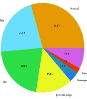
The Histopathological findings of colonoscopic biopsies. Histopathological findings of colonoscopic biopsies are shown in Table 3, where normal findings showed that 31 patients (29.5%) compared to 23 patients (21.9%) who had been diagnosed with non-specific colitis, followed by IBD, 22 (21%). The juvenile polyp (JP) was reported in 15 patients (14.3%). The findings are summarized in Table 3. Results are presented as a pie chart in Figure 1.
Table 3.
Frequencies of Histopathological Findings of Colonoscopic Biopsies a
| Histopathological Finding | Values |
|---|---|
| Normal | 31 (29.5), (95% CI: 21.3 - 39.2%) |
| Non-specific colitis | 23 (21.9), (95% CI: 14.7 - 30.8%) |
| IBD (first diagnosis and follow-up) | 22 (21.0), (95% CI: 13.7 - 30.2%) |
| Ulcerative colitis | 12 |
| Crohn’s disease | 10 |
| Juvenile polyp | 15 (14.3), (95% CI: 8.5 to 22.4%) |
| Non-specific ileitis | 4 (3.8) |
| Haemorrhoids | 2 (1.9) |
| Others (Proctitis, Solitary rectal ulcer, Angiodysplasia) | 8 (7.6) |
| Total | 105 (100) |
a Values are expressed as No. (%), (95% CI: CI %).
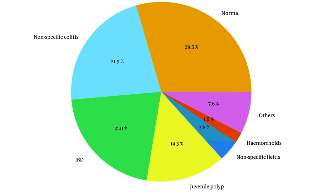
Figure 1.
Chart presenting frequencies of histopathological findings
Association between presenting symptoms and final Diagnosis Among those patients who presented with BPR, polyps were the most common diagnostic finding (15 out of 30 patients (50%). While those 59 patients who presented with suspected IBD (which were as follows: Ten patients with BPR, 27 with diarrhoea, 13 with abdominal pain and weight loss, 6 with anaemia, 3 with perianal fistulas), nonspecific colitis was the commonest diagnosis for them (38.9%), followed by IBD diagnosis (37.3%). On the other hand, 73.3% of 15 patients who presented with miscellaneous symptoms such as abdominal pain (10 patients) or constipation with attacks of bleeding (5 patients), had normal colonoscopy.
A ‘suspected IBD’ cluster was defined a priori as the presence of ≥ 2 symptoms suggestive of chronic intestinal inflammation (chronic diarrhea, rectal bleeding, abdominal pain, anemia, weight loss, or growth failure). This reflects the clinical decision-making process that typically leads to referral for colonoscopy in children with suspected IBD. Table 4 shows the association between presenting symptoms and final diagnosis. Diagnoses are based on histopathology. Patients with overlapping symptoms were categorized according to their primary presenting indication.
Table 4.
Association Between Presenting Symptoms and Final Diagnosis a
| Presenting Symptom Cluster and Diagnosis | Values |
|---|---|
| BPR (excluding IBD cases) (n = 30) | |
| Polyp | 15 (50) |
| Solitary rectal ulcer | 1 (3.3) |
| Proctitis | 3 (10) |
| Angiodysplasia | 1 (3.3) |
| Normal | 10 (33.3) |
| Total | 30 |
| Suspected IBD (BPR, chronic/bloody diarrhea, anemia, weight loss) (n = 59) | |
| IBD | 22 (37.3) |
| Non-specific colitis | 23 (38.9) |
| Non-specific ileitis | 4 (6.8) |
| Normal | 10 (17) |
| Total | 59 (100) |
| Miscellaneous (abdominal pain, constipation with bleeding) (n = 15) | |
| Proctitis | 2 (13.3) |
| Solitary ulcer with hemorrhoids | 1 (6.7) |
| Hemorrhoids | 1 (6.7) |
| Normal | 11 (73.3) |
| Total | 15 (100) |
Abbreviations: BPR, bleeding per rectum; IBD, inflammatory bowel disease.
a Values are expressed as No. (%).
Finally, among 15 patients diagnosed with polyps, 14 of them had a solitary colonic polyp while 1 patient had 2 synchronous polyps at multiple sites. Of the total 17 polyps in those 15 children, 12 were located in the rectosigmoid colon (Table 5).
Table 5.
Locations of the 17 Colonic Polyps in the Fifteen Children with Polyps a
| Location | Values |
|---|---|
| Rectum | 8 (47) |
| Sigmoid | 4 (23.6) |
| Cecum | 2 (11.8) |
| Ascending colon | 2 (11.8) |
| Transverse colon | 1 (5.8) |
a Values are as expressed as No. (%).
4. Discussion
To our knowledge, this is the first systematic study of pediatric colonoscopy in Iraq. Pediatric endoscopy services in Iraq remain limited, with relatively few specialized centers and constrained access to pediatric-trained gastroenterologists and anesthesiologists. This lack of infrastructure often delays diagnosis and treatment of chronic gastrointestinal conditions.
The aim of this study was to describe the demographic and clinical characteristics of children undergoing colonoscopy in Iraq and to observe how indications relate to final diagnoses. We found that rectal bleeding, diarrhea, and abdominal pain were the most frequent indications, while colonic inflammation (non-specific colitis and IBD) and juvenile rectal polyps were the predominant diagnoses. Local data on pediatric colonoscopy are scarce, and it remains unclear how age affects clinical presentation and diagnostic yield in this population.
One hundred and five colonoscopies were performed on patients with an average age of 8 years; most of the patients were between the ages of 5 and 10 years, with 35 children older than 10 years. A slight female predominance was observed among the patients. Similar to a Chinese study done in 2010 in which the mean age was 9.2 ± 4.2 years (12). Colonoscopy in children below 5 years occurs much less frequently, as found by Isoldi et al. (13), which is similar to the observation of this study; only 15 (14.3%) patients were less than 5 years old.
This study observed that successful cecal intubations were achieved in 100 out of the 105 patients (i.e. 96%). This is consistent with the findings of many previous studies; in Pasquarella et al. study, a colonoscopy was completed in 91% of patients (14), and 96.4% in a Korean study by Lee et al. (15). The diagnostic yield was relatively high, which may partly reflect the careful pre-selection of children with significant symptoms (e.g., bleeding, chronic diarrhea, weight loss) in a tertiary referral center. It may also reflect a narrower diagnostic spectrum compared with centers that perform colonoscopy for broader indications. It is important to note that neither Cap-assisted colonoscopy (CAC) nor the water immersion technique was utilized in this study, as these methods are currently not available in our center. Standard pediatric colonoscopy techniques were employed, including careful insufflation with air, gradual passage through the colon, and the use of appropriately sized pediatric colonoscopes. Despite these limitations, successful cecal intubation was achieved in 96% of patients, comparable to international studies (14, 15).
Previous studies have reported high bleeding per rectum as an indication for colonoscopy. Tam et al. (2010) conducted a multicenter study in China, reporting 55.7% of indications (12). In a Saudi study by Alrashidi et al., bleeding per rectum constituted 53.5% of indications for colonoscopy procedures (16), while in our study bleeding per rectum represented 40 (38.1%), less than observed in these studies due to the larger study samples taken. In this study, diarrhea and abdominal pain were the second most common indications for performing colonoscopy; this is in agreement with studies done in Japan and another in Sydney (17, 18).
The histopathological findings recorded in this study show that while 31 patients (29.5%) had normal outcomes, IBD (primary diagnosis and in patients with follow-up) was found in 22 patients (21%). Inflammatory bowel disease was found to be an important finding in other studies as a cause of prolonged rectal bleeding (19, 20). Non-specific colitis was found to be a frequent diagnosis, as it also occurred in 23 patients. The term non-specific colitis (NSC) refers to an inflammatory condition of the colon that microscopically lacks the characteristic features of any specific form of colitis and is commonly seen in histopathological reports following colonoscopic biopsies. The presence of chronic inflammatory cell infiltrate in the colonic mucosa can be seen at some stage in the natural history of colitis due to many etiologies, while cryptitis would usually suggest IBD (21). Similar to the findings of this study, Nambu et al. study reported occurrence of non-specific colitis in 37 out of 275 patients, identifying it as a cause of rectal bleeding (22); our sample is less than that taken in this study.
Regarding the association between presenting symptoms and final diagnosis, in the present study, bleeding per rectum, diarrhea, and abdominal pain were the most common presentations for pediatric colonoscopy referral, corroborating the findings of a previous study by Park, who found that the main causes leading to colonoscopy were bleeding per rectum (56.0%), abdominal pain (27.5%), and diarrhea (3.0%) (20). Seventy-four patients out of 105 patients had a positive diagnosis, including mostly IBD, juvenile polyp (JP), and nonspecific colitis, whereas no abnormalities were observed in 31 patients. Inflammatory bowel disease mostly presented with abdominal pain, bleeding per rectum, or diarrhea. These findings are approximate to several other studies in Japan (18), in which IBD was the most common diagnosis, about 44%, in patients who presented with BPR, abdominal pain, and diarrhea, followed by NSC then JP, and also comparable to recent work such as Morita et al. (23), in which diagnostic colonoscopy in pediatric patients yielded IBD, followed by NSC and polyps, among the leading diagnoses. Similarly, Abdul Aziz et al. (19) reported that IBD and polyps were the most prevalent diagnoses in pediatric colonoscopy.
In children, rectal bleeding is the most common presentation of colonic polyps. Most polyp tumors discovered in children are virtually invariably benign. In this study, most patients with juvenile (refers to the histological type of polyp, not the age) polyps had bleeding per rectum, which is in tandem with findings of previous studies, as in Parga et al. (24), in which the prevalence of polyps was 14.43%. Also, similar to the findings of this study, polyps have been reported in 23 out of 79 patients in Tam et al. study (12). In our study, the majority of polyps were in the rectosigmoid region. A Chinese study by Parga et al. (25) showed that polyps were mainly solitary and located in the rectosigmoid region. A recent study by Kim et al. similarly found that pediatric colorectal polyps were predominantly located in the distal colon. Moreover, a survey of pediatric endoscopists in Korea reported that more than 90% of solitary polyps were found in the left colon (26, 27).
An interesting finding in our cohort was the absence of patients with colonic lymphonodular hyperplasia (CLNH) or eosinophilic colitis (EC), which have been reported in other pediatric series. The lack of CLNH may reflect the age distribution of our population, as this condition is more frequently observed in younger children (< 5 years), who represented only 14.3% of our cohort. Similarly, eosinophilic colitis could have been underdiagnosed, as histopathological evaluation of mucosal eosinophilia can be subtle and may require more extensive sampling than routinely performed in standard colonoscopies. A relatively high number of patients (n = 31, 29.5%) had no definitive histopathological diagnosis. This could be explained by several factors: Mild or patchy disease not captured in biopsies, transient inflammatory conditions, or functional gastrointestinal disorders.
This study is limited by its single-center design and relatively small sample size. Additionally, the lack of standardized long-term follow-up data limits assessment of disease progression, particularly for non-specific colitis. Future multicenter studies are needed to better understand regional epidemiology, risk factors, and outcomes, and to establish local reference data for pediatric colonoscopy in Iraq.
The main strength of our study is its prospective design and comprehensive inclusion of all children undergoing colonoscopy during the study period, offering valuable local epidemiologic data. Limitations include the single-center setting, relatively small sample size, and lack of long-term follow-up, which may restrict generalizability and the assessment of outcomes over time. Nevertheless, these findings fill a critical gap in pediatric gastroenterology in Iraq and provide a foundation for future multi-center studies.
4.1. Conclusions
Pediatric colonoscopy remains a valuable diagnostic tool in children with lower gastrointestinal symptoms. In our cohort, the most frequent indications were rectal bleeding and chronic diarrhea, while the most common diagnoses were colonic inflammation (IBD and nonspecific colitis) and colorectal polyps. Total colonoscopy with terminal ileum intubation is particularly important in patients with suspected IBD, as it allows more accurate assessment of disease extent and severity. While our findings highlight the diagnostic yield of colonoscopy in this setting, they should be interpreted within the context of a single-center study with a selected patient population.