
1. Background
Congenital malformations remain a major cause of prenatal and neonatal mortality worldwide (1-3). According to the World Health Organization, congenital anomalies are structural and functional abnormalities present at birth and often arise during the perinatal period (4). The prevalence of these anomalies varies across populations, with reported rates ranging from 1% to 8% (5-7).
Among congenital anomalies, central nervous system (CNS) malformations are among the most commonly identified categories, largely owing to advances in high-resolution sonographic technology (8). In many countries, routine anomaly screening is performed between 18 and 22 weeks of gestation to detect these conditions (9, 10). However, in our national context, screening is performed earlier because of ethical and legal considerations related to pregnancy termination (11).
Approximately 60% to 90% of congenital anomalies are detected during routine second-trimester anomaly scans. The estimated incidence of CNS malformations ranges from 2 to 10 per 1000 live births (1, 12). Early identification through perinatal screening is crucial for predicting pregnancy outcomes and enabling informed decision-making regarding the management and possible continuation of pregnancies affected by CNS malformations (1, 13, 14).
The prognosis and expected outcomes of pregnancies affected by congenital malformations are of great importance for expectant parents. Timely and accurate prenatal diagnosis enables healthcare professionals to provide comprehensive counseling. This process supports informed parental decisions about continuing or terminating the pregnancy while considering medical, ethical, and legal factors.
2. Objectives
This study aimed to assess the prevalence of prenatally diagnosed CNS malformations in educational hospitals serving as regional referral centers for fetal medicine. Although these findings provide valuable context-specific insights, their generalizability to populations with different demographic characteristics or healthcare-system characteristics may be limited, underscoring the need for cautious interpretation.
3. Methods
3.1. Study Design and Setting
This retrospective cohort study was conducted at Yas Hospital Complex, affiliated with the Tehran University of Medical Sciences, Department of Fetal-Maternal Medicine, between December 2021 and December 2024. Yas Hospital Complex serves as a regional referral center for fetal medicine. It is recognized by legal authorities and patients as a trusted institution for sonographic evaluation in the context of medically indicated pregnancy termination.
3.2. Participants
All pregnant women who visited Yas Hospital Complex during the study period were included. Participants were 18 to 45 years of age and underwent prenatal sonography. Eligibility required a prenatally diagnosed CNS malformation identified by ultrasound. Exclusion criteria were cases in which a second specialist could not validate the initial diagnosis and pregnancies for which the research team could not establish follow-up contact with the patient or obtain relevant information from the patient’s medical records.
3.3. Sonographic Evaluation and Diagnostic Validation
All sonographic examinations were performed by an attending perinatologist using the GE Voluson E6 ultrasound system. Transabdominal imaging was performed with a 2 - 5 MHz probe, and transvaginal assessments were performed with a 4 - 7 MHz probe, as clinically indicated. All anomaly scans were conducted according to the International Society of Ultrasound in Obstetrics and Gynecology (ISUOG) guidelines for routine second-trimester screening, typically performed between 18 and 22 weeks of gestation.
When a CNS malformation was confirmed, patients were referred for secondary evaluation by an independent perinatologist or radiologist to ensure diagnostic accuracy. If both physicians agreed on the diagnosis, it was considered validated. If the anomaly was considered lethal or associated with severe morbidity, the patient was subsequently referred for legal and ethical assessment regarding the possibility of medically indicated pregnancy termination.
3.4. Data Collection and Genetic Evaluation
Demographic and clinical data were collected for all patients, including maternal age, Body Mass Index (BMI), and detailed obstetric history. Information on screening results, including nuchal translucency measurements and noninvasive prenatal testing (NIPT), was extracted from registry forms and medical records. Prenatal sonographic findings and diagnoses were also systematically documented.
When genetic evaluation was indicated, parents were counseled regarding available diagnostic options, and informed consent was obtained before testing. Amniocentesis was performed, followed by either conventional karyotyping or chromosomal microarray analysis (CGH array), depending on the clinical context and suspected anomaly.
3.5. Follow-Up and Outcome Definitions
Patients were followed by telephone or in-person clinic visits, and documentation was retained. Pregnancy outcomes were classified as termination (legal termination, illegal termination, or reduction in twins), spontaneous abortion, or live birth. Legal termination of pregnancy was permitted under the supervision of a forensic medicine organization for certain fetal malformations, provided that the gestational age was less than 19 weeks; these procedures were typically conducted in public hospitals. Illegal termination occurred without the required forensic approval, particularly when gestational age exceeded 19 weeks or when the reason for termination was not approved by the organization; this was often observed in private healthcare settings.
Fetal reduction involved the termination of one fetus in a multiple pregnancy, such as dichorionic diamniotic twins, because of a known abnormality. This procedure was performed under ultrasound guidance using a 22-G spinal needle to inject 15% potassium chloride into the fetal thoracic cavity or heart to induce asystole in the affected fetus.
In cases of live birth, data were collected on the mode of delivery, gestational age at birth, and neonatal intensive care unit (NICU) admission. Neonatal outcomes were documented during a scheduled clinical evaluation conducted by a hospital-affiliated pediatrician within the first postnatal month. Documentation from this visit, including neurodevelopmental assessment, was used to determine early neonatal status and classify outcomes accordingly.
3.6. Ethical Considerations
This study was conducted in accordance with the Declaration of Helsinki and was approved by the Ethics Committee of Tehran University of Medical Sciences under approval code IR.TUMS.MEDICINE.REC.1400.880. Data were collected through retrospective chart review and structured follow-up contacts according to the approved protocol. All patient information was de-identified before analysis, stored on secure institutional servers, and accessible only to authorized study personnel to ensure confidentiality.
3.7. Statistical Analysis
Data were analyzed using IBM SPSS Statistics, version 26.0.0.0. Both quantitative and qualitative variables were analyzed. Categorical variables were analyzed using the Fisher exact test, and quantitative variables were compared using the Mann-Whitney test. Binary logistic regression was used to assess the study outcome. Missing data were handled using multiple imputation by chained equations with 20 imputations. Differences were considered statistically significant at P < 0.05.
4. Results
A total of 398 patients with documented fetal malformations on prenatal sonography were evaluated during the study period. Among these, 140 cases were initially classified as CNS malformations. Five cases were subsequently excluded according to the predefined entry criteria because ultrasound confirmation of the diagnosis by a second physician was not available. Consequently, CNS malformations constituted the most prevalent category, identified in 135 cases (33.9%). Pregnancy outcome data were unavailable for 17 patients (12.5%) because of loss to follow-up (Figure 1).
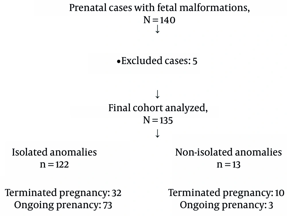
Figure 1.
Study flow diagram
The mean maternal age was 30.49 ± 5.92 years (range, 18 - 43 years). The mean maternal BMI was 26.03 ± 4.14 kg/m2 (range, 18.69 - 41.66 kg/m2). Among the study population, 90.2% of patients reported no consanguineous relationship with their spouse. A history of diabetes mellitus, type unspecified, was documented in 7.4% of cases. In addition, 5.9% of patients had a previous child with congenital malformations. Chronic hypertension was present in 3.0% of patients, and 9.6% had a history of hypothyroidism (Table 1).
Table 1.
Demographic and Clinical Characteristics of All CNS Cases (N = 135) a
| Variables | Value |
|---|---|
| Maternal age, y | 30.49 ± 5.92 |
| Body mass index, kg/m2 | 26.03 ± 4.14 |
| Consanguineous relationship, yes | 13 (9.6) |
| History of a child with an anomaly, yes | 8 (5.9) |
| Diabetes mellitus, yes | 10 (7.4) |
| Chronic hypertension, yes | 4 (3.0) |
| Hypothyroidism, yes | 13 (9.6) |
a Values are expressed as mean ± SD or No. (%).
The mean gestational age at the time of malformation diagnosis was 23 ± 7.4 weeks (range, 11 - 38 weeks). The live birth rate was 54.1%, whereas 25.9% of pregnancies were terminated legally, 3.0% were terminated illegally, and 3 DCDA (dichorionic diamniotic) twin pregnancies underwent reduction to a single twin (Table 2). Intrauterine fetal demise occurred in 3.0% (n = 3) of cases, and the neonatal death rate was 3.7% (n = 5). Notably, 9.6% of malformations were identified during the first trimester.
Table 2.
Pregnancy Outcomes in All CNS Cases (N = 135) a
| Pregnancy Outcomes | Isolated anomaly | Non-isolated anomaly | Total |
|---|---|---|---|
| Vaginal delivery | 16 (13.1) | 1 (7.6) | 17 (12.6) |
| Cesarean section | 57 (46.7) | 2 (15.3) | 59 (43.7) |
| Legal termination of pregnancy | 25 (20.4) | 10 (76.9) | 35 (25.9) |
| Illegal termination of pregnancy | 4 (3.2) | 0 (0) | 4 (3.0) |
| Reduction procedure | 3 (2.4) | 0 (0) | 3 (2.2) |
| Missing outcome data | 17 (13.9) | 0 (0) | 17 (12.5) |
| Total | 122 (100) | 13 (100) | 135 (100.0) |
a Values are expressed as No. (%).
The most common CNS malformation was ventriculomegaly, with 74 cases (54.8%), and mild ventriculomegaly alone accounted for nearly 39% of all anomalies. Ventriculomegaly is defined as dilation of the lateral cerebral ventricles measuring ≥ 10 mm at the level of the atrium. It is typically classified into three categories based on ventricular width: Mild (10 - 11.9 mm), moderate (12 - 14.9 mm), and severe (≥ 15 mm) (15). The next most frequent anomalies were agenesis of the corpus callosum and mega cisterna magna. Most other anomalies occurred in ≤ 6 cases, with vein of Galen aneurysm being the rarest anomaly, identified in 1 case (Table 3). Of the total cases studied, 94 (69.6%) had a single CNS malformation, whereas 41 had multiple CNS abnormalities detected on prenatal ultrasound.
Table 3.
Distribution of Fetal Anomalies in All CNS Cases (N = 135)
| Fetal Malformation | No (%) |
|---|---|
| Mild ventriculomegaly | 53 (39.3) |
| Moderate ventriculomegaly | 5 (3.7) |
| Severe ventriculomegaly | 16 (11.8) |
| Agenesis of the corpus callosum | 9 (6.7) |
| Hypoplasia of cerebellar vermis | 5 (3.7) |
| Mega cisterna magna | 7 (5.2) |
| Holoprosencephaly | 4 (3.0) |
| Encephalocele | 4 (3.0) |
| Anencephaly | 5 (3.7) |
| Microcephaly | 4 (3.0) |
| Myelomeningocele | 6 (4.4) |
| Sacrococcygeal teratoma | 2 (1.5) |
| Scoliosis | 4 (3.0) |
| Cysts | 7 (5.1) |
| Aneurysm | 1 (0.7) |
| Other anomalies | 3 (2.2) |
| Total | 135 (100) |
Three patients in the cohort had a nuchal translucency (NT) measurement exceeding 3 mm. One fetus was diagnosed with anencephaly, and another was diagnosed with holoprosencephaly; both pregnancies were legally terminated. The third case involved mild ventriculomegaly. The patient declined amniocentesis, carried the pregnancy to term, and delivered at 38 weeks. The neonate was reported to be clinically normal at the one-month follow-up.
Noninvasive prenatal testing was performed in 15 patients, and all results were reported as low risk for common chromosomal aneuploidies. Among the 15 patients who underwent amniocentesis, 13 (86.7%) had a normal fetal karyotype. One fetus was diagnosed with trisomy 13, associated with cerebellar hypoplasia and severe ventriculomegaly; this pregnancy was legally terminated. Another case revealed triploidy, accompanied by severe ventriculomegaly and complex congenital heart disease, which also resulted in legal termination. Overall, a normal karyotype was the most common finding among patients who underwent invasive testing.
Associated anomalies involving other organ systems were identified in 13 patients (9.6%). Among these, cardiac and renal malformations were the most frequently observed coexisting anomalies in fetuses with CNS malformations. Cases were categorized as CNS isolated when abnormalities were confined to the central nervous system. In contrast, the non-isolated group included fetuses with CNS anomalies associated with malformations in other organ systems.
When the CNS-isolated and non-isolated groups were compared, there were no differences in age, BMI, or gravidity; however, gestational age at diagnosis (24.9 vs. 17.9 weeks; P = 0.004) and at termination of pregnancy (32.7 vs. 21.5 weeks; P = 0.001) were older in the CNS-isolated group. Consanguineous relationships were more common in the non-isolated group, and this difference was statistically significant (30.8% vs. 7.4%; P = 0.023). Other factors, including diabetes, hypertension, hypothyroidism, and a history of a child with an anomaly, did not show statistically significant differences (Table 4). In terms of outcomes, the non-isolated group had a higher rate of abortions, and this comparison reached statistical significance (P = 0.003) (Table 5). The NICU admission rate was 66.7% in the non-isolated group and 21.9% in the isolated group; however, the difference was not significant (P = 0.138).
Table 4.
Comparison of Demographic and Clinical Characteristics Between Isolated and Non-Isolated Anomaly Groups (N = 135) a
| Variables | Isolated Anomaly (n = 122) | Non-Isolated Anomaly (n = 13) | P-Value b |
|---|---|---|---|
| Maternal age, y | 30.36 ± 5.79 | 31.61 ± 7.21 | 0.409 |
| Body mass index, kg/m2 | 26.15 ± 4.22 | 24.83 ± 3.10 | 0.554 |
| Consanguineous relationship, yes | 9 (7.4) | 4 (30.8) | 0.023 |
| History of a child with an anomaly, yes | 8 (6.6) | 0 (0.0) | 1.000 |
| Diabetes mellitus, yes | 9 (7.4) | 1 (7.7) | 1.000 |
| Chronic hypertension, yes | 4 (3.3) | 0 (0.0) | 1.000 |
| Hypothyroidism, yes | 13 (10.7) | 0 (0.0) | 0.614 |
a Values are expressed as mean ± SD.
b P-values were computed using the Mann-Whitney test and Fisher exact test.
Table 5.
Correlation Between Anomaly Type and Pregnancy Outcomes in All Followed CNS Cases (N = 118) a
| Pregnancy Outcomes | Isolated Anomaly b | Non-isolated Anomaly | Total | P-Value |
|---|---|---|---|---|
| Not aborted | 73 (69.5) | 3 (23.1) | 76 (64.4) | 0.003 c |
| Abortion | 32 (30.5) | 10 (76.9) | 42 (35.6) | |
| Total | 105 (100) | 13 (100) | 118 (100) |
a Values are expressed as No. (%).
b Missing data were 17.
c P-values were computed using pooled binary logistic regression.
In the non-isolated group, 10 patients (76.9%) underwent legal abortion, whereas 3 continued their pregnancies. One patient with severe ventriculomegaly and a cardiac malformation delivered by repeat cesarean section at 38 weeks; the male neonate died 2 days after birth. Another patient with severe ventriculomegaly, hydrocephaly, and ventricular septal defect (VSD) delivered at 30 weeks following induced termination; the neonate died 1 day after birth. The third patient had mild ventriculomegaly and multicystic kidney disease; the male infant was born at 33 weeks, weighed 1500 g, and was delivered by repeat cesarean section because of preterm labor. He survived and remained intubated until 1 month after birth.
Three cases of dichorionic diamniotic twin pregnancies were managed, each complicated by a fetal malformation. In all 3 cases, selective fetal reduction was performed via intracardiac potassium chloride injection.
In 1 case, the affected twin had hydrocephaly; following reduction, the co-twin also demised. In another case, the malformed twin had spina bifida, and in the third case, the affected twin had holoprosencephaly. Both pregnancies progressed to term after reduction.
Overall, ventriculomegaly was the predominant CNS anomaly, most cases were isolated, and pregnancy outcomes varied according to systemic involvement.
5. Discussion
In our cohort of 135 fetuses diagnosed with CNS malformations, these anomalies were the most frequently encountered congenital abnormalities during the study period. These observations are consistent with the findings of Yang et al., who also identified CNS malformations as the predominant category of congenital disorders (16).
Among the various CNS anomalies observed, mild and moderate ventriculomegaly were the most prevalent. According to Carta et al., ventriculomegaly is among the most commonly diagnosed fetal CNS abnormalities, with an estimated prevalence ranging from 0.3 to 1.5 per 1000 pregnancies (15, 17). In our study, 53 cases were classified as mild ventriculomegaly (39.3%) and 5 as moderate ventriculomegaly (3.7%). Notably, all cases of mild and moderate ventriculomegaly resulted in live birth, reflecting a 100% success rate.
These outcomes contrast with the findings of Moens et al., who reported that 29.3% of mild and 51.2% of moderate ventriculomegaly cases led to pregnancy termination (15). Their study also documented live birth rates of 93.1% in mild cases and 78.6% in moderate cases. The observed discrepancies may stem from variations in clinical decision-making, referral patterns, or underlying etiologies across different populations. Tan et al. also investigated fetal CNS malformations and identified ventriculomegaly as the most frequently observed anomaly in their cohort (1).
The mean gestational age at diagnosis in our cohort was 23 ± 7.4 weeks (range, 11 - 38 weeks), closely aligning with the findings of Tan et al., who reported a mean of 24.65 ± 7.37 weeks (range, 10 - 39 weeks) (1). Given the legal restrictions in Iran regarding gestational age for termination, the timing of anomaly detection has critical implications. In our cohort, only 9.6% of CNS malformations were identified during the first trimester. This contrasts with the report by Hu et al., which found that approximately one-third of CNS malformations were detected in the first trimester (18). This discrepancy may reflect differences in sample size and the nature of sonographic assessments; in our population, first-trimester scans were primarily limited to crown-rump length and NT measurements, without dedicated anomaly screening. These findings underscore the importance of implementing early anomaly scans, as timely detection provides parents with the necessary time window to consider legal termination. Notably, in our cohort, 3.0% of abortions were performed outside the legal framework (18, 19).
In our cohort, genetic evaluation with amniocentesis was performed in only 15 patients (11%), of whom 13 (86%) had a normal karyotype. One fetus was diagnosed with trisomy 13, and another was diagnosed with triploidy. By comparison, Tan et al. reported that 30% of their patients underwent chromosomal analysis, with abnormal karyotypes identified in 49% of those with CNS malformations. Edwards syndrome and Patau syndrome were the most frequently observed abnormalities in that study (1). Similarly, Goetzinger et al. analyzed 587 cases of CNS malformation, among which 85% underwent karyotyping; trisomy 21 and trisomy 18 were the most common findings (8). The markedly lower rate of genetic testing in our population likely reflects financial constraints, as the cost of genetic assessment is borne directly by patients and is not covered by insurance. Furthermore, parents who elect pregnancy termination often decline genetic evaluation, contributing to the limited uptake observed in our study (1).
In our analysis, adverse outcomes were predominantly observed among patients with multisystem malformations; notably, 76.9% of these cases resulted in legal termination of pregnancy (P = 0.003). This observation is consistent with the findings of Moens et al., who reported that termination decisions were more frequent in non-isolated cases of ventriculomegaly (15). By contrast, Tan et al. demonstrated that the presence of additional or extracranial anomalies did not significantly influence parental decisions regarding pregnancy termination (1).
5.1. Conclusions
In conclusion, our study found that the mean gestational age at diagnosis of fetal CNS malformations was 23 weeks. This finding underscores the importance of early detection and comprehensive evaluation through first-trimester anomaly scans. The burden of CNS malformations on families is substantial, and our findings, along with those from other studies, emphasize the need to reduce this burden (20). Doing so may improve outcomes in subsequent pregnancies and reduce the occurrence of illegal abortions. When considering legal abortions in cases of CNS malformations, gestational age must be taken into account. In particular, a higher gestational age, especially as it approaches viability, should be a key factor in the decision-making process for the standing committee when abortion may be warranted.
Based on our findings, the number of patients who received genetic assessment was relatively small, and the predominant karyotype observed was normal. This highlights the importance of genetic evaluation, particularly the use of CGH-array technology, because many cases of CNS malformation present with a normal karyotype, while array testing may reveal abnormalities.
5.2. Study Limitations
This study had several limitations. First, 12% of outcome data were missing because of loss to follow-up. Second, subgroup comparisons of clinical outcomes between isolated and non-isolated CNS malformations were limited by the small sample size. In addition, the physicians who performed sonography or evaluated neonates differed among cases, and some patients with a fetus or neonate with malformation refused additional visits or communication.