The anatomical structure of the chest, which consists of air-containing alveoli, muscles, soft tissues, and bones (with major variations in tissue density), makes low-dose CT possible. The X-ray exposure dose is proportional to the square of tube voltage; therefore, reducing the tube voltage can significantly decrease the radiation dose. In a previous study, the radiation dose was reduced by 37% at 100 kV compared to 120 kV (
13). Additionally, Pan et al. (
14) and Meng et al. (
15) found that reducing the tube voltage could significantly reduce the radiation dose. However, reduction of tube voltage led to a decrease in direct photon flow, which had a direct effect on image noise and streak artifacts and could influence the diagnostic value of the image (
16). To improve the image quality, the tube current needs to be increased; therefore, it is not advisable to simply reduce the tube voltage to decrease the radiation dose.
The radiation dose has a linear relationship with the tube current. Reduction of tube current can effectively decrease the radiation dose. In a study by Zhang et al. (
17), the radiation dose was reduced by 87.6%, 75.2%, and 62.8% at 20, 40, and 60 mA, respectively compared to 100 mA; simultaneously, the image noise increased, while the SNR reduced (
18). In another study, the radiation dose nearly doubled when the tube current was reduced from 200 mA to 50 mA (
19); therefore, reduction of radiation dose cannot be achieved by simply decreasing the tube current. Generally, it is an important and arduous task to reach the best combination of tube voltage and tube current while making a diagnosis.
The ASIR technology selectively removes noise by improving the original data reconstruction algorithm, thereby ensuring image quality when scanning at lower doses (
20-
23). The Philips introduced iDose
4, a fourth-generation iterative reconstruction technique based on a dual-space multi-model, which can effectively improve the image spatial resolution and density resolution, remove noise and suppress low-dose artifacts, and sustain the fidelity of CT images by maintaining the structural information through the frequency noise spectrum. Some studies have reported that iDose
4 iterative reconstruction can be applied to low-dose CT scans (
24-
27). The radiology branch of the Chinese Medical Association recommends that the scanning conditions for low-dose CT (LDCT) should be 100 - 120 kV and < 30 mA, using the new generation of nonlinear iterative reconstruction algorithms. In this study, a Philips 256-Layer Brilliance iCT Scanner was used, and a fixed tube current of 30 mA combined with the iDose
4 iterative reconstruction technique at a low tube voltage (80 kV) was considered as the low-dose scanning parameter.
The thymus gland is found at the upper end of the sternum between the right and left lung lobes, below the thyroid gland. The transverse diameter of the thymus is greater than its long diameter during fetal life and becomes narrower and thicker after birth due to pressure on the chest cavity. It is relatively heavy at birth and continues to develop with age until it gradually deteriorates during adolescence (
28-
30). Because of the presence of thymic structures, at a low tube voltage of 80 kV, when the control group used automatic tube current modulation, the radiation dose increased due to the increased attenuation of localized image to X-rays, and the equipment automatically increased the tube current to ensure image quality.
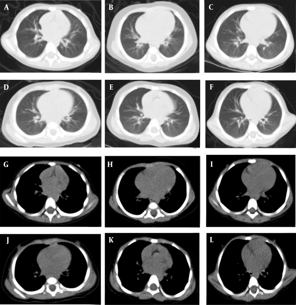
In the present study, a fixed tube current of 30 mA was used for the experimental group, which did not change with the attenuation coefficient. Combined with the iDose4 iterative reconstruction technique, the radiation dose was maintained at a lower level, and the image quality was improved without affecting the diagnostic accuracy. Accordingly, the radiation dose was lower in the experimental group compared to the control group in all age subgroups. The 0-1-year-old infants showed lower lung inflation compared to older infants and had the largest relative thymus weight and attenuation coefficient. Therefore, the 0-1-year-old subgroup exhibited the most significant ED reduction in the experimental group compared to the control group.
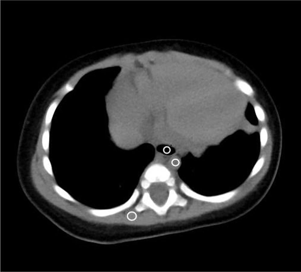
To ensure the stability of data, the CT values of the erector spinae, trachea, and descending aorta were measured at the level of the tracheal ramus for each child (
31). In this study, there was a significant difference in the SNR of the erector spinae in the 0-1-year-old subgroup. The SNR was significantly higher in the control group compared to the experimental group, while the SNR of the descending aorta and trachea showed no significant differences. The SNRs of the erector spinae and descending aorta were significantly different in the 1-2-year-old subgroup; the SNR of the control group was significantly higher than that of the experimental group, while the SNR of the trachea showed no significant differences. There were significant differences in the SNR of the erector spinae, trachea, and descending aorta in the 2-3-year-old subgroup; the SNR was significantly higher in the control group compared to the experimental group. According to this finding, the image noise increased as the radiation dose decreased. With advancing age, the difference in the image SNR of each tissue gradually became significant, while the difference in the trachea image noise was not significant in the 0-2-year-old subgroup, thereby ensuring the image quality for lung tissues containing air. Although the image noise increased in the 2-3-year-old subgroup, the difference in subjective image quality scores was not significant between the experimental and control groups and did not affect the diagnostic accuracy (
32).
The participants of this study were divided into three age subgroups considering the differences in their body size at different ages. According to the literature, the corresponding lowest dose index (DRI) was selected for each age subgroup to make the experiment more scientific, reasonable, and practical. In this study, for the experimental group, the flat-scan phase was used in enhanced CT imaging, even if the image did not meet the diagnostic requirements due to dose reduction; also, in the enhanced phase, conventional scanning conditions were used to ensure diagnosis. In this study, the CTDIvol and DLP, automatically generated by the computer, were collected to calculate the ED as a radiation dose parameter. The ED responds to non-uniform radiation doses received by different parts of the body and is derived from the weighting factor K according to different ages in different parts of the body to ensure the scientific accuracy of data.
The present study had some shortcomings. First, it mainly evaluated the lung tissue structure and lesions, without evaluating structures, such as the mediastinum and bone tissue. Second, the sample size was small, and a larger population is needed to confirm the results. Third, only infants and children aged 0 - 3 years were included in this study, and no other age subgroups were examined; therefore, other age subgroups can be added to validate the results. Finally, this study only included a low-dose plain phase and did not include a booster phase.
In conclusion, a fixed low tube current (30 mA) at a low tube voltage (80 kV), combined with the iDose4 iterative reconstruction technique, for lung CT scans in infants and children aged 0 - 3 years could reduce the radiation dose, while meeting the diagnostic requirements.