


1. Background
2. Objectives
3. Patients and Methods
3.1. General Data
3.2. Equipment and Method
3.3. Image Analysis
3.3.1. Subjective Assessment
3.3.2. Objective Evaluation
![A, The computed tomography (CT) values and image noise (SD) values of common hepatic artery (region of interest [ROI]; 2.0 mm<sup>2</sup> ± 0.5 mm<sup>2</sup>), abdominal aorta ROI (2.0 mm<sup>2</sup> ± 0.5 mm<sup>2</sup>), liver parenchyma ROI (20.0 mm<sup>2</sup> ± 2.0 mm<sup>2</sup>) and erector spine muscle ROI (20.0 mm<sup>2</sup> ± 2.0 mm<sup>2</sup>) were measured at the level of common hepatic artery during the arterial phase. B, The CT values and image noise (SD) values of portal vein main ROI (2.0 mm<sup>2</sup> ± 0.5 mm<sup>2</sup>), liver parenchyma ROI (20.0 mm<sup>2</sup> ± 2.0 mm<sup>2</sup>), and erector spine muscle ROI (20.0 mm<sup>2</sup> ± 2.0 mm<sup>2</sup>) were measured at the portal phase. C, The CT values and image noise (SD) values of hepatic vein ROI (2.0 mm<sup>2</sup> ± 0.5 mm<sup>2</sup>), hepatic liver parenchyma ROI (20.0 mm<sup>2</sup> ± 2.0 mm<sup>2</sup>), and erector spine muscle ROI (20.0 mm<sup>2</sup> ± 2.0 mm<sup>2</sup>) were measured at the exit of the left, middle, and right hepatic vein.](https://brieflands.com/journals/ijradiology/articles/138586/figures/ijradiology-138586-g001-F1-preview.webp "A, The computed tomography (CT) values and image noise (SD) values of common hepatic artery (region of interest [ROI]; 2.0 mm<sup>2</sup> ± 0.5 mm<sup>2</sup>), abdominal aorta ROI (2.0 mm<sup>2</sup> ± 0.5 mm<sup>2</sup>), liver parenchyma ROI (20.0 mm<sup>2</sup> ± 2.0 mm<sup>2</sup>) and erector spine muscle ROI (20.0 mm<sup>2</sup> ± 2.0 mm<sup>2</sup>) were measured at the level of common hepatic artery during the arterial phase. B, The CT values and image noise (SD) values of portal vein main ROI (2.0 mm<sup>2</sup> ± 0.5 mm<sup>2</sup>), liver parenchyma ROI (20.0 mm<sup>2</sup> ± 2.0 mm<sup>2</sup>), and erector spine muscle ROI (20.0 mm<sup>2</sup> ± 2.0 mm<sup>2</sup>) were measured at the portal phase. C, The CT values and image noise (SD) values of hepatic vein ROI (2.0 mm<sup>2</sup> ± 0.5 mm<sup>2</sup>), hepatic liver parenchyma ROI (20.0 mm<sup>2</sup> ± 2.0 mm<sup>2</sup>), and erector spine muscle ROI (20.0 mm<sup>2</sup> ± 2.0 mm<sup>2</sup>) were measured at the exit of the left, middle, and right hepatic vein.")
A, The computed tomography (CT) values and image noise (SD) values of common hepatic artery (region of interest [ROI]; 2.0 mm2 ± 0.5 mm2), abdominal aorta ROI (2.0 mm2 ± 0.5 mm2), liver parenchyma ROI (20.0 mm2 ± 2.0 mm2) and erector spine muscle ROI (20.0 mm2 ± 2.0 mm2) were measured at the level of common hepatic artery during the arterial phase. B, The CT values and image noise (SD) values of portal vein main ROI (2.0 mm2 ± 0.5 mm2), liver parenchyma ROI (20.0 mm2 ± 2.0 mm2), and erector spine muscle ROI (20.0 mm2 ± 2.0 mm2) were measured at the portal phase. C, The CT values and image noise (SD) values of hepatic vein ROI (2.0 mm2 ± 0.5 mm2), hepatic liver parenchyma ROI (20.0 mm2 ± 2.0 mm2), and erector spine muscle ROI (20.0 mm2 ± 2.0 mm2) were measured at the exit of the left, middle, and right hepatic vein.
3.4. Statistical Analysis
4. Results
4.1. General Data
| Information | Group | |
|---|---|---|
| Control Group | Experimental Group | |
| Male-to-female ratio | 20:11 | 16:12 |
| X2 value | 0.336 | |
| P-value | 0.562 | |
| BMI (kg/m2) | 17.687 ± 3.417 | 16.182 ± 2.733 |
| t value | 1.855 | |
| P-value | 0.069 | |
| Average age (mo) | 6.9 ± 2.9 | 6.2 ± 2.8 |
| t value | 0.851 | |
| P-value | 0.398 | |
Abbreviation: BMI, body mass index.
a Values are expressed as mean ± SD.
4.2. Subjective Evaluation of Image Quality
| Group (Scanning Phase) | Physician A Acores | Physician B Scores | Kappa Value | ||||||
|---|---|---|---|---|---|---|---|---|---|
| 1 Points | 2 Points | 3 Points | 4 Points | 1 Points | 2 Points | 3 Points | 4 Points | ||
| Experimental group | |||||||||
| Arterial phase | 0 | 0 | 1 | 27 | 0 | 0 | 2 | 26 | 1.000 |
| Portal vein phase | 0 | 0 | 2 | 26 | 0 | 0 | 2 | 26 | 1.000 |
| Venous phase | 0 | 0 | 1 | 27 | 0 | 0 | 0 | 28 | 1.000 |
| Control group | |||||||||
| Arterial phase | 0 | 0 | 0 | 31 | 0 | 0 | 1 | 30 | 1.000 |
| Portal vein phase | 0 | 0 | 1 | 30 | 0 | 0 | 1 | 30 | 1.000 |
| Venous phase | 0 | 0 | 0 | 31 | 0 | 0 | 2 | 29 | 1.000 |
4.3. Objective Evaluation of Image Quality
| Object of Observation | SNR Value | t-Value | P-Value | CNR Value | t | P-Value | ||
|---|---|---|---|---|---|---|---|---|
| Control Group | Experimental Group | Control Group | Experimental Group | |||||
| Common hepatic artery | 17.287 ± 3.305 | 13.608 ± 3.509 | 4.147 | < 0.001 | 14.931 ± 3.291 | 11.444 ± 3.467 | 3.962 | < 0.001 |
| Aorta abdominalis | 23.153 ± 7.268 | 15.305 ± 3.886 | 5.091 | < 0.001 | 20.628 ± 6.569 | 13.444 ± 4.014 | 4.982 | < 0.001 |
| Hepatic portal vein | 12.638 ± 3.624 | 9.505 ± 1.832 | 4.121 | < 0.001 | 9.608 ± 3.185 | 7.186 ± 1.731 | 3.572 | 0.001 |
| Hepatic vein | 12.954 ± 3.755 | 8.371 ± 1.732 | 5.911 | < 0.001 | 9.347 ± 3.068 | 5.937 ± 1.379 | 5.405 | < 0.001 |
Abbreviations: SNR, signal-to-noise ratio; CNR, contrast-to-noise ratio.
a Values are expressed as mean ± SD.
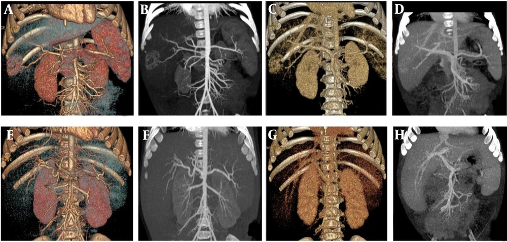
A, B, C, and D are volume rendering (VR) and maximum intensity projection (MIP)min images of the arterial phase and portal veins phase in the control group under scanning conditions. E, F, G, and H are VR and MIPmin images of the arterial phase and portal veins phase in the experimental group under scanning conditions.
4.4. Radiation Dose Comparison
| Group (Parameter) | Control Group | Experimental Group | t-value | P-Value |
|---|---|---|---|---|
| Arterial phase | ||||
| CTDIvol (mGy) | 2.435 ± 0.928 | 1.311 ± 0.442 | 5.843 | < 0.001 |
| DLP (mGy/cm) | 59.861 ± 22.287 | 32.957 ± 11.200 | 5.761 | < 0.001 |
| ED (mSv) | 0.898 ± 0.334 | 0.495 ± 0.168 | 5.764 | < 0.001 |
| Portal vein phase | ||||
| CTDIvol (mGy) | 2.461 ± 0.994 | 1.311 ± 0.441 | 5.643 | < 0.001 |
| DLP (mGy/cm) | 60.048 ± 23.985 | 32.489 ± 11.162 | 5.557 | < 0.001 |
| ED (mSv) | 0.901 ± 0.360 | 0.488 ± 0.167 | 5.56 | < 0.001 |
| Venous phase | ||||
| CTDIvol (mGy) | 2.435 ± 0.928 | 1.311 ± 0.442 | 5.843 | < 0.001 |
| DLP (mGy/cm) | 60.184 ± 22.251 | 32.996 ± 11.214 | 5.828 | < 0.001 |
| ED (mSv) | 0.903 ± 0.334 | 0.495 ± 0.168 | 5.831 | < 0.001 |
Abbreviations: CTDIvol, volume CT dose index; DLP, dose-length product; ED, effective dose.
a Values are expressed as mean ± SD.


