Several reports have demonstrated the diagnostic accuracy of SWE (
2,
8,
12,
19,
20). However, radiologists usually use this technique in addition to conventional US. Thus, we investigated the corrected results of the additional use of SWE to conventional US compared with those of conventional US alone. Moreover, we analyzed the differences between E
max and E
mean, which are the most commonly used parameters.
The current study revealed that the additional use of SWE to conventional US was clinically useful and markedly improved the specificity, PPV and accuracy. However, this approach also resulted in slight diminutions of sensitivity and NPV.
Sensitivity and NPV of conventional US in this study were both 100%. A large number of studies have demonstrated that breast US is already highly sensitive (
21-
24). However, the specificity of US is relatively low, and a high rate of benign biopsies based on US has also been reported (
20,
25-
27). In the present study, the additional use of SWE to conventional US reduced the rate of benign lesion biopsy by 46.0% to 85.7%. This result is similar to those others have reported (
8,
28,
29). In BI-RADS category 3 cases, biopsies are frequently performed at the request of the clinician or patient. Additionally, certain category 3 or 4A breast lesions exhibit overlapping benign and malignant features that may induce false-negative results. Histopathology indicated that 100% of category 3 lesions and 97% of category 4A lesions were benign. This high negative biopsy rate indicated that the decision to perform a biopsy is most often required for category 3 and 4A breast lesions. It has been hypothesized that use of E
max and E
mean cutoff values to reassess lesions that are incorrectly assessed as category 3 or 4A based on conventional US would reduce the numbers of false-negative and false-positive cases. In the present study, application of E
max and E
mean cutoff values to category 3 and 4A lesions resulted in improvements in specificity and accuracy (
Table 1). Therefore, the current results indicate that the use of quantitative SWE with cutoff values, in combination with conventional US, may facilitate the differentiation of breast lesions, particularly those categorized as 3 or 4A based on conventional US alone. Both E
max and E
mean values were effective diagnostic parameters.
Regarding the assessment of elasticity, SWE provides several parameters. Maximum (E
max), mean (E
mean) and minimum (E
min) stiffness represent the general stiffness of the mass. Moreover, elasticity ratio (Eratio) indicates the relative stiffness of the mass to that of fat tissue and standard deviation (SD) indicates the internal heterogeneity of the mass. All of these parameters can help to improve the diagnostic accuracy of US. However, results regarding the identities of the most useful parameters vary across individual studies. Evans et al. reported that E
mean is more useful than E
max and SD (
13). Berg et al. reported optimal diagnostic performance with E
max (
12) and some studies have reported that SD alone exhibits excellent performance (
11). In SWE, the parameters are obtained from a fixed ROI, which is generally the stiffest area, and SD does not reflect the heterogeneity of the entire lesion (
19). Therefore, based on the combination of these studies, we focused on E
max and E
mean.
The present study determined an optimal E
mean cutoff value of 60.7 kPa. This value was lower than that reported by Chang et al. (
8) (80.17 kPa) but similar to the values reported in other studies (
9,
13). Similarly, the optimal cutoff value for E
max determined in the present study (81.3 kPa) was consistent with the findings of previous studies (
9,
12).
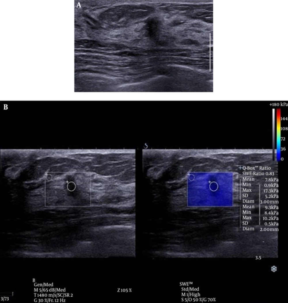
There was one false negative case in this study (
Figure 3). This case involved a 5 mm sized invasive ductal carcinoma (IDC). Although it has been reported that the size of a breast mass does not influence the diagnostic performance of elastography (
30), smaller malignant masses tend to be early stage breast cancers that are composed of softer tissues than larger invasive masses, and this pattern may lead to false negative SWE resulted based on E
max (
15). Additional studies of the application of elastography to small sized, early stage breast cancer on elastography are anticipated in the future.
Conventional ultrasound (US) and shear wave elastography (SWE) of the breast of a 46-year-old female patient. A, conventional US imaging determined a true-positive result. An irregular, taller-than-wide orientated, hypoechoic lesion was identified and classified as category 4C on ultrasound, according to the breast imaging reporting and data system. B, SWE determined a false-negative result. The lesion exhibited a mean elasticity of 7.8 kPa and a maximum of 17.3 kPa. This lesion was diagnosed as invasive ductal carcinoma.
The present study showed statistical difference of average E
max only in deep location. Several investigators indicated that the elasticity of deep located lesions could be affected by the chest wall (
15,
16). However, E
mean showed no statistical significant difference according to lesion depth. This is probably due to the small sized study group in deep location. Further evaluation might be necessary with a large study group about lesion depth.
There are several limitations to the present study. Notably, this study was a small, single-center study with a relatively low number of observers and patients with cancer. Therefore, a larger sample of patients with cancer is required to adequately assess the use of SWE for the diagnoses of soft tumors. Additionally, the present study was performed by radiologists who had only recently been introduced to the center and were thus, relatively inexperienced. This issue may have influenced the performance of the diagnostic imaging techniques. Therefore, with increased experience in elastography, an improved understanding of its value in the assessment of breast lesions may be obtained.
In conclusion, application of SWE appears to increase the diagnostic accuracy of conventional US in the diagnosis of breast lesions. Furthermore, for category 3 or 4A lesions as assessed based on conventional US, use of SWE may reduce the rate of benign biopsies. Additionally, both Emax and Emean values were effective diagnostic parameters and there was no significant difference between these two parameters.


![Receiver operating characteristic (ROC) curves for mean and maximum elasticity values. The area under the ROC curve is 0.8908 [standard error (SE), 0.0423; 95% confidence interval (CI), 0.80805 - 0.97382] for mean elasticity and 0.8884 (SE, 0.0434; 95% CI, 0.80293 - 0.97306) for max elasticity. These results indicated that the mean and maximum elasticity values were of equivalent benefit for differentiating benign and malignant lesions (P = 0.5028).](https://brieflands.com/journals/ijradiology/articles/13486/figures/iranjradiol-14-1-13486-i002-preview-preview.webp "Receiver operating characteristic (ROC) curves for mean and maximum elasticity values. The area under the ROC curve is 0.8908 [standard error (SE), 0.0423; 95% confidence interval (CI), 0.80805 - 0.97382] for mean elasticity and 0.8884 (SE, 0.0434; 95% CI, 0.80293 - 0.97306) for max elasticity. These results indicated that the mean and maximum elasticity values were of equivalent benefit for differentiating benign and malignant lesions (P = 0.5028).")