Cover image of the article: Detection of a Rare Complication of Congenital Cystic Adenomatoid Malformation in a Recipient Twin With Twin-Twin Transfusion Syndrome Using Prenatal Magnetic Resonance Imaging

Image Credit:I J Radiol

Detection of a Rare Complication of Congenital Cystic Adenomatoid Malformation in a Recipient Twin With Twin-Twin Transfusion Syndrome Using Prenatal Magnetic Resonance Imaging

Authors

Yu-Peng Liu1, 2, Yang-Kai Fan1, 2, Yi-Lan Lin1, 2,*, Chih-Ping Chen3, 4, Wen-Ko Su1, 2
1Department of Radiology, Mackay Memorial Hospital, Hsinchu, Taiwan
2Mackay Junior College of Medicine, Nursing and Management, Taipei, Taiwan
3Department of Obsterics and Gyneco, Mackay Memorial Hospital, Taipei, Taiwan
4Department of Medical Research, Mackay Memorial Hospital, Taipei, Taiwan
*Corresponding Author: Corresponding author: Yi-Lan Lin, Department of Radiology, Mackay Memorial Hospital, Hsinchu, Taiwan. Tel: +886-36119595, Fax: +886-225239437, E-mail: Email: a5338@ms7.mmh.org.tw

Iranian Journal of Radiology:Vol. 13, issue 2; e13180
Published online:Apr 27, 2016
Article type:Letter
Received:Jun 25, 2013
Accepted:Apr 15, 2014
How to Cite:Liu Y, Fan Y, Lin Y, Chen C, Su W. Detection of a Rare Complication of Congenital Cystic Adenomatoid Malformation in a Recipient Twin With Twin-Twin Transfusion Syndrome Using Prenatal Magnetic Resonance Imaging. I J Radiol. 2016;13(2):e13180. doi: https://doi.org/10.5812/iranjradiol.13180

Dear Editor,
We would like to report a case of recipient twin with twin-twin transfusion syndrome (TTTS) that developed a very rare complication of congenital cystic adenomatoid malformation (CCAM) detected by magnetic resonance imaging (MRI) but not by ultrasonography (US). MRI was found to be more efficient than prenatal US for the detection of CCAM lesions.
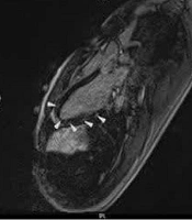
A 30-year-old, gravida 3, para 2 woman who had developed TTTS at 24 weeks’ gestation was referred to our hospital for evaluation of complications in the recipient twin owing to the death of the donor twin. The patient and her husband were healthy and denied any family history of genetic disorders or congenital malformations. The US findings of the recipient twin were normal (Figure 1), and no associated fetal anomalies, except polyhydramnios, were observed. Single-shot fast spin-echo sequence MRI was performed at the 25th gestational week for further evaluation of fetal complications. Findings of the brain were normal, but several small wedge-shaped hyperintense lesions were observed along the periphery of both right and left lungs (Figure 2A-C). The characteristics of these lesions were most consistent with CCAM. The parents decided to terminate the pregnancy. At 26 weeks of gestation, the fetus was delivered with a body weight of 698 g, a normal karyotype of 46 XX and the diagnosis of CCAM was confirmed by subsequent autopsy.
A gravida 3, para 2 pregnant woman with twin-twin transfusion syndrome at 24 weeks’ gestation and death of one of the fetuses. Axial ultrasonography of the chest shows no abnormality.
Figure 1.
A gravida 3, para 2 pregnant woman with twin-twin transfusion syndrome at 24 weeks’ gestation and death of one of the fetuses. Axial ultrasonography of the chest shows no abnormality.
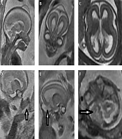
MRI of the chest of the fetus at 25 weeks gestation. A, Axial; B, Coronal; C, Sagittal views.
Figure 2.
MRI of the chest of the fetus at 25 weeks gestation. A, Axial; B, Coronal; C, Sagittal views.
Recipient twin in TTTS may be complicated by brain or heart damage, and rarely CCAM (1, 2). The use of US in cases of TTTS has been well documented, and US is the primary imaging modality in these pregnancies (3). This case highlights the contribution of MRI to the prenatal diagnosis of lung lesions that cannot be detected by US. Therefore, MRI is a more valuable tool for the evaluation of fetal complications such as CCAM in the case of TTTS and it has benefits over US (1, 2). Six cases of CCAM in the recipient twin with TTTS have been reported in the literature that have been detected by prenatal MRI while they had normal US findings (1, 2). On MRI, these lesions may appear as minor wedge-shaped hyperintense lesions along the lung periphery (1), or, as in our case, appear as severe, diffuse, small ill-defined hyperintense lesions on both lungs (2). TTTS is a complication of monochorionic/diamniotic twin pregnancies. It is characterized by an imbalance in blood flow through placental anastomoses causing potentially significant morbidity and mortality in both twins (1, 3). CCAM is characterized by adenomatoid proliferation of the cysts in the lung that resemble bronchioles. The pathogenesis of CCAM remains unknown. Increased cell proliferation, decreased apoptosis, and local disturbances in the levels of normal lung growth factors have been suggested as mechanisms underlying CCAM (4). Imbalance in the blood flow in the recipient with TTTS, causing local disturbances in the levels of normal lung growth factors may play an important role in the development of CCAM in the recipient twin. MRI can serve as a helpful and complementary tool to US for the detection of complications in fetuses with TTTS.
Footnotes

  • Authors’ Contributions:Yu-Peng Liu, first author and corresponding author; Yang-Kai Fan and Yi-Lan Lin, MRI acquisition; Chih-Ping Chen, case collection; Wen-Ko Su, revision of the manuscript and final approval.

References

  • 1.
    Kline-Fath BM, Calvo-Garcia MA, O'Hara SM, Crombleholme TM, Racadio JM. Twin-twin transfusion syndrome: cerebral ischemia is not the only fetal MR imaging finding. Pediatr Radiol. 2007;37(1):47-56. [PubMed ID: 17063351]. https://doi.org/10.1007/s00247-006-0337-5.
  • 2.
    Liu YP, Chang TY. Congenital cystic adenomatoid malformation in twin-twin transfusion syndrome: fetal MR imaging. Pediatr Radiol. 2007;37(9):941. [PubMed ID: 17593361]. https://doi.org/10.1007/s00247-007-0530-1.
  • 3.
    Quintero RA, Morales WJ, Allen MH, Bornick PW, Johnson PK, Kruger M. Staging of twin-twin transfusion syndrome. J Perinatol. 1999;19(8 Pt 1):550-5. [PubMed ID: 10645517].
  • 4.
    Fromont-Hankard G, Philippe-Chomette P, Delezoide AL, Nessmann C, Aigrain Y, Peuchmaur M. Glial cell-derived neurotrophic factor expression in normal human lung and congenital cystic adenomatoid malformation. Arch Pathol Lab Med. 2002;126(4):432-6. [PubMed ID: 11900567].

Copyright

Copyright © 2016, Tehran University of Medical Sciences and Iranian Society of Radiology. This is an open-access article distributed under the terms of the Creative Commons Attribution-NonCommercial 4.0 International License (http://creativecommons.org/licenses/by-nc/4.0/) which permits copy and redistribute the material just in noncommercial usages, provided the original work is properly cited.

Similar Articles

9
Sep
2026
Concurrent Acute Myocarditis in Monozygotic Twins: A Case Report with Similar CMR Findings

Concurrent Acute Myocarditis in Monozygotic Twins: A Case Report with Similar CMR Findings

Arezoo Khosravi,
Mohsen Sadeghi Ghahrodi,
Zahra Alizadeh Sani,
Seyed Javad Davari Sani

Khosravi A, Sadeghi Ghahrodi M, Alizadeh Sani Z, Davari Sani SJ. Concurrent Acute Myocarditis in Monozygotic Twins: A Case Report with Similar CMR Findings. Int Cardiovasc Res J. 2026;20(1):e171799. doi: https://doi.org/10.69107/icrj-171799

30
Dec
2007
Acardiac Parabiotic Twin: A Case Report

Acardiac Parabiotic Twin: A Case Report  

Shirin Bavarian

Bavarian S. Acardiac Parabiotic Twin: A Case Report  . I J Radiol. 2007;4(4):e79086. doi:

12
Apr
2017

Conjoineted Twins Sonography

Ahmad Soltani Shirazi

Soltani Shirazi A. Conjoineted Twins Sonography. I J Radiol. 2017;14(Special Issue):e48195. doi: https://doi.org/10.5812/iranjradiol.48195

28
Sep
2020
A Case of Apert Syndrome: Early Detection of Temporal Lobe Abnormalities and Scalp Cyst in Fetal Brain MRI

A Case of Apert Syndrome: Early Detection of Temporal Lobe Abnormalities and Scalp Cyst in Fetal Brain MRI

Ahmad Reza Tahmasebpour,
Behnaz Moradi,
Mohammad Ali Kazemi

Tahmasebpour AR, Moradi B, Kazemi MA. A Case of Apert Syndrome: Early Detection of Temporal Lobe Abnormalities and Scalp Cyst in Fetal Brain MRI. Inn J Pediatr. 2020;30(6):e103841. doi: https://doi.org/10.5812/ijp.103841

22
Jul
2015
Twin Reversed Arterial Perfusion (TRAP) Sequence; Characteristic Gray-Scale and Doppler Ultrasonography Findings

Twin Reversed Arterial Perfusion (TRAP) Sequence; Characteristic Gray-Scale and Doppler Ultrasonography Findings

Ayla Buyukkaya,
Guven Tekbas,
Ramazan Buyukkaya

Buyukkaya A, Tekbas G, Buyukkaya R. Twin Reversed Arterial Perfusion (TRAP) Sequence; Characteristic Gray-Scale and Doppler Ultrasonography Findings. I J Radiol. 2015;12(3):e14979. doi: https://doi.org/10.5812/iranjradiol.12(3)2015.14979

More by these authors

Yu-Peng LiuPubMedScholar
Yang-Kai FanPubMedScholar
Yi-Lan LinPubMedScholar
Chih-Ping ChenPubMedScholar
Wen-Ko SuPubMedScholar
Share
Cited by
Metrics