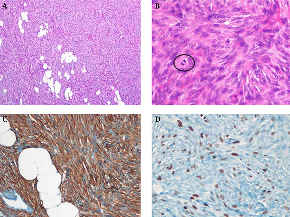
DFSP is a rare cutaneous malignancy that arises from dermis and subcutaneous fat layer. The reported incidence is around 5 cases per 1 million persons annually and is most commonly found in adults aged 20 and 50 years (
1). Although the tumor is a low-to-intermediate grade sarcoma, it can infiltrate into the surrounding deeper tissue with a propensity for local recurrence, especially after inadequate excision. While metastasis is rare, it can occur in distant sites, especially the lung (
2-
4). Furthermore, differentiation into higher-grade sarcomas with higher metastatic potential tends to occur in poorly treated recurrent tumors (
1).
DFSP most commonly appears on the trunk (50 - 60%), extremities (25%) and in the head and neck (10 - 15%). Although breast involvement is quite unusual, it has been reported in the literature with imaging features (
1).
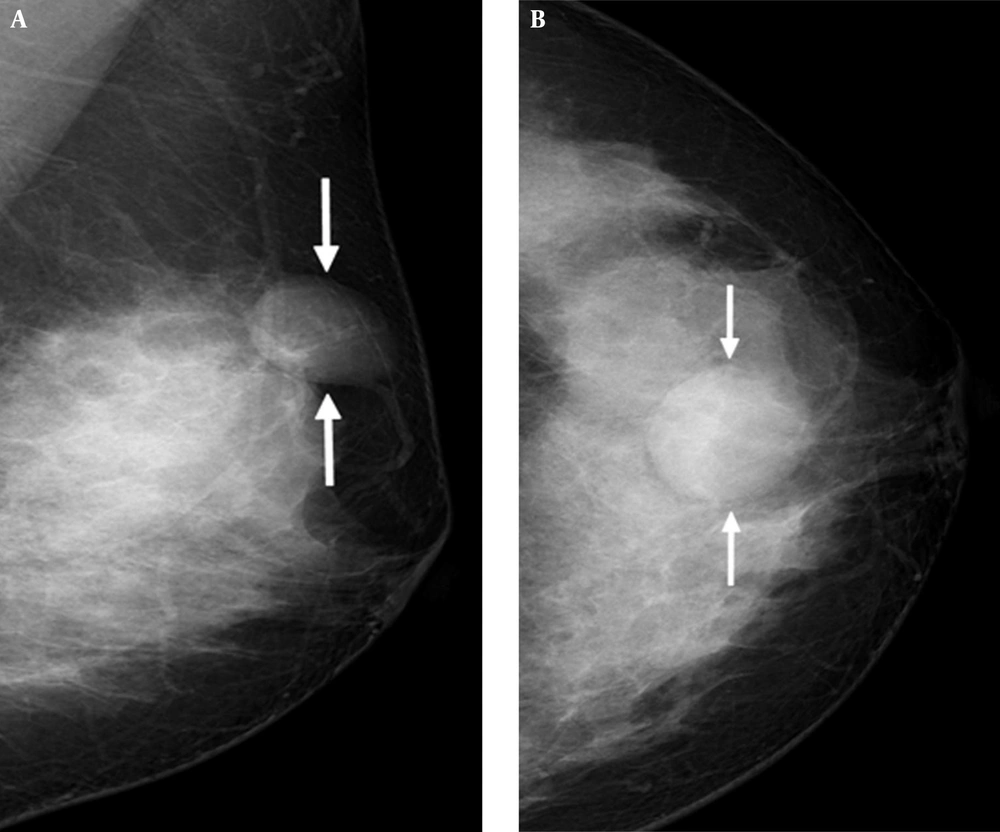
The reported mammographic findings of DFSP are nonspecific, i.e. a skin-based or intramammary-located, oval, circumscribed mass without calcification that mimics a benign lesion (
1,
2,
5). These features are consistent with our case, in which a circumscribed, equal- density, oval mass without calcification was observed in superficially subcutaneous fat layer of the upper central portion of the left breast.
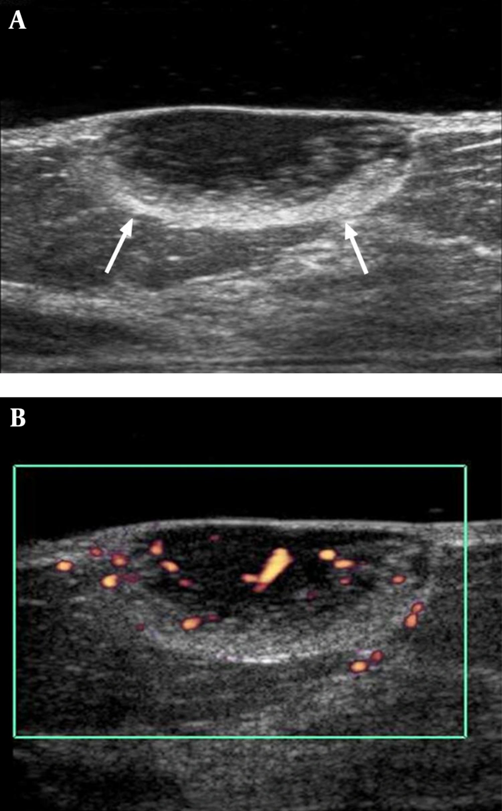
The characteristic ultrasonographic features of DFSP of breast were a circumscribed, hypoechoic, subcutaneous mass with increased internal blood flow in Color Doppler image (
4,
6,
7). Especially, hypervascularity of the tumor helps it to be differentiated from other benign breast lesions. Although a breast abscess or complicated epidermal inclusion cyst such as infection or rupture can appear as a hypoechoic mass with a thick echogenic periphery and increased vascular flow (
8). However, our patient had no clinical symptoms associated with infection such as pain, redness, a sense of heating or fever. Another distinct ultrasonographic feature of DFSP of the breast is a peripheral hyperechoic band like rim in the subcutaneous tissue, which represents a mixture of tumor cells and fibrous tissue infiltrating the subcutaneous fat (
2).
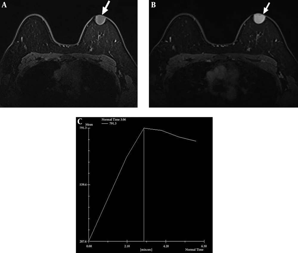
Although the signal intensity of DFSP on breast MR imaging is nonspecific, MRI is the most helpful imaging modality for evaluating the tumor extent and anatomical relationships with surrounding structures. Compared to ultrasound, MRI represents more accurately the tumor size and the boundary because of excellent contrast with surrounding tissue. MR appearance of breast DFSP is a well-circumscribed, intermediate low-intensity tumor on both T1 and T2-weighted images with washout-type kinetics and strong enhancement (
4). We also observed a low signal intensity mass on T1-weighted images with initially rapid strong enhancement with washout kinetics in our case.
Although DFSP has a high tendency for local recurrence after insufficient surgical excision, metastasis is rare. However, distant metastasis may occur at the lung, bone or regional lymph nodes after repeated local recurrence (
9,
10). FDG-PET is a useful tool for predicting tumor recurrence and distant metastasis, detecting multicentric tumor origin and providing relevant anatomical information (
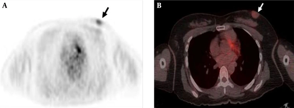
11). A few reports have described PET-CT findings of DFSP including mild uptake in the skin of the trunk and head or more marked uptake at metastatic sites in the lung, retroperitoneum or muscles (
12,
13).
Folpe et al. (
14) reported a highly significant relationship between the highest PET SUV values and cellularity and proliferative indices in sarcomas. In the present case, the tumor showed mild FDG uptake with a SUVmax of 1.8. Although a correlation between FDG uptake and histologic grade in DFSP has not been reported in the literature, we assume that low SUV of the DFSP of the breast was attributable to low cellularity and proliferative indices. To our knowledge, this is the first case showing 18F-FDG PET findings of breast DFSP.
In conclusion, breast DFSP is a rare sarcoma that may simulate a primary breast mass both clinically and radiographically. We described a case of breast DFSP and presented radiologic features that can potentially help provide a differential diagnosis between DFSP and primary benign breast lesions. Mammography, ultrasound, MRI and FDG-PET findings of a subcutaneously located, highly vascular, non-inflammatory tumor with a wide base to the skin might be suggestive of DFSP.