


1. Introduction
Nodular fasciitis is a benign lesion that develops due to proliferation of fibroblastic and myofibroblastic cells within subcutaneous tissue (1-3), commonly found in soft tissues (4). The lesion is seen more frequently in the upper limbs and trunk of young to middle-aged individuals, so development in the breast area is rare (4). The definitive etiology of this lesion is unclear (4-6), but it develops within the breasts of females aged 36 to 48 (1-3). This tumor-like lesion consists of myofibroblastic cells, which, are seeded through a mucopolysaccharide stroma in a circular pattern with a feathery appearance.
Nodular fasciitis is well known to dermatologists and pathologists, due to its preferential occurrence in subcutaneous tissues and importance in the differential diagnosis of soft-tissue tumors. Nodular fasciitis is rarely encountered in other fields of clinical practice. In this case report, we describe a patient with nodular fasciitis in the breast tissue that mimicked a breast cancer manifestation.
2. Case Presentation
The patient was a 43 year-old female who had detected a mass within the upper-lateral quadrant of her left breast 20 days earlier. A past medical history of thyroidectomy, hyperlipidemia and diabetes mellitus existed. Due to a harsh mastalgia, some diagnostic sonographies had been conducted on her breasts during the previous year. No evidence of malignancy was reported, and only fibrocystic changes were detected. Therefore, a follow-up schedule including annual sonography and physical examination was recommended to the patient. Two months after the last survey, the patient detected a mass within the aforementioned site of her left breast.
In examination, the mass was tiny, without any sharp margins, and tender to palpation. No evidence of active painful sensation or discharge from the nipple was detected. Complementary studies including sonography, mammography and excisional biopsy were established and the following findings were reported:
In the sonogram, a 10 mm diameter hypoechoic mass was detected in the 12 o’clock position within the left breast. The first sonography of the lesion [with a BIRADS category of 2] showed a suspicious mass and the second sonography [in which the BIRADS category increased to 4] revealed the mass is growing (Figure 1). Considering the position of the lesion, mammography showed no evidence of mass, but some evidence of fibroglandular condensation were obvious (Figure 2).
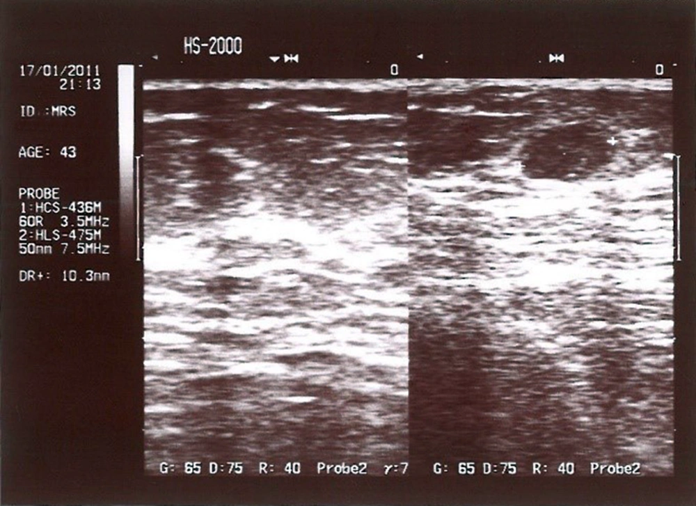
Figure 1.
A 43 year-old woman with a mass in upper-lateral quadrant of left breast. Breast sonography shows a hypoechoic mass.
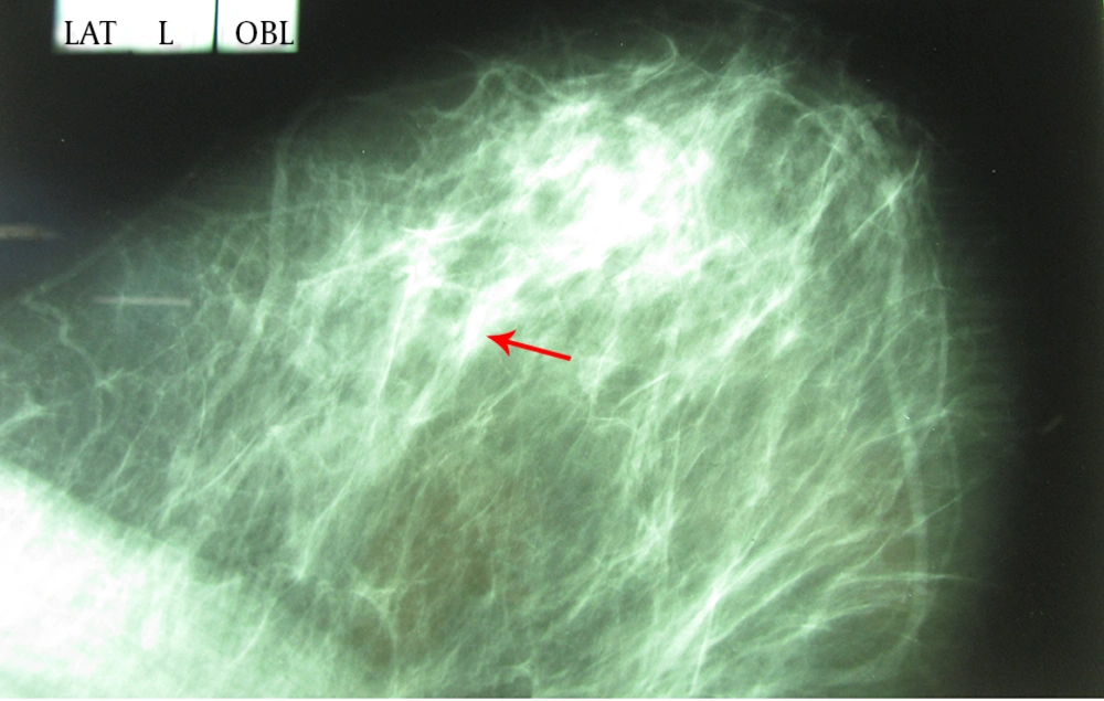
Figure 2.
BIRADS category 4 in mammography of the left breast (mass is marked in the mammography)
Finally, according to the pathologic study, macroscopic and microscopic findings are listed below:
Macroscopic appearances: a gray-cream tissue with the size of 2 × 1.5 × 1 cm contained a creamy nodular lesion with 1.5 cm in diameter and loose formidability.
Microscopic features: the tissue was composed of fusiform fibroblastic cells with the bright ellipsoid-like nuclei, elevated nucleolus and also mitotic formations were obvious.
Low cellular and high cellular zones with hyaline fibrosis and erythrocyte accumulation beside a light lymphocytic infiltration existed.
Considering all of these features in addition to adipocytic accumulation within margins of this lesion, suggested the definitive diagnosis of nodular fasciitis (Figures 3 and 4).
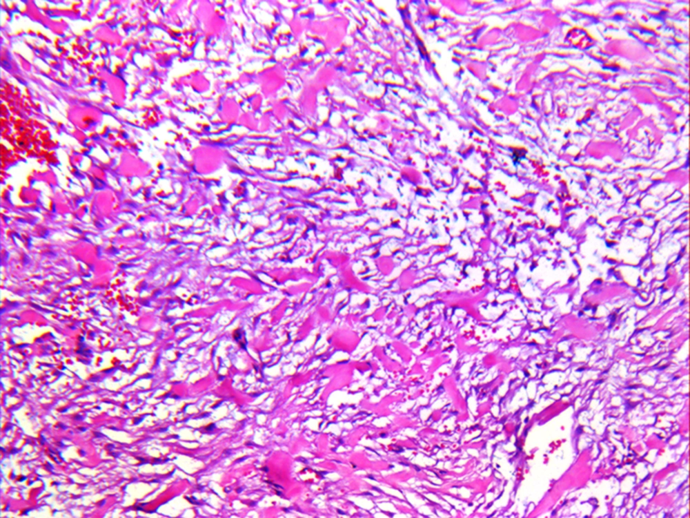
Figure 3.
Microscopic appearance of the mass (H&E staining, ×10)
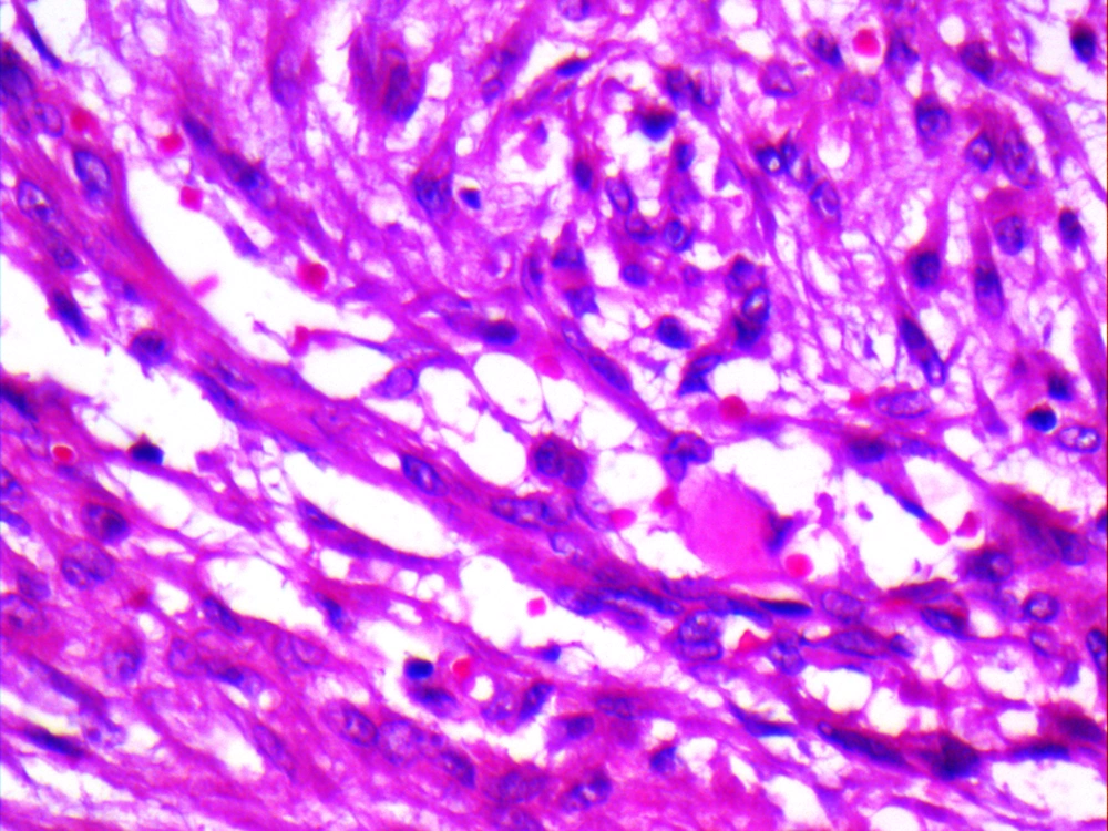
Figure 4.
Microscopic appearance of the mass (H&E staining, ×40)
3. Discussion
Nodular fasciitis rarely develops within the breast (5) and commonly presents with subcutaneous manifestations (6). The incidence ratio of nodular fasciitis between males and females is 1:1 (4, 6).
Nodular fasciitis is clinically and pathologically similar to breast cancer (7). However, in this lesion, nipple discharge is absent and the mass grows rapidly (1-3). For the patient in our example, the mass growth had progressed within only 20 days. In 36% of cases, nodular fasciitis develops in the forearm and 20% in the head and neck region, but the occurrence of this lesion within the breast is quite rare. As shown in Table 1, in our wide internet search, there were only 19 cases of this kind from 1988 until now (4, 6)
Table 1. Summary of Different Cases
| Case | Reference | Year | Age/Gender | Size | Treatment |
|---|---|---|---|---|---|
| 1 | Kontogeorgos et al. (8) | 1988 | - | - | Excised |
| 2 | B. Maly and A. Maly (9) | 2001 | 15-year-old woman | 2 | Excised |
| 3 | Dahlstrom et al. (2) | 2001 | 38-year-old woman | 1.2 | Excised |
| 4 | Polat et al. (6) | 2002 | 66-year-old woman | - | Excised |
| 5 | Tulbah et al. (10) | 2003 | 18-year-old woman | - | Excised |
| 6 | Brown and Carty (11) | 2003 | 65-year-old woman | 5.5 | observed |
| 7 | Porter et al. (12) | 2006 | 75-year-old woman | - | - |
| 8 | Porter et al. (12) | 2006 | 52-year-old woman | - | - |
| 9 | Hayashi et al. (5) | 2007 | 41 -year-old woman | 1.5 | Excised |
| 10 | Squillaci et al. (13) | 2007 | 40-year-old woman | 3.5 | Excised |
| 11 | Yamamoto et al. (14) | 2009 | 18-year-old woman | 0.8 | Excised |
| 12 | Hayashi et al. (5) | 2012 | 25-year-old woman | 0.5 | Excised |
| 13 | Paker et al. (15) | 2013 | 17-year-old woman | 1.5 | Excised |
| 14 | Son et al. (16) | 2013 | 41-year-old woman | 1.1 | Excised |
| 15 | Kakimoto et al. (17) | 2014 | 78-year-old woman | 1.5 | Excised |
| 16 | Samardzic et al. (18) | 2014 | 68-year-old woman | 4 | Excised |
| 17 | Yamamoto S. et al. (14) | 2014 | 35-year-old woman | 3.5 | Excised |
| 18 | Choi et al. (19) | 2014 | 35-year-old woman | 1.5 | Excised |
| 19 | Rhee et al. (20) | 2014 | - | - | - |
In Table 1, we describe cases reported from 2010 till 2014. All of the cases that introduced nodular fasciitis from 1980 until the present totaled 150, but the presence of nodular fasciitis in the breast tissue was rare.
Differential diagnosis between nodular fasciitis and breast cancer is difficult when using radiography or mammography, so the definitive diagnosis is established using accurate histopathology (1-3). In sonography, this lesion manifests as an ellipsoid mass. However, we cannot discriminate nodular fasciitis from breast cancer with this technique (7, 8). Nodular fasciitis develops within the breasts of middle-aged females from 36 to 48 year-old (1-3). Considering this age range for incidence of nodular fasciitis, it could be thought that our patient is in the middle of this range. Fifty percent of all lesions are located within superficial fascias whereas others are within deep ones, correlated with muscles, tendons, vessels, neuronal sheaths and periosteums (4, 6). The size of the mass is commonly around 1 to 3 cm and rarely is more than 5 cm (4, 6).
Finally, considering the extreme similarity between nodular fasciitis and breast cancer, the use of cytologic techniques could reduce unnecessary biopsies and enable physicians to decide whether interventional surgical procedure is more suitable. This also reduces the risk of iatrogenic harm to the patient (1-3).

