SFT is a rare mesenchymal neoplasm accounting for < 2% of all soft tissue tumors (
3). It most commonly presents during the fourth to seventh decades of life with equal incidence rates by sex (
4). It has been known to occur most frequently in the pleural cavity, but it has also been described in other sites - such as the retroperitoneum, abdominal cavity, proximal extremity, and head and neck - which could have occurred owing to the increased recognition of this entity (
3). Among the extrapleural sites, SFTs in the breast have only been reported in 23 cases, which is rare (
3-
9). Moreover, involvement of the male breast is rarer, and only a few cases have been reported (
4,
5).
The typical clinical feature of breast SFT is a slowly enlarging nontender mass, and the symptoms differ depending on the location. When the SFT is in the lungs, chest pain and cough may arise. Abdominal pain and distension may be present when the SFT grows in the abdominal cavity. Intracranial SFT can cause headaches, while no other special symptom except a palpable mass is present in a chest wall SFT (
10). This was also observed in our case.
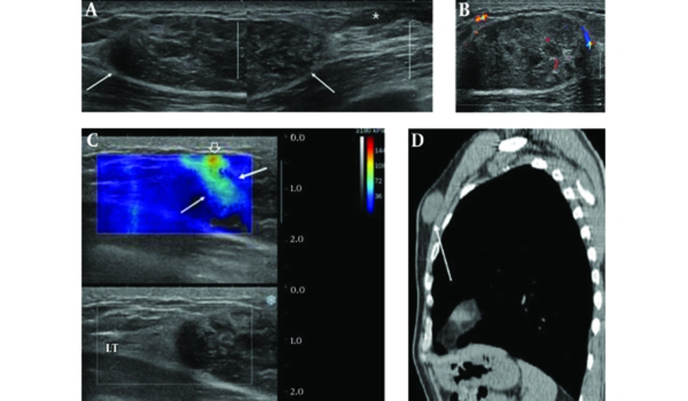
The known ultrasonographic features of breast SFT are nonspecific and similar to a benign solid mass, such as a well-circumscribed, oval-shaped, isoechoic, or hypoechoic mass with vascularity (
3-
8). In our case, the breast SFT was depicted as well-circumscribed, oval-shaped, and heterogeneously hypoechoic mass with peripheral vascularity on color Doppler US.
Elastography, a method for imaging tissue stiffness, is the most noteworthy of the new technologies in recent diagnostic ultrasound systems that improve the specificity for solid masses in the breast. It can help differentiate between benign and malignant masses. According to Barr RG et al., who published the ultrasound elastography guideline, if BI-RADS 3 lesion has characteristics of a malignancy on elastography (strain or shear-wave), such as high stiffness, the lesion should be upgraded and biopsied (
11). In our case, the mass showed high shear wave speed color-coding red on the SWE. It was upgraded to BI-RADS 4; therefore, it had to be biopsied. To the best of our knowledge, there have been no articles describing elastographic findings of the breast SFT.
SFT can be hypodense or hyperdense, with respect to muscle depending on the collagen content, with heterogeneous enhancement on CT. On magnetic resonance image (MRI), the SFT is usually isointense on T1-weighted images and variable on T2-weighted images with vigorous enhancement (
2). Because of these nonspecific radiologic findings and rarity of the tumor, it is difficult to distinguish SFT from other benign soft tissue tumors. Therefore, the exact diagnosis is usually made after surgical resection and immunohistochemical analysis.
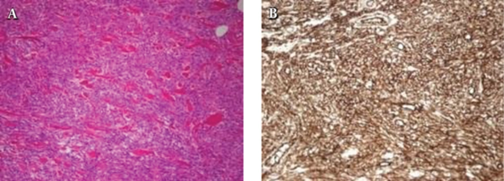
Histologically, SFTs have fibroblast-like spindle-shaped cells and collagen bands. They have high vascularity and a tendency to undergo myxoid degeneration. Differential diagnosis of SFTs includes other bland-looking monomorphic spindle cell lesions in the breast, such as MFB, fibromatosis, nodular fasciitis, hemangiopericytomas, and benign peripheral nerve sheath tumors (
8). Among these, MFBs and SFTs share many histological findings, which makes it difficult to differentiate them from each other. However, MFBs have immunoreactivity to muscle antigens, such as a-SMA and desmin, while SFTs have immunoreactivity to CD34 (
9). In our case, the pathologic result by core needle biopsy was MFB, but the final diagnosis was SFT based on the detailed immunohistochemical examination. Therefore, small tissue sampling by core biopsy alone may have limited diagnostic value for this rare entity. Recently, nuclear expression of STAT6 (signal transducer and activator of transcription 6, interleukin-4 induced) has been found to a highly sensitive and almost perfectly specific immunohistochemical marker for SFT, and it can be helpful to distinguish this tumor type from histological mimics (
12).
Although most SFTs are benign lesions, approximately 10% - 20% of SFTs are malignant, and even benign SFTs have indeterminate malignant potential. Malignant SFTs are correlated with local recurrence and metastasis and the risk of metastasis has been reported to be as high as 25% (
3,
4). Histologically malignant lesions are characterized by hypercellularity, with moderate to marked atypia, necrosis, and mitotic activity of more than four mitoses per 10 high-power fields and infiltrative margins (
3). Up to now, two malignant breast SFTs have been reported in the literature. These showed clear and focal lobulated boundary on mammography or well-defined margin with pathologic uptake on positron emission tomography with 2-deoxy-2-(fluorine-18) fluoro-D-glucose integrated with computed tomography (
7,
13).
In conclusion, we presented a case of breast SFT with a well-defined heterogeneously hypoechoic mass with intralesional blood flows on color Doppler sonography that showed hard elasticity on SWE. Because of the difficulty in its differentiation from other soft tissue tumors due the nonspecific imaging findings and its unpredictable behavior, complete surgical resection with clear margins should be performed, and immunohistochemistry is essential for exact diagnosis.