


1. Introduction
Lipomatosis of the nerve was first described in 1953 by Mason (1). It was previously known as neural fibrolipoma, intraneural lipoma, perineural lipoma, or fibrolipomatous hamartoma (2). In 2002, the world health organization (WHO) suggested the term lipomatosis of the nerve for this entity (3). Lipomatosis of the nerve is characterized by an abnormal infiltration of the nerve by fatty and fibrous tissue; thus, the affected nerve becomes thicker and simulates a mass lesion. It may be associated with macrodactyly, sometimes enough to cause localized gigantism (1, 4, 5). Although most of the cases present in youth, it may be seen in any age (5). Lipomatosis usually affects the nerves of the upper extremities, especially median nerves. The involvement of nerves of the lower extremity is extremely rare (2). Based on our literature review, only few cases with or without macrodactyly have been reported (4-6).
Although both ultrasound and computerized tomography are useful in diagnosis, magnetic resonance imaging (MRI) with high soft tissue resolution is the gold standard. Lipomatosis of the sciatic nerve has unique MRI findings that may prevent unnecessary, contraindicated biopsies. MRI provides the diagnosis of lipomatosis of the nerve with confidence, without requiring histopathologic correlation (2, 6). In this report, the MRI and diffusion-weighted MRI findings of a case of giant sciatic nerve lipomatosis are presented.
2. Case Presentation
A 60-year-old female patient was referred to this clinic to obtain a contrast-enhanced MRI with a prediagnosis of palpable posterior thigh mass and buttock pain. Her chief complaint was swelling in the posterior aspect of the distal left thigh that became larger in the last few years. Contrast-enhanced MRI and diffusion-weighted imaging (DWI), which are part of the tumor protocol, were performed. Axial, coronal T1-weighted, fat-suppressed T2-weighted, and contrast-enhanced fat-suppressed axial and coronal T1-weighted images were obtained using a 1.5-Tesla system (Achieva; Philips, the Netherlands) (Figure 1).
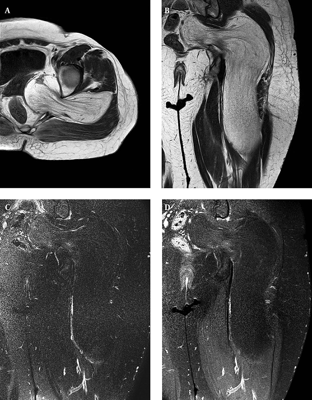
Figure 1.
A 60-year-old female patient with a palpable posterior thigh mass and buttock pain. T1-weighted axial (A) and coronal (B) images show a giant space-occupying lesion that is located in the sciatic nerve course. The tumor is isointense to the subcutaneous fat and there is fine fibrillar appearance inside of it. No signal increase on fat-suppressed T2-weighted image is seen (C). Postcontrast fat-suppressed T1-weighted image (D) shows no intensive enhancement except for faint signal increase at the fibrillar appearance that may represent inherent neural fiber enhancement.
The DWI was obtained before contrast administration with a single-shot spin-echo echo-planar imaging (EPI) technique (repetition time, 4500 ms; echo time, 105 ms; directions of the motion-probing gradients, three orthogonal axes; b value, 0 and 1000 seconds/mm2; field of view, 220 mm; matrix size, 128 × 80 - 128; section thickness, 5 mm with 0.2 - 1.0-mm intersection gaps; NSA and SENSE with a reduction factor of 1 - 1.5). A 7.5 × 25 cm, well-circumscribed giant mass that originated from the osseous pelvis and extended to the gluteal region was detected that located in the sciatic nerve course. Its intensity was similar to those of subcutaneous fat on all pulse sequences. There was a fine fibrillar appearance inside of it. There was no deformity or destruction of the adjacent osseous structures. The mass showed no enhancement after contrast administration.
On the DWI and apparent diffusion coefficient (ADC) mapping, the lesion demonstrated a low signal, isointense to subcutaneous fat, due to a fat-saturated pulse that was used to exclude chemical-shift artifacts. The typical MRI findings and the sciatic nerve course of the lesion allowed the diagnosis of lipomatosis of the sciatic nerve (Figure 2).
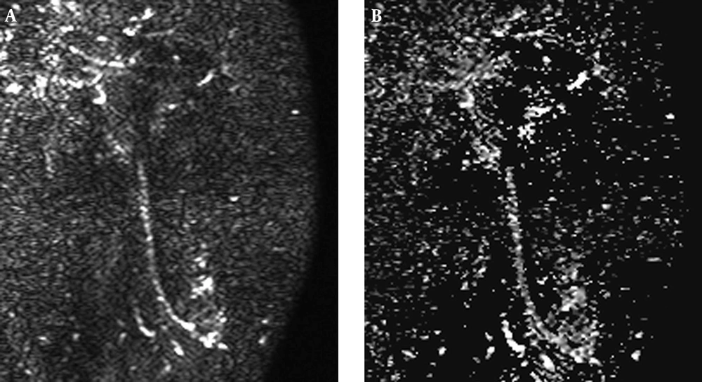
Figure 2.
The lesion is isointense to the subcutaneous fat on diffusion weighted imaging (A) and apparent diffusion coefficient map (B).
Since the patient had suffered an increasing limitation of movement caused by the giant, space-occupying lesion, an internal neurolysis was performed with microsurgical techniques.
3. Discussion
The MRI and diffusion-weighted MRI findings of a case with giant sciatic nerve lipomatosis were presented. The involvement of nerves of the lower extremity is extremely rare, and, to the best of our knowledge, diffusion-weighted MRI findings have not been reported. The abnormal fatty tissue that surrounds the nerve fibers and fusiform enlargement of the nerve are seen as spaghetti-like in longitudinal section and as coaxial cable-like in cross-section; they are usually pathognomonic for lipomatosis of the nerve on T1-weighted images. T2-weighted images do not typically show an increased signal (2, 4, 6). With fat suppression techniques, the high signal of the adipose tissue disappears almost completely. After an intravenous contrast injection, enhancement is not present. However, there may be faint enhancement due to the compression of surrounding structures. In a fusiform mass with these typical findings, the diagnosis is clear; there is no need for further diagnostic work up or surgical biopsy.
A conventional MRI helps characterize the lesions, but it provides low specificity in the differential diagnosis of soft tissue tumors because many of the lesions exhibit nonspecific characteristics. A DWI used in association with a conventional MRI improves the diagnostic confidence. A DWI allows quantitative and qualitative analyses of tissue cellularity and cell membrane integrity, and it has been widely used for tumor detection and characterization (7). It has been reported that a DWI can differentiate benign from malignant soft tissue tumors (8, 9). In this particular case, the described conventional MRI findings were characteristic for lipomatosis; although it is rare in this location with these dimensions. The MRI confirmed the diagnosis. The DWI, which is part of the routine protocol, did not add much information. While performing the DWI, in all of the images, a fat-saturated pulse was used to exclude the chemical-shift artifacts and to reduce the signal in this tumor, confirming its lipomatous nature.
The first case of sciatic nerve lipomatosis was found in Marom and Helms’ study, which was published in 1999 (6). They reviewed the imaging characteristics of 10 cases of nerve lipomatosis in a retrospective study. In 1 of the 10 cases, a 75-year-old male had sciatic nerve lipomatosis without macrodactyly. The size of the tumor was reported to be 28 × 15 × 120 millimeters. Wong et al. reported two cases of sciatic nerve lipomatosis in 2006 (4). The first case was a 68-year-old male with sciatic neuropathic symptoms. An MRI of the left thigh showed a fusiform enlargement of the sciatic nerve, which began at the sciatic notch and extended distally about 10 cm; thus, an MRI diagnosis sciatic nerve lipomatosis without macrodactyly was made. The second case was a 34-year-old female with the diagnosis of sciatic nerve lipomatosis with macrodactyly. Fandridis et al. reported a 26-year-old patient who suffered from sciatic nerve symptoms in 2009 (5). They found a 12 cm sciatic nerve lipomatosis without macrodactyly that started just distal to the ischial tuberosity.
To the best of our knowledge, only these three cases of sciatic nerve lipomatosis without macrodactyly have been reported. This case is the fourth case; it has the largest dimensions and showed the most severe mass effect.
As a final statement about lipomatosis of the nerve, it should be understood that nerve biopsy is usually contraindicated, and it may cause motor and sensory deficits. Therefore, correct interpretation of the MRI is crucial for the diagnosis.

