Most studies have investigated normal breast Hb absorption coefficients, but some data indicated abnormal breast hemoglobin spectroscopy in two types of vessels via NIR light and its comparison with a normal breast. The present study suggests that assessing the absorption coefficient of Hb in major and minor vessels is a valuable method for the diagnosis of abnormal breast tissue.
Previous theoretical and software studies have revealed that the absorption coefficient increases significantly at shorter wavelengths following the Hb spectrum’s improved detection conditions generally being achieved during strong absorption (
24). Thus, a reduction of the illumination wavelength, which leads to an increase in the absorption properties, could provide better contrast and spatial resolution for optical imaging. In addition, the amount of oxygen in the Hb plays an important role on the absorption coefficient.
The blood Hb absorption coefficient in healthy women was 0.04 ± 0.02 cm
-1, while the Hb concentration of abnormal blood was two times as high in pre-menopausal women and four times as high in post-menopausal women than it was in healthy breasts (
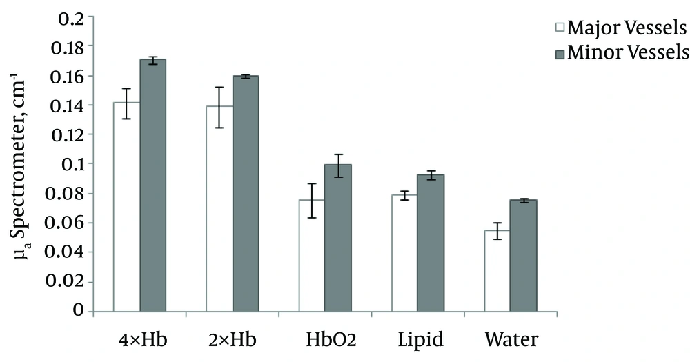
11). Therefore, the Hb concentration may effectively be used as a biomarker for the early detection of breast disease due to hormone variants in different age groups of women. The present study selected the 2× and 4× concentrations of Hb to express the susceptibility of data with this method in different ranges of women in age groups in both pre- and post-menopause. The results showed that the absorption coefficient at 2× and 4× Hb concentrations in abnormal breast blood were significantly distinguishable from normal Hb (P < 0.001).
In the study, the homogeneous phantom μa of oxy- and deoxy-Hb were 0.024 cm
-1 and 0.21 cm
-1, respectively (
25), but the μa of oxy- and deoxy-Hb in our study were 0.075 cm
-1 and 0.17 cm
-1, respectively, in the major vessels.
In a review study, the mean absorption coefficient in oxy-Hb was 0.1 cm
-1; at diffuse optical mammography (600 - 1100 nm), the range of normal breast tissue was 0.02 - 0.12 cm
-1 (
4). In the present study, the absorption coefficients in different components of healthy breast tissue (Hbo
2, lipids, water) ranged from 0.05 - 0.099 cm
-1.
A method used to integrate the sphere technique and inverse Monte Carlo simulations at 633 nm with a 98% oxygen saturation showed a 0.021 ± 0.002 cm
-1 absorption coefficient (
26). In this study, the absorption coefficients at 637 nm in oxy-Hb were 0.075 and 0.99 cm
-1 in the major and minor vessels, respectively, which may have been due to the different oxygen saturations of the Hb. The aforementioned studies indicated that there is controversy in μa for oxy- and deoxy-Hb at different concentrations of Hb. There is no evidence of major or minor vessels.
The chromosphere materials, such as oxy- and deoxy-Hb, are at the origin of the absorbing structures since they account for the blood absorption coefficient. The studies on water and lipid absorption coefficients revealed μa that were ∼10
-2 cm
-1 (
27,
28). In this study, the μa values for water and lipids were 0.055 and 0.079 cm
-1, respectively.
A comparison between the components of the breast and the Hb absorption coefficients showed that the water μa was lower than that of lipids and also lower than Hbo
2 and Hb (
4,
29-
31). In the present study with the SM, the water μa in minor vessels was 0.075 ± 0.003, while lipids were 0.093 ± 0.007 and Hbo
2 were 0.099 ± 0.017. Almost all previous studies used only spectroscopy data for their absorption coefficient calculations, but this study determined the Hb concentration with SM and PM. As a result, we showed that there was no significant difference between data from SM and PM, and both data were significant for the separation of oxy- and deoxy-Hb in the breast. The simultaneous study of two types of vessels, major and minor, provided a different μa and showed the effectiveness of the diameter of vessels in the detection of normal and abnormal breast properties. In the previously mentioned studies, there were no investigations on the diameter of vessels or its effect.
The SM and PM had the ability to detect both normal and abnormal breast blood Hb absorption coefficients via NIR transmission. There is one application of optical imaging that can take advantage of its high sensitivity to the Hb concentration of cancerous tissue without suffering from the limited specificity of this measure. In fact, a non-ionizing and non-invasive technology, such as intrinsic-contrast-based diffuse optical imaging, which can be safely performed repeatedly on the same patient at regular time intervals, is particularly suited for monitoring individual responses to a cancer diagnosis in breast screening, especially for younger women.
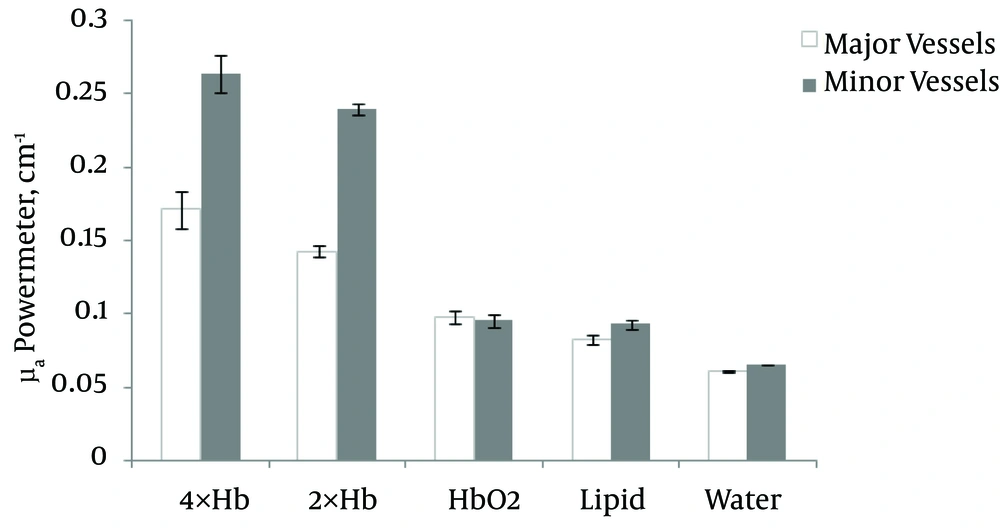
The PM and SM are used for measuring the NIR light output of the breast blood vessel Hb and can differentiate between major and minor vessels and also between different concentrations of Hb and other fluids. One of this study’s novelties is its consideration of the diameter of vessels as an effective factor in early detection using spectrometry or PM assay. The NIR recording results were obtained from the right and left sides of the upper and lower parts of the breast. Therefore, any locations of variation on the breast are clear. The average differentiates can be investigated between water, lipids, 2×, 4×, and oxy-Hb using a SM. However, a PM showed obvious differences between 2×, 4×, and oxy-Hb in the major and minor vessels. SM sensitivity and specificity values in the diagnosis of normal and abnormal Hb were 89.4% and 85.7%, respectively, whereas the PM sensitivity and specificity values were 100% and 100%, respectively, which indicates a high diagnostic capability. This study showed a significant effect of the vessel diameter on μa Hbo2, 2× Hb, and 4× Hb in major and minor vessels. In addition, the usefulness of the data of different concentrations of Hb for the pre-or post-menopause age groups of women was ascertained.
Our data showed that the relations between absorption coefficients in measured fluids in a breast phantom containing major and minor vessels were as follows: water ≤ lipids < Hbo2 < 2× Hb < 4× Hb. Significant differences in NIR absorption properties have been found between normal (Hbo2) and abnormal (2× Hb or 4× Hb) breasts. Thus, the PM and the SM are both reliable devices for measuring different concentrations of Hb via NIR diffusion in a breast phantom.
Finally, assessment of the Hb absorption coefficient is a useful method for the non-invasive and early differentiation of healthy and abnormal breast tissue.