


1. Introduction
2. Case Presentation
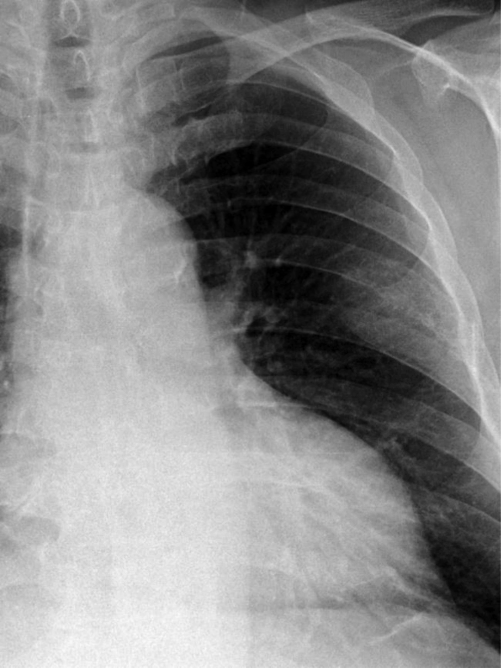
A 63-year-old man presenting with an incidentally detected rib mass on a plain chest radiograph diagnosed as hemangioma. Plain radiograph of the chest illustrates a moderately defined mass in the left upper hemithorax arising from the sixth rib.
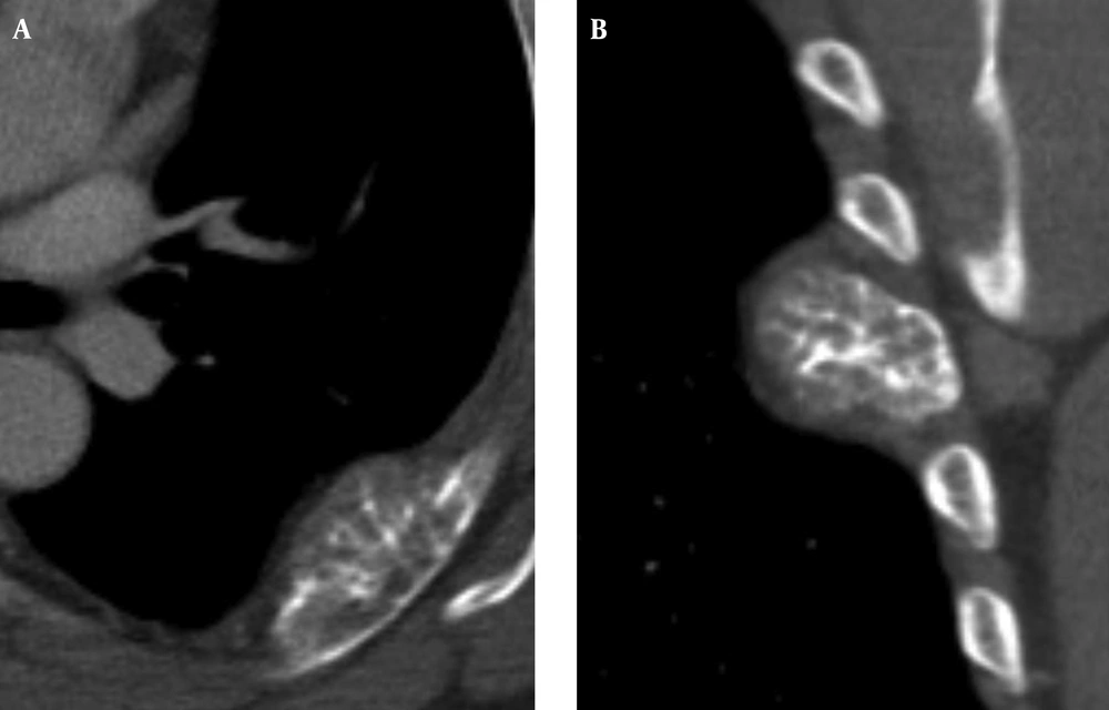
Axial contrast-enhanced chest CT image (A) and coronal reformation image (B) show an osteolytic eccentric expansive mass in the posterolateral part of the left sixth rib with sunburst calcification and focal cortical disruption.
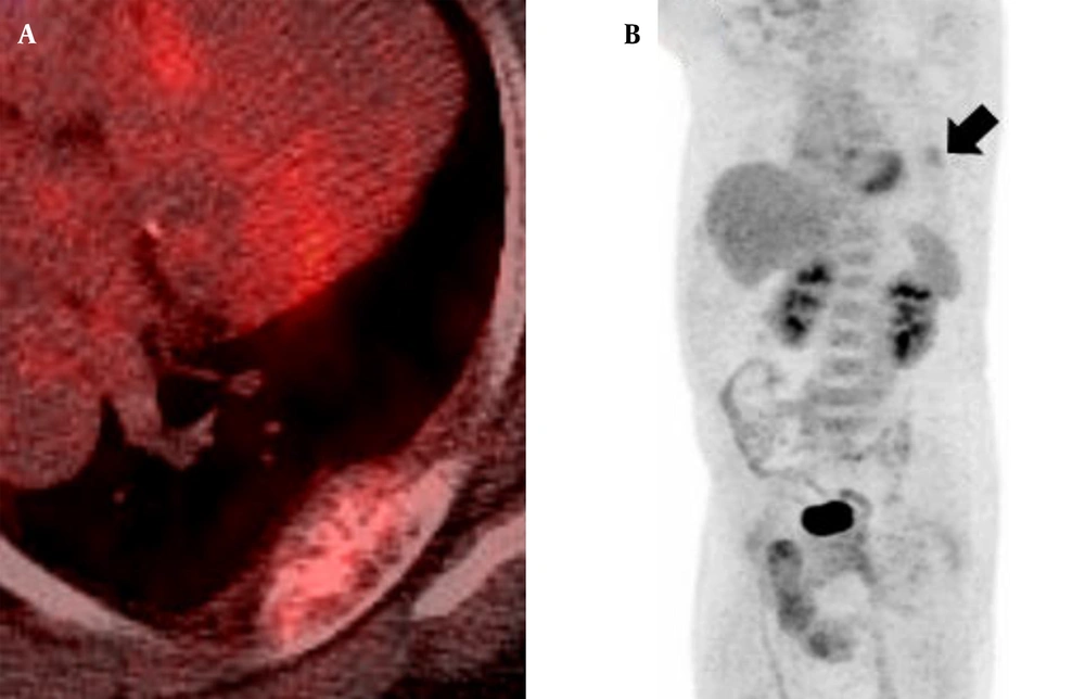
Axial 18F-FDG PET/CT fusion image (A) and MPI image (B) demonstrate non-discrete focal 18F-FDG uptake (SUVmax 2.4) in the left sixth rib (arrow) without any other abnormal 18F-FDG uptake lesion.
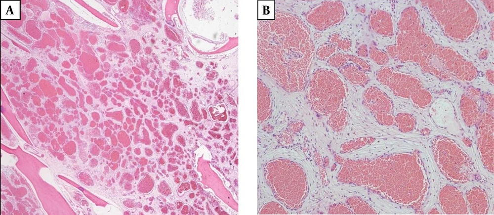
Microscopic findings of the resected mass. A, Normal bone tissues are interspersed within the mass (hematoxylin-eosin, original magnification ×20). B, The mass consists of thin-walled blood vessels with single layer of endothelial-cell lining containing red blood cells (hematoxylin-eosin, original magnification ×100).

