


1. Background
2. Objectives
3. Patients and Methods
3.1. Patients
| Venography | No. (%) |
|---|---|
| CTV only | 29 (59.2) |
| MRV only | 8 (16.3) |
| CTV + DSA | 5 (10.2) |
| CTV + MRV | 4 (8.2) |
| CTV + MRV + DSA | 2 (4.1) |
| MRV + DSA | 1 (2.0) |
| Total | 49 (100) |
Abbreviations: CTV, CT venography; DSA, digital subtraction angiography; MRV, MR venography.
3.2. CTV Protocol
3.3. MR and MR Venography Protocols
3.4. Image Analysis
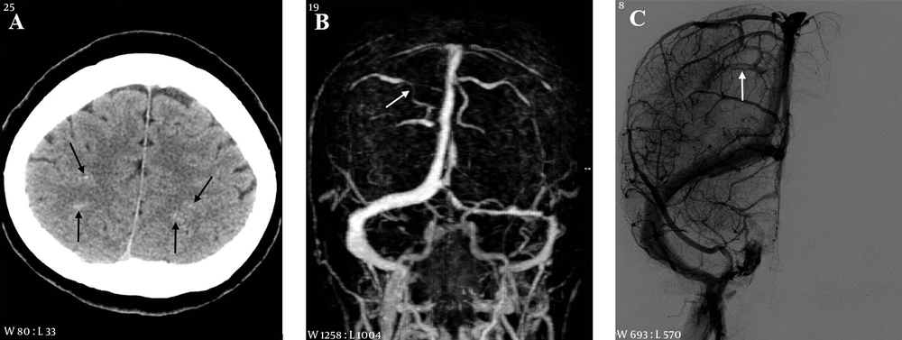
A 53-year-old female with headache and right weakness 3 days before imaging. A, CT scan shows subarachnoid hemorrhage in the cortical sulci of both cerebral convexities (arrows); B, MR venography reveals segmental narrowing in a right cortical vein (arrow); C, On digital subtraction angiography (DSA), there is a filling defect in a right cortical vein (arrow).
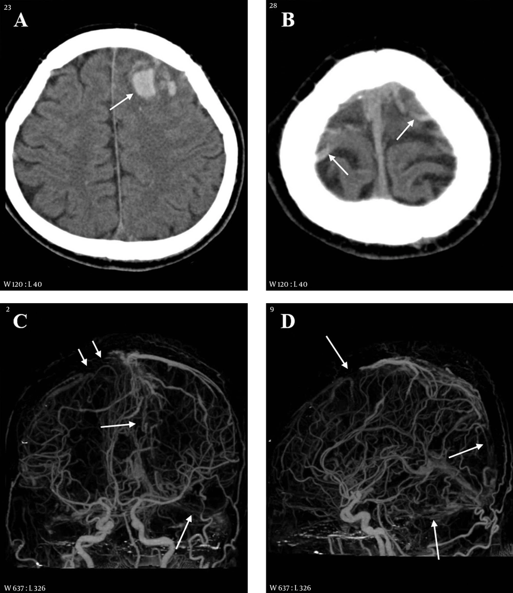
A 38-year-old male with headache, altered mentality and right weakness 2.5 hours before imaging. A, CT scan shows multiple hemorrhages in the left frontal lobe and adjacent cortical sulci (arrow); B, CT scan of the cerebral vertex level reveals hyperdense cortical veins (arrows) at both cerebral convexities; C and D, On CT venography frontal (C) and lateral (D) projection images, the cortical vein (short arrows) and superior sagittal and transverse sinuses (long arrows) are extensively thrombosed.
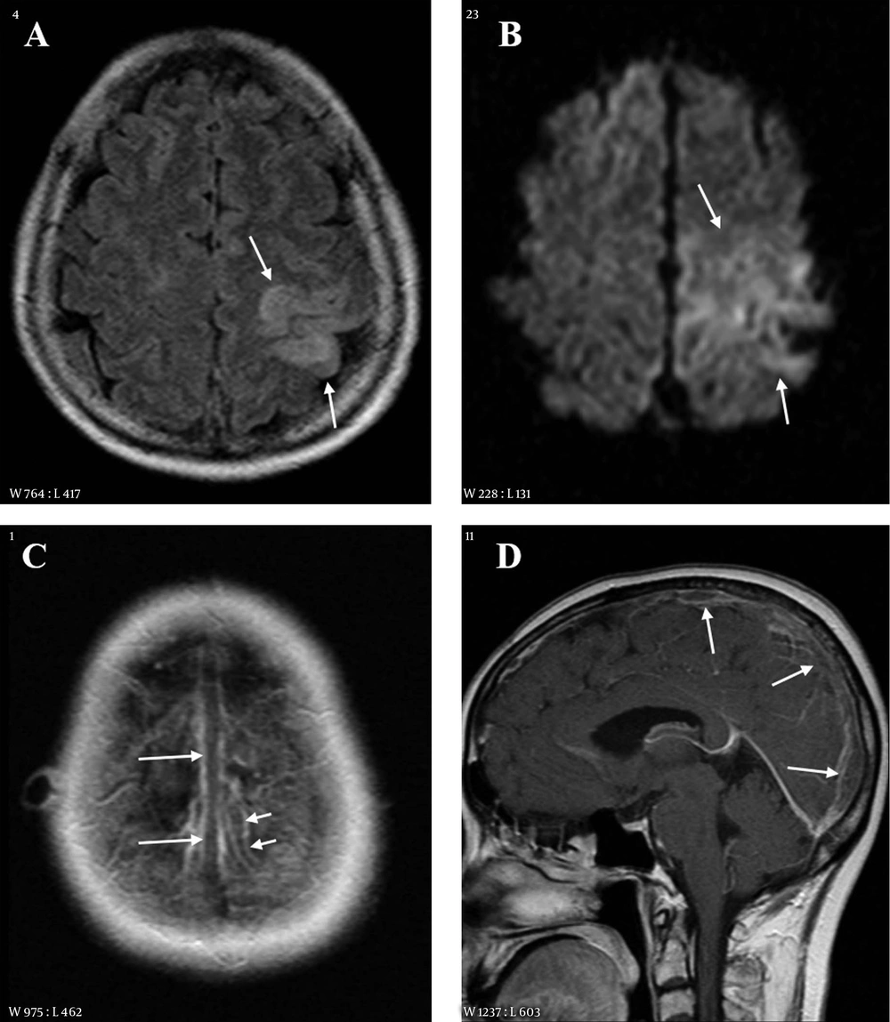
A 55-year-old female with right weakness and seizure 10 hours before imaging. A, Fluid attenuated inversion recovery (FLAIR) image shows a hyperintense lesion in the left parietal lobe; B, Diffusion weighted imaging (DWI) also reveals the lesion as hyperintensity. The extent of lesion is larger than that on FLAIR; C and D, Contrast enhanced axial (C) and sagittal (D) images show filling defects in the superior sagittal sinus (long arrows) and in a cortical vein of the left parietal area (short arrows).
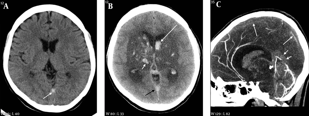
A 35-year-old female with headache and seizure 3 days and 1 hour before imaging, respectively. A, Initial CT scan obtained at an outside hospital shows a hyperdense lesion in the straight sinus area; B, On follow-up CT taken after seizure, there is a large hypodense lesion involving both basal ganglia and thalami. Multiple hemorrhages are associated within the lesion (white short arrow) and in both lateral ventricles (white long arrow). A hyperdense lesion is also seen in the straight sinus area (black arrow); C, Sagittal reformation CT venography image reveals extensive thrombosis in the superior sagittal (long arrows) and straight sinuses (short arrows).
3.5. Statistical Analysis
4. Results
| Total (n = 49) | CV positive (n = 31) | CV negative (n = 18) | P value | |
|---|---|---|---|---|
| Age, year, mean ± SD | 48.6 ± 20.8 | 48.2 ± 20.5 | 49.4 ± 21.8 | 0.848 |
| Male, No. (%) | 22 (44. 9) | 13 (41.9) | 9 (50.0) | 0.767 |
| Time from symptom onset (hour) median, range | 11.0 (0.5 - 480) | 13.0 (0.5 - 432) | 5.6 (1 - 480) | 0.590 |
| Symptoms and signs | ||||
| Headache | 30 (61.2) | 18 (58.1) | 12 (66.7) | 0.762 |
| Focal neurologic deficit | 24 (49.0) | 21 (67.7) | 3 (16.7) | 0.001 |
| Seizure | 20 (40.8) | 13 (41.9) | 7 (38.9) | 1 |
| Altered consciousness | 9 (18.4) | 4 (12.9) | 5 (27.8) | 0.259 |
| Psychotic symptom | 1 (2.0) | 0 (0) | 1 (5.6) | 0.367 |
| mRS at discharge (median, range) | 0 (0 - 6) | 0 (0 - 6) | 0 (0 - 6) | |
| 0 - 2 | 37 (75.5) | 24 (77.4) | 13 (72.2) | 0.738 |
| 3 - 6 | 12 (24.5) | 7 (22.6) | 5 (27.8) | 0.738 |
Abbreviations: CV, cortical vein; mRS, modified rankin scale; CVT, cerebral venous thrombosis; SD,standard deviation.
| Sites | Total (n = 49) | CV positive (n = 31) | CV negative (n = 18) | P value |
|---|---|---|---|---|
| CV | 31 (63.3) | 31 (100) | 0 (0) | < 0.001 |
| SSS | 30 (61.2) | 23 (74.2) | 7 (38.9) | 0.032 |
| TS | 26 (53.1) | 12 (38.7) | 14 (77.8) | 0.016 |
| SS | 21 (42.9) | 11 (35.5) | 10 (55.6) | 0.234 |
| StS | 10 (20.4) | 3 (9.7) | 7 (38.9) | 0.025 |
Abbreviations: CV, cortical vein; SSS, superior sagittal sinus; TS, transverse sinus; SS, sigmoid sinus; StS, straight sinus, CVT, cerebral venous thrombosis.
aValues are expressed as No.(%).
| Findings | Total | CV positive | CV negative | P value |
|---|---|---|---|---|
| Intracranial hemorrhage (n = 49) | 33 (67.3) | 24 (77.4) | 9 (50.0) | 0.063 |
| ICH | 20 (40.8) | 14 (45.2) | 6 (33.3) | 0.550 |
| SAH | 19 (38.8) | 16 (51.6) | 3 (16.7) | 0.018 |
| SDH | 4 (8.2) | 2 (6.5) | 2 (11.1) | 0.618 |
| IVH | 3 (6.1) | 0 (0) | 3 (16.7) | 0.044 |
| Parenchymal hyperintensity (n = 38) | 24 (63.2) | 18 (69.2) | 6 (50.0) | 0.296 |
Abbreviations: CV, cortical vein; CVT, cerebral venous thrombosis; ICH, intracerebral hemorrhage; IVH intraventricular hemorrhage; SAH, subarachnoid hemorrhage; SDH, subdural hemorrhage.
aValues are expressed as No.(%).


