Contrast-Enhanced Ultrasound Characteristics of Bladder Leiomyoma: A Case Report

Author(s):
Yu YangYu Yang1,*
1Department of Ultrasound, Beijing Friendship Hospital, Capital Medical University, Beijing, China
*Corresponding Author: Department of Ultrasound, Beijing Friendship Hospital, Capital Medical University, P.O. Box: 100050, No.95, Rong’an Rd, Beijing, China. Email: [email protected]

IJ Radiology:Vol. 16, issue 1; e68855
Published online:Aug 13, 2018
Article type:Case Report
Received:Mar 24, 2018
Accepted:Jul 16, 2018
How to Cite:Yang Y. Contrast-Enhanced Ultrasound Characteristics of Bladder Leiomyoma: A Case Report. I J Radiol. 2019;16(1):e68855. doi: https://doi.org/10.5812/iranjradiol.68855

Abstract

Bladder leiomyoma is a rare benign neoplasm that may mimic a malignant lesion in medical images. We report a woman with bladder leiomyoma who was correctly diagnosed by contrast-enhanced ultrasound (CEUS). The patient was referred to our hospital after a bladder neoplasm was detected during a health checkup. CEUS showed fast wash-in and slow wash-out in the tumor, similar to a normal bladder wall. To conclude, bladder leiomyoma has its specific characteristics on CEUS, and this modality may be a good option for diagnosis of bladder leiomyoma.

1. Introduction

Bladder leiomyoma is a rare benign neoplasm that is most commonly seen in women in the third through sixth decades of life. It may be endovesical, intramural, or extravesical (1, 2). Ultrasound is the preferred modality for imaging of bladder neoplasms. However, bladder leiomyomas may mimic malignant lesions, and depending on the location, is often correctly diagnosed only after surgical removal (3).

2. Case Presentation

A 58-year-old woman was referred to our hospital for a bladder neoplasm that had been detected during a routine health examination. She had no obstructive or irritative urinary symptoms, and laboratory workup was normal. Sonographic examination was performed using an EPIQ 5 ultrasound machine (Philips Medical Systems, Netherlands) equipped with a transvaginal probe (C10-3v). The examination, performed with the patient in the lithotomy position, revealed a 28 mm × 16 mm × 25 mm, well-defined, hypoechoic sessile mass in the posterior wall of the bladder (Figure 1). Contrast-enhanced ultrasound (CEUS) was used to detect the tumor blood flow. Informed consent was obtained before CEUS was performed. Contrast agent (SonoVue; Bracco, Milan, Italy) was mixed with 5 mL normal saline and a 2.4-mL bolus of this mixture was administered via the median cubital vein, followed immediately by a 5-mL normal saline flush. Real-time continuous ultrasound examination under a low mechanical index was used to match up this microbubble contrast agent.
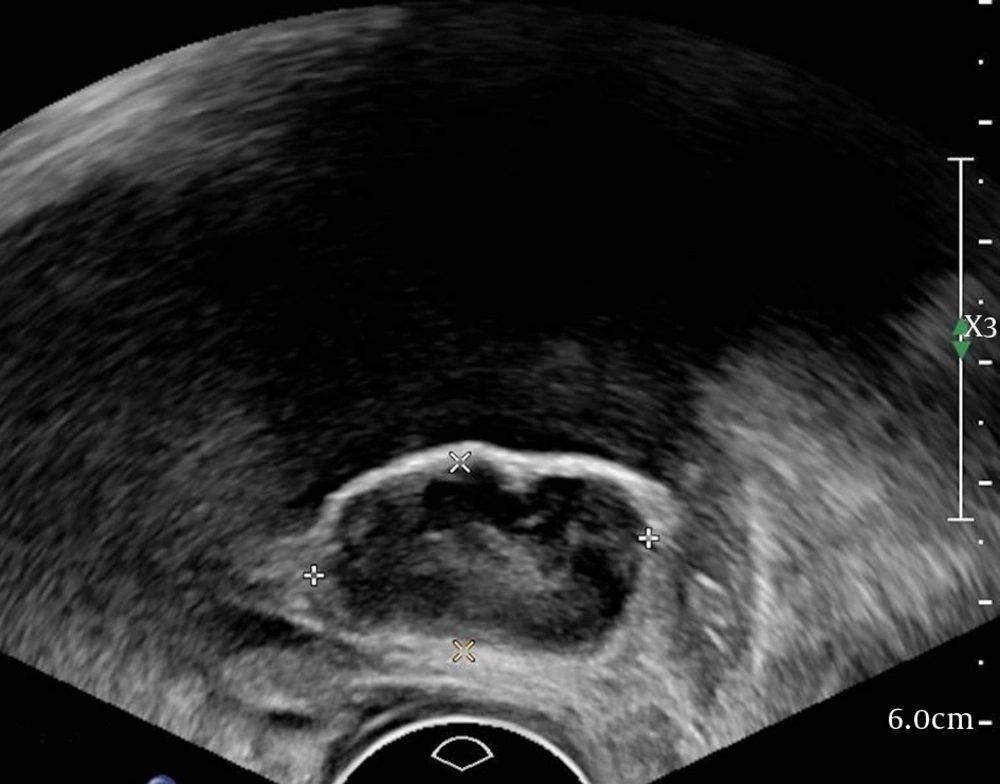
A 58-year-old woman with bladder neoplasm. Sagittal transvaginal sonogram shows a well-defined, hypoechoic mass in the posterior wall.
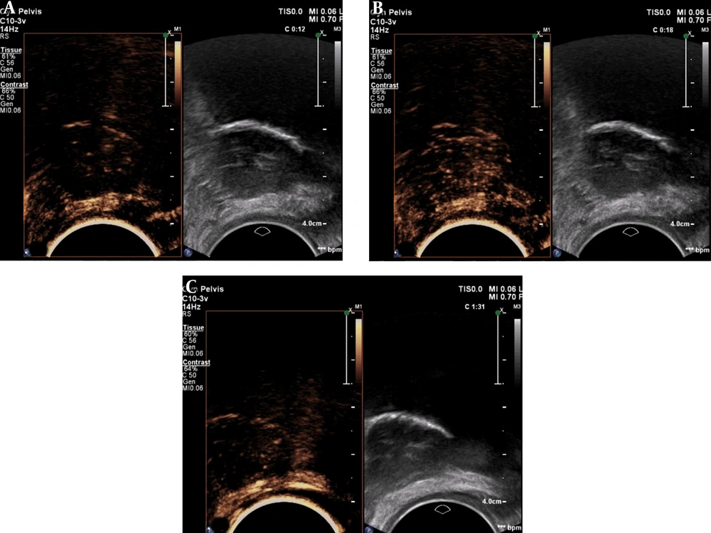
Figure 1.

A 58-year-old woman with bladder neoplasm. Sagittal transvaginal sonogram shows a well-defined, hypoechoic mass in the posterior wall.

Contrast-enhanced ultrasound in the same patient. A, Contrast agent wash-in at 12 seconds; B, Maximum enhancement signal at 18 seconds; C, A small amount of microbubbles is still present at 91 seconds.
Figure 2.

Contrast-enhanced ultrasound in the same patient. A, Contrast agent wash-in at 12 seconds; B, Maximum enhancement signal at 18 seconds; C, A small amount of microbubbles is still present at 91 seconds.

The Philips Ultrasound QVue DICOM Viewer was used for analysis of CEUS images (Figure 2). Contrast agent appeared in the mass 12 seconds after intravenous injection (Figure 2A), and the mass enhanced homogeneously. At 18 seconds, the peak time, the mass showed mild enhancement in comparison to the adjacent bladder wall (Figure 2B). Then, the contrast was washed out gradually. A small amount of contrast was still present at 91 seconds (Figure 2C).
Transurethral resection was performed. Cystoscopy showed the mass arising from the posterior wall close to the neck of the bladder. The epithelial lining in the area appeared normal. Immunohistochemical staining showed the mass to be Actin (+), Desmin (+), S-100 (−), CD34 (−), and Ki-67 (< 5%+), confirming the diagnosis leiomyoma.

3. Discussion

Bladder leiomyoma is a rare benign tumor and there are only a few reports of the pattern of enhancement in these tumors on contrast-enhanced CT and MRI (4). There are no reports of CEUS findings in bladder leiomyoma. Ultrasound examination, especially transvaginal examination, can be advantageous for diagnosis of bladder leiomyoma because of its high soft tissue resolution. Moreover, CEUS using SonoVue can give an accurate picture of tumor blood flow as this microbubble contrast agent has a strictly intravascular distribution (5).
Drudi et al. (6), showed that contrast agent is washed out of urothelial cell carcinoma (UCC) within 58 seconds of intravenous administration; whereas, in the normal bladder wall, it persists beyond 80 seconds. In our patient, microbubbles were still seen at 91 seconds, which is consistent with the normal bladder wall of Drudi et al.’s findings. Wang et al. also reported that most UCCs show fast wash-in and fast wash-out, relative to the corresponding bladder wall or prostate (7).
The ideal treatment for bladder leiomyoma has not yet been established. However, some authors have suggested that an inference can be drawn from the wide experience with uterine leiomyomas. Bladder leiomyomas have many histopathological features in common with uterine leiomyomas (1). The probability of uterine leiomyoma transforming to sarcoma is small, and there have been no reports of malignant transformation in bladder leiomyoma. Therefore, a wait-and-watch approach seems reasonable (8, 9). Correct identification of bladder leiomyoma by medical imaging will thus help avoid unnecessary surgery.
Based on the experience of this case, CEUS appears to be a good option for noninvasive diagnosis of bladder leiomyoma.


Footnotes

  • Authors’ Contributions:Yu Yang carried out the final preparation of the manuscript.

  • Conflict of Interests:The authors have no conflict of interest to report.

  • Funding/Support:This work was supported by Beijing Excellent Talent Program [grant number 2006000021469G228].

References

  • 1.
    Knoll LD, Segura JW, Scheithauer BW. Leiomyoma of the bladder. J Urol. 1986;136(4):906-8. [PubMed ID: 3761457].
  • 2.
    Campbell EW, Gislason GJ. Benign mesothelial tumors of the urinary bladder: review of literature and a report of a case of leiomyoma. J Urol. 1953;70(5):733-41. [PubMed ID: 13109927].
  • 3.
    Park JW, Jeong BC, Seo SI, Jeon SS, Kwon GY, Lee HM. Leiomyoma of the urinary bladder: a series of nine cases and review of the literature. Urology. 2010;76(6):1425-9. [PubMed ID: 20947147]. https://doi.org/10.1016/j.urology.2010.02.046.
  • 4.
    Mouli S, Casalino DD, Nikolaidis P. Imaging features of common and uncommon bladder neoplasms. Radiol Clin North Am. 2012;50(2):301-16. vi. [PubMed ID: 22498444]. https://doi.org/10.1016/j.rcl.2012.02.001.
  • 5.
    Nicolau C, Bunesch L, Peri L, Salvador R, Corral JM, Mallofre C, et al. Accuracy of contrast-enhanced ultrasound in the detection of bladder cancer. Br J Radiol. 2011;84(1008):1091-9. [PubMed ID: 21123306]. [PubMed Central ID: PMC3473820]. https://doi.org/10.1259/bjr/43400531.
  • 6.
    Drudi FM, Di Leo N, Maghella F, Malpassini F, Iera J, Rubini A, et al. CEUS in the study of bladder, method, administration and evaluation, a technical note. J Ultrasound. 2014;17(1):57-63. [PubMed ID: 24616745]. [PubMed Central ID: PMC3945194]. https://doi.org/10.1007/s40477-013-0032-y.
  • 7.
    Wang XH, Wang YJ, Lei CG. Evaluating the perfusion of occupying lesions of kidney and bladder with contrast-enhanced ultrasound. Clin Imaging. 2011;35(6):447-51. [PubMed ID: 22040789]. https://doi.org/10.1016/j.clinimag.2010.11.001.
  • 8.
    Katz RB, Waldbaum RS. Benign mesothelial tumor of bladder. Urology. 1975;5(2):236-8. [PubMed ID: 1114563].
  • 9.
    Bai SW, Jung HJ, Jeon MJ, Jung DJ, Kim SK, Kim JW. Leiomyomas of the female urethra and bladder: a report of five cases and review of the literature. Int Urogynecol J Pelvic Floor Dysfunct. 2007;18(8):913-7. [PubMed ID: 17333443]. https://doi.org/10.1007/s00192-006-0257-9.

Copyright

Copyright © 2018, Author(s). This is an open-access article distributed under the terms of the Creative Commons Attribution-NonCommercial 4.0 International License (http://creativecommons.org/licenses/by-nc/4.0/) which permits copy and redistribute the material just in noncommercial usages, provided the original work is properly cited.

Similar Articles

30
Jun
2009

Leiomyoma of the Urinary Bladder - A Case Report and Brief Review of Literature

Seyed Sajjad Nazir,
Aakifa Maqbool,
Muneer Khan

Nazir S, Maqbool A, Khan M. Leiomyoma of the Urinary Bladder - A Case Report and Brief Review of Literature. Nephro-Urol Mon. 2009;1(1):. doi:

15
Dec
2012

Transvesical Enucleation of Multiple Leiomyoma of Bladder and Urethra

Alireza Ghadian,
Seyyed Yousef Hoseini

Ghadian A, Hoseini SY. Transvesical Enucleation of Multiple Leiomyoma of Bladder and Urethra. Nephro-Urol Mon. 2012;5(1):709-711. doi: https://doi.org/10.5812/numonthly.5122

15
Dec
2012

Giant Ureteral Fibroepithelial Polyp Presenting As a Bladder Mass Resected Ureteroscopically: A Case Report

Ali Momenzadeh,
Farhad Sarrafzadeh,
Mohammad-Hossein Nourbala,
Amin Saburi,
Zeinab Telkabadi

Momenzadeh A, Sarrafzadeh F, Nourbala M, Saburi A, Telkabadi Z. Giant Ureteral Fibroepithelial Polyp Presenting As a Bladder Mass Resected Ureteroscopically: A Case Report. Nephro-Urol Mon. 2012;5(1):706-708. doi: https://doi.org/10.5812/numonthly.4933

12
May
2014

Large Clitoral Leiomyoma in a Forty-Two Years Old Premenopausal Woman

Santosh Kumar,
Swati Agrawal,
Kumar Jayant,
Sriharsha Ajjoor Shankargowda

Kumar S, Agrawal S, Jayant K, Ajjoor Shankargowda S. Large Clitoral Leiomyoma in a Forty-Two Years Old Premenopausal Woman. Nephro-Urol Mon. 2014;6(3):e17022. doi: https://doi.org/10.5812/numonthly.17022

27
Nov
2016

Quantitative Dynamic Contrast-Enhanced Magnetic Resonance Imaging in Symptomatic Uterine Fibroids and Normal Uterus: A Feasibility Study

Soheila Majd,
Basri Johan Jeet Abdullah,
Sazilah Ahmad Sarji,
Chai Hong Yeong,
Noor Azmi Mat Adenan

Majd S, Johan Jeet Abdullah B, Ahmad Sarji S, Hong Yeong C, Azmi Mat Adenan N. Quantitative Dynamic Contrast-Enhanced Magnetic Resonance Imaging in Symptomatic Uterine Fibroids and Normal Uterus: A Feasibility Study. I J Radiol. 2017;14(2):e36213. doi: https://doi.org/10.5812/iranjradiol.36213

Download PDF2.39 MB

Crossmark

Crossmark

Checking

Share on
Cited by
Metrics

Ordering Reprints

Articles are published under the Creative Commons license stated on each article. No permission or royalty fee is required for uses permitted by that license. CCC handles optional bulk and customized reprint orders. Any quotation covers production and delivery services only, not copyright permission. > Request Reprints from CCC
