Giant Ureteral Fibroepithelial Polyp Presenting As a Bladder Mass Resected Ureteroscopically: A Case Report

Author(s):
Ali MomenzadehAli Momenzadeh1, Farhad SarrafzadehFarhad Sarrafzadeh2, Mohammad-Hossein NourbalaMohammad-Hossein Nourbala1, Amin SaburiAmin Saburi3,*, Zeinab TelkabadiZeinab Telkabadi1
1Nephrology and Urology Research Center, Baqiyatallah University of Medical Sciences, Tehran, IR Iran
2Department of Internal Medicine, Faculty of Medicine, Kerman University of Medical Sciences, Kerman, IR Iran
3Chemical Injury Research Center, Baqiyatallah University of Medical Sciences, Tehran, IR Iran

Nephro-Urology Monthly:Vol. 5, issue 1; 706-708
Published online:Dec 15, 2012
Article type:Case Report
Received:Mar 19, 2012
Accepted:Apr 22, 2012
How to Cite:Momenzadeh A, Sarrafzadeh F, Nourbala M, Saburi A, Telkabadi Z. Giant Ureteral Fibroepithelial Polyp Presenting As a Bladder Mass Resected Ureteroscopically: A Case Report. Nephro-Urol Mon. 2012;5(1):706-708. doi: https://doi.org/10.5812/numonthly.4933

Abstract

1. Introduction

Primary ureteral neoplasms are very rare in adults and are responsible for less than 1% of all genitourinary neoplasms (1). Only one-fifth of them are benign, and the mesodermal benign tumor such as Ureteral Fibroepithelial polyp (UFP) is more common (2). The UFP is usually located on the proximal portion of the ureter and is covered by a layer of normal urothelium (3, 4). generally speaking, it is not significantly symptomatic unless it makes obstruction (partial or complete) which characterized by obstructive or irritability symptoms such as supra-pubic or flank pain, hematuria (usually microscopic), frequency and dysuria (3). The releasing symptoms depend on polyp size and situation. Imaging diagnostic studies show ambiguous space occupying lesions commonly which was certainly diagnosed and treated by excisional biopsy (endoscopic or surgical) and histopathological report (5). We report a giant ureteral fibroepithelial polyp in adult patients representing a bladder mass which was resected ureteroscopically at the first time from Iran.

2. Case Presentation

A medical center diagnosed dysuria with obscure lower abdominal pain (during avoiding, specially) in a 44-year old female patient two weeks ago. Microscopic hematuria results from urine and blood sample laboratory analysis. She had no remarkable history of renal stone, urinary tract infections or any urinary stigmas. Physical examination has shown mild tenderness in right postvertebral angel without any other significant evidence. In hydronephrosis, a small effect has been diagnosed in the urinary bladder (US) which located on the right ureterovesical junction without any evidence of hydronephrosis. Computed tomography (CT) scan and IVP have revealed a small vegetative mass with 50Ă—18 mm diameter situated near to trigon (Figure 1). Cystoscopy showed a pedunculated polyploidy tumor establishing on the right ureterovesical orifice and then the lesion was resected completely. Histopathology report was compatible with polyploidy cystitis without malignant components. Forty days later, the patient was admitted in this center due to dysuria and right flank pain. Except microscopic hematuria in urine cytology, another laboratory result consisting blood biochemical data were in normal ranges and urine cultures were negative. Renal US displayed moderate to severe hydronephrosis. She was undergone Re-Cystoscopy with diagnosis of urinary bladder mass. Cystoscopy showed a mass with a lobulated appearance sticking outside of the right vesicoureteral orifice. The fragmentally mass resection revealed the base of the tumor (90 mm in length and 10 mm in diameter) situated in the lower portion of ureter. The polyp stalk was coagulated and excised with claspers forceps and then the whole right ureteroscopy was assessed perfectly. Double-J stent was resided to prevent the ureteral stricture and periodically was reexamined by ultrasound or IVP until removal (3 weeks later). Histopathological analysis and retrospectively previous specimen re-assessment have confirmed a ureteral fibroepithelial polyp (Figure 2). There were no evidences of polyp recurrence or ureteral stenosis in 12 months after the double-J stent discontinuation and the patient was asymptomatic.
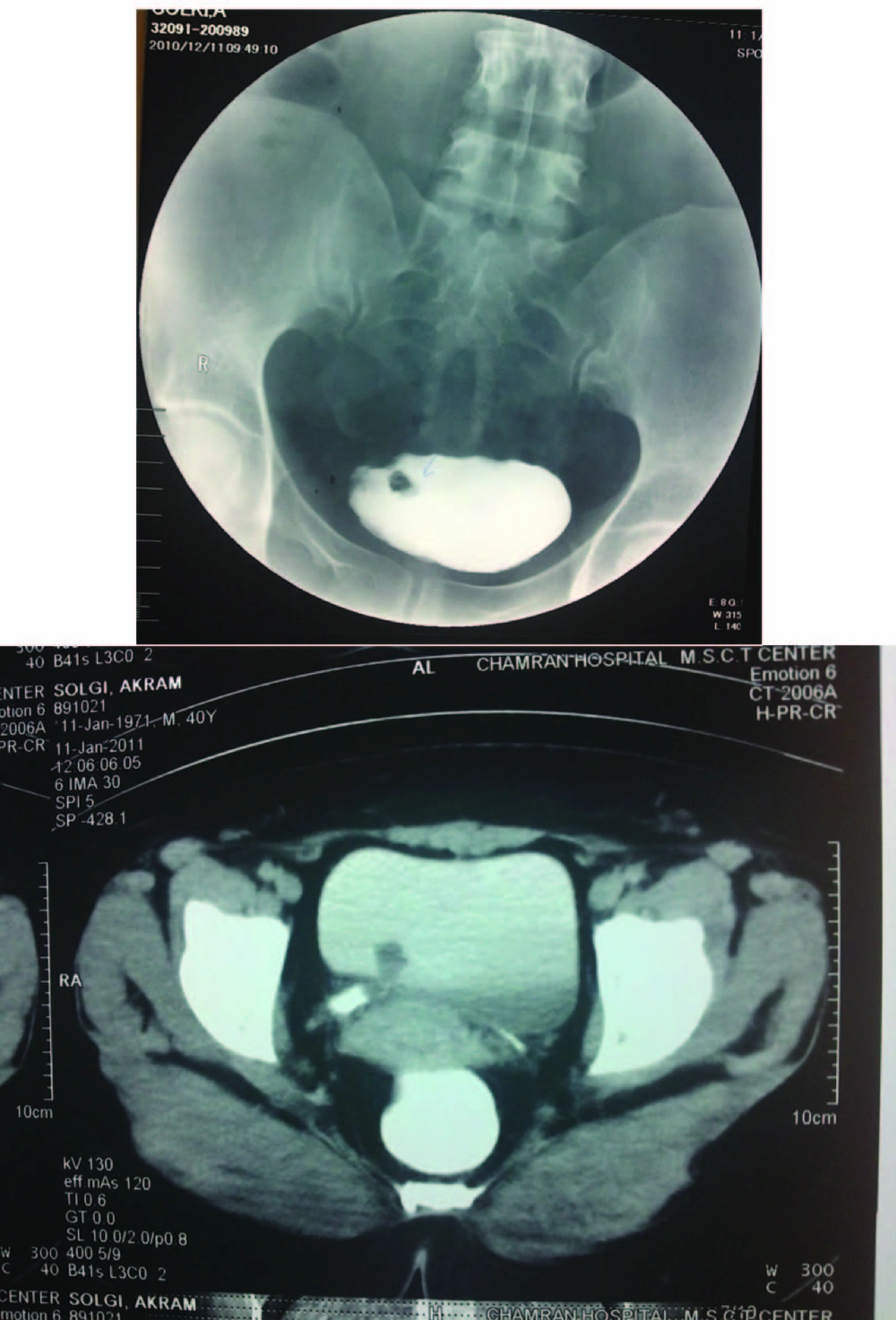
IVP Findings; A Small Round Shape Mass Near to Trigon Which Was Prolapsed Into Bladder
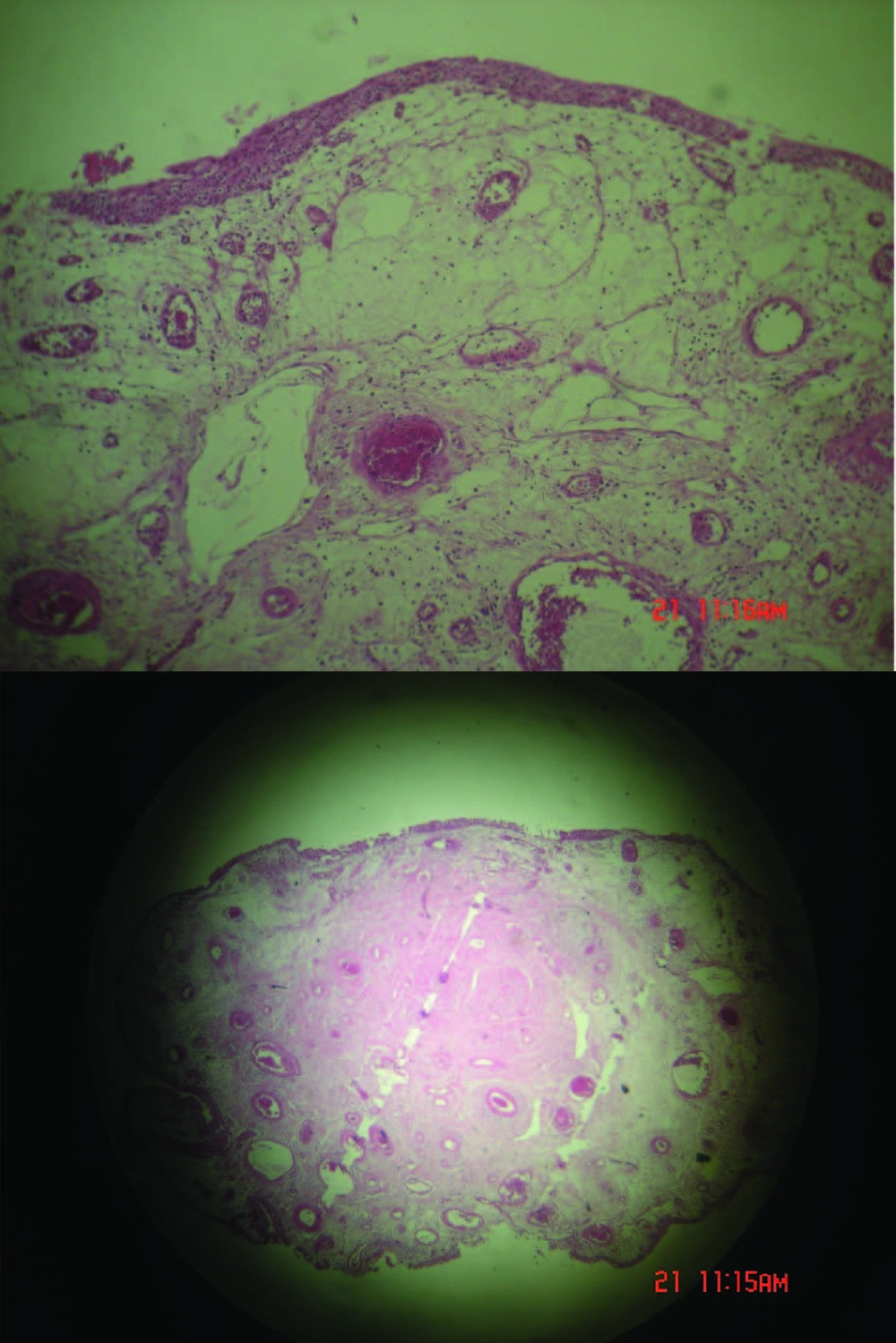
Figure 1

IVP Findings; A Small Round Shape Mass Near to Trigon Which Was Prolapsed Into Bladder

Pathologic Figures
Figure 2

Pathologic Figures

3. Discussion

Neoplasms which originated from ureter are rare and commonly malignant. However, UFPs are the most common non-malignant neoplasms; they are still rare clinically and primarily seen in males and on the left side (1). UFPs grossly seem as finger-like projection originating from the sub-mucosa of the ureter which attached to a single base (6). UFPs are composed of vascular and fibrous tissue covered with normal transitional epithelium (7). The clinical presentations are different depending on lesion location in ureter (1). UFP can be lengthened enough prolapsed into the bladder cavity and it can induce surgeons' aberrant (8). Management plan has different location and complication (5). Endoscopic plan of large UFPs is an acceptable treatment option with minimal adverse complications and durable treatment outcomes (5, 6, 9). Open resection approved a ureteral polyp originating from the lower ureter elongated into the bladder, which mimicked vesicle mass (7, 8).
We report a case of large UFP presenting a vegetative bladder mass excised ureteroscopically. Reviewing the English literature illustrated multiple reports of UFP, but ureteral fibroepithelial polyp elongated into the bladder cavity was rare which mimicked bladder mass resected ureteroscopically (3, 8-15). UFP was not reported from Iran and it was the first report. The authors recommend further assessment for any mass juxtaposed vesicoureteral junction and also suggest ureteroscopically approach to distal UFPs.

Acknowledgments

Footnotes

References

  • 1.
    Piovesan A, Torricelli F, Borges L, ChambĂ´ J, Mesquita J, Srougi M. Ureteral fibroepithelial polyps in a pregnant woman: case report. Sao Paulo Med J. 2009;127(4):238-40. [PubMed ID: 20011930]. https://doi.org/10.1590/S1516-31802009000400010.
  • 2.
    Tato Rodriguez J, Lema Grille J, Cimadevila Garcia A, Blanco Parra M, Lamas Cedron P, Rios Tallon L, et al. [A fibroepithelial polyp of the ureter. A report of 2 new cases]. Actas Urol Esp. 1997;21(4):420-5. [PubMed ID: 9265418].
  • 3.
    Baltogiannis D, Kafetsoulis A, Giannakis D, Saito M, Miyagawa I, Sofikitis N. Fibroepithelial polyp of distal ureter with periodic prolapse into bladder. Urol Int. 2008;80(3):338-40. [PubMed ID: 18480645]. https://doi.org/10.1159/000127355.
  • 4.
    Ruiz-Lopez MJ, Ramirez-Garrido F, Nogueras-Ocana M, Munoz-Hoyos A, Martin A. Recurrent ureteric fibroepithelial polyp in a child. Eur J Pediatr. 2004;163(2):124-5. [PubMed ID: 14716557]. https://doi.org/10.1007/s00431-002-1052-3.
  • 5.
    Lam JS, Bingham JB, Gupta M. Endoscopic treatment of fibroepithelial polyps of the renal pelvis and ureter. Urology. 2003;62(5):810-3. [PubMed ID: 14624899]. https://doi.org/10.1016/S0090-4295(03)00691-5.
  • 6.
    Yagi S, Kawano Y, Gotanda T, Kitagawa T, Kawahara M, Nakagawa M, et al. Endoscopic treatment of a long fibroepithelial ureteral polyp. Int J Urol. 2001;8(8):467-9. [PubMed ID: 11555017]. https://doi.org/10.1046/j.1442-2042.2001.00343.x.
  • 7.
    Lai TK, Chung CH, Chin AC, Szeto PS, Wong WC, Chan RY, et al. Magnetic resonance imaging for ureteral fibroepithelial polyp. Hong Kong Med J. 2008;14(5):408-10. [PubMed ID: 18840916].
  • 8.
    Yeh CC, Lin HC, Che CC, Wu HC. Ureteral fibroepithelial polyp prolapsed into the bladder cavity suspending a bladder stone. J Urol. 2001;165(5):1627-8. [PubMed ID: 11342937]. https://doi.org/10.1016/S0022-5347(05)66368-9.
  • 9.
    Coloma del Peso A, Bocardo Fajardo G, Teba del Pino F, Fernandez Gonzalez I, Brime Menendez R, Fernandez Borrell A, et al. Endoscopic treatment of a giant fibroepithelial polyp of the ureter. Arch Esp Urol. 2010;63(4):305-8. [PubMed ID: 20511688].
  • 10.
    Numakura K, Tsuchiya N, Inoue T, Horikawa Y, Kakinuma H, Togashi H, et al. [A case of fibroepithelial polyp of the ureter resected ureteroscopically]. Hinyokika Kiyo. 2005;51(4):277-81. [PubMed ID: 15912790].
  • 11.
    Sano K, Terashima K, Aida N, Ijiri R, Tanaka Y, Nosaka S. [A case of ureteral polyp prolapsing into bladder in a 3-year-old boy]. Hinyokika Kiyo. 1998;44(10):737-8. [PubMed ID: 9850841].
  • 12.
    Macksood MJ, Roth DR, Chang CH, Perlmutter AD. Benign fibroepithelial polyps as a cause of intermittent ureteropelvic junction obstruction in a child: a case report and review of the literature. J Urol. 1985;134(5):951-2. [PubMed ID: 4057382].
  • 13.
    Luthans F, Avolio BJ, Walumbwa FO, Li W. The psychological capital of Chinese workers: Exploring the relationship with performance. Manage Organiz Rev. 2005;1(2):249-71. https://doi.org/10.1016/S1607-551X(09)70202-5.
  • 14.
    Schiotz HA. Fibroepithelial polyp of the ureter. A case history and discussion. Acta Obstet Gynecol Scand. 1990;69(3):267-8. [PubMed ID: 2220351]. https://doi.org/10.3109/00016349009028692.
  • 15.
    Terai A, Terachi T, Yoshida S. Prolapsed benign polyp of ureter associated with giant ureteral calculus. Hinyokika Kiyo. 1989;35(5):871-3. [PubMed ID: 2801381].

Similar Articles

30
Jun
2009

Leiomyoma of the Urinary Bladder - A Case Report and Brief Review of Literature

Seyed Sajjad Nazir,
Aakifa Maqbool,
Muneer Khan

Nazir S, Maqbool A, Khan M. Leiomyoma of the Urinary Bladder - A Case Report and Brief Review of Literature. Nephro-Urol Mon. 2009;1(1):. doi:

15
Dec
2012

Transvesical Enucleation of Multiple Leiomyoma of Bladder and Urethra

Alireza Ghadian,
Seyyed Yousef Hoseini

Ghadian A, Hoseini SY. Transvesical Enucleation of Multiple Leiomyoma of Bladder and Urethra. Nephro-Urol Mon. 2012;5(1):709-711. doi: https://doi.org/10.5812/numonthly.5122

12
Dec
2015

Bilateral Ureteroceles Along With Horseshoe Kidney in an Adult: A Case Report and Review of Literature

Mojtaba Ameli,
Amir Jarhi,
Alireza Mahmoudabadi

Ameli M, Jarhi A, Mahmoudabadi A. Bilateral Ureteroceles Along With Horseshoe Kidney in an Adult: A Case Report and Review of Literature. Thrita J Neu. 2015;4(4):e33549. doi: https://doi.org/10.5812/thrita.33549

27
Dec
2012

Ureterocele Associated with Renal Agenesia Presented as a Pelvic Mass in an Adult

Mohammad Ghasem Mohseni,
Seyed Reza Hosseini,
Alborz Salavati,
Shahrzad Dadgari

Mohseni MG, Hosseini SR, Salavati A, Dadgari S. Ureterocele Associated with Renal Agenesia Presented as a Pelvic Mass in an Adult. I J Radiol. 2013;10(1):45-47. doi: https://doi.org/10.5812/iranjradiol.10262

5
Nov
2014

Delivery of Huge Bladder Stone in a Thirty-Five-Year-Old Man

Alireza Farshi,
Reza Sari Motlagh,
Rasole Jafari Arismani

Farshi A, Sari Motlagh R, Jafari Arismani R. Delivery of Huge Bladder Stone in a Thirty-Five-Year-Old Man. Nephro-Urol Mon. 2014;6(6):e20574. doi: https://doi.org/10.5812/numonthly.20574


Crossmark
Crossmark
Checking
Share on
Cited by
Metrics

Ordering Reprints

Articles are published under the Creative Commons license stated on each article. No permission or royalty fee is required for uses permitted by that license. CCC handles optional bulk and customized reprint orders. Any quotation covers production and delivery services only, not copyright permission. > Request Reprints from CCC 

Search Relations

Author(s):

Related Articles