


Full Text
Full text is available in PDF
IJ Radiology
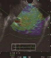
In general population, chronic liver disease is a great public health problem. Liver fibrosis, a common pattern in the usual causes of chronic liver disease, involves the gathering of collagen and other macromolecules inside the extracellular matrix. Fibrosis tends to proceed, leading to liver dysfunction, portal hypertension, and eventually cirrhosis. Liver biopsy is the gold standard for the diagnosing of liver fibrosis, and unfortunately it is invasive, expensive, with sampling variability and probable complications (mortality rate is procedure-related: one in 10,00012,000). For patients with chronic liver disease, it is important to delineate the degree and grading of fibrosis progression. Ultrasonography (US) provides ideal noninvasive assessment for diffuse liver disease because of its low cost and wide availability. Conventional B-mode US is good for the diagnosis of advanced cirrhosis, but is insensitive for fibrosis. Elastography may be employed as a substitute diagnostic technique for fibrosis. The METAVIR scoring system assigns fibrosis to five stages of F0: no fibrosis, F1: portal and periportal fibrosis with no septum, F2: portal and periportal fibrosis with rare septa, F3: portal and periportal fibrosis with many septa, F4: cirrhosis). In elastography, a mechanical force (compression or vibration) is applied to the soft tissues, and a conventional imaging technique such as US or MR imaging is used to create a map of soft-tissue deformation. There are two types of elastography: static and dynamic elastography. Static elastography is now a more common type of elastographic imaging. In this method, a single and very small compressive force is used by pressing with the ultrasound probe, typically and then the displacement of tissue is calculated. Static elastography has been widely used with superficial organs. It is more difficult to apply in deeper organs, and also the qualitative figures have not been displayed to have value for detection of liver fibrosis by using a vibratory stimulus to produce shear waves (which slowly pass through tissues) to be tracked by ultrasound. The spreading rate of shear waves depends upon the shear modulus of tissues, which is usually proportional to the Youngs modulus directly. Hence, quantifying tissue stiffness is possible by using this method. The first ultrasound system to apply this method is the Fibroscan (by Echosens) which can evaluate liver fibrosis. Shear waves are sent into the liver by a piston at the center of the probe, and the shear waves velocity is used to assess the shear modulus. This tool has proved feasible for the noninvasive diagnosis of moderate and severe fibrosis, but mild changes are not detected. Development of other systems to apply acoustic radiation force imaging (ARFI) (such as the Siemens S2000) by pushing on organs with a high-powered ultrasound pulse is under way. Currently, there are three main techniques to evaluate liver stiffness: transient elastography (FibroScan by Echosens), acoustic radiation force impulse (ARFI) imaging, and shear-wave (SW) elastography . Among these methods, transient elastography offers the capacity to estimate mean tissue stiffness noninvasively. Several recent studies have proved that the measurements of liver stiffness with transient elastography are well correlated with the METAVIR stages of fibrosis. In addition, transient elastography is helpful as it can be performed repeatedly. However, it is not a real-time technique because the sonographic images are not visible during the measurement. In patients with hepatosteatosis and increased body mass index (BMI), the reproducibility of transient elastography is substantially reduced as the modality of ultrasonography itself has restrictions for visualizing the liver clearly in these patients. Real-time sonoelastography is another method for the evaluation of tissue elasticity. It uses a B-mode ultrasound, combining with elastography with the conventional US scanner. This device estimates the relative hardness of the organs and displays this data as real-time color images. It can show tissue elasticity images and conventional B-mode images at the same time. There are many reports and approaches to evaluate liver stiffness without liver biopsy. Among them, the FibroScan appears useful.
© 2014, Author(s). This open-access article is available under the Creative Commons Attribution 4.0 (CC BY 4.0) International License (https://creativecommons.org/licenses/by/4.0/), which allows for unrestricted use, distribution, and reproduction in any medium, provided that the original work is properly cited.

Yue Z, Hong D, Peili F, Yuli Z, Yuan Z, et al. Non-Invasive Assessment of Liver Fibrosis in a Rat Model: Comparison of Shear Wave Elastography with Serologic Tests. I J Radiol. 2018;15(3):e60665. doi: https://doi.org/10.5812/iranjradiol.60665
Gutkowski K, Hartleb M. Usefulness of Non-Invasive Tools in Liver Fibrosis Assessment. Hepat Mon. 2008;8(1):. doi:
Yao D, Jiang H, Qin S. Preliminary Study on the Evaluation of Liver Fibrosis Using Endoscopic Ultrasound Elastography and Transabdominal Ultrasound Transient Elastography Combined with Strain Ratio. Hepat Mon. 2024;24(1):e138304. doi: https://doi.org/10.5812/hepatmon-138304
Zarei F, Moini M, Abedi M, Ravanfar Haghighi R, Zeinali-Rafsanjani B. Liver Fibrosis Assessment Using Transient Elastography by FibroScan and Shear Wave Elastography by Sonography: A Comparative Cross-sectional Study in an Outpatient Liver Clinic. I J Radiol. 2021;18(4):e112589. doi: https://doi.org/10.5812/iranjradiol.112589

Zaleska-Dorobisz U, Inglot M, Pawlus A, Patyk M, Markiewicz BD, et al. The Evaluation of Liver Fibrosis in the Real-Time Shear Wave Elastography Technique in Chronic Hepatitis B and C Patients: How Many Measurements Are Necessary to Assign Patients to METAVIR Score Properly?. Hepat Mon. 2018;18(6):e61189. doi: https://doi.org/10.5812/hepatmon.61189

Checking
Last Update: 2 weeks ago
Last Update: 2 weeks ago
Last Update: 2 weeks ago
Last Update: 4 days ago
Ordering Reprints
Articles are published under the Creative Commons license stated on each article. No permission or royalty fee is required for uses permitted by that license. CCC handles optional bulk and customized reprint orders. Any quotation covers production and delivery services only, not copyright permission. > Request Reprints from CCC
Author(s):