


1. Background
2. Objectives
3. Materials and Methods
3.1. Animals
3.2. SWE Procedure
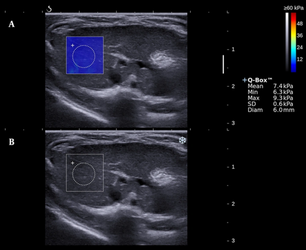
An elastogram of the liver stiffness based on shear wave elastography (SWE). A, Gray scale image. B, Elastogram with a 0.6 cm diameter circular region (Q-BOX) positioned in the homogeneous color map of 2 × 2cm. The measured hepatic elasticity values are displayed on the right.
3.3. Serological Tests
3.4. Pathological Examination
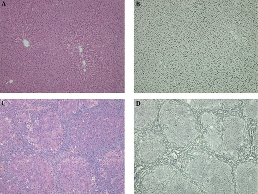
Hematoxylin-eosin (HE) staining (A and C) and reticular fiber staining (B and D) of two specimens. Normal hepatic architecture can be seen in the control group (A and B). Fibrous bridges between centrilobular-centrilobular, centrilobular-periportal and periportal-periportal areas can be seen in a histologically proven F4 rat, and the fibers separate liver parenchyma into pseudolobules (C and D).
3.5. Statistical Analysis
4. Results
4.1. Animal Results
4.2. Reliability of shear wave elastography measurements
4.3. Correlation between liver stiffness and fibrosis stages
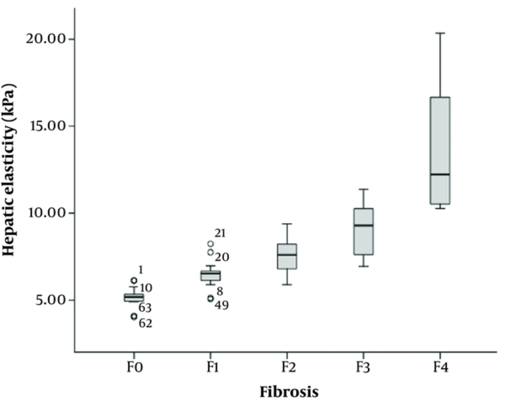
Boxplot of the distribution of hepatic elasticity measured by shear wave elastography (SWE) indicates the discrete value.
| Fibrosis stage | Number (Percentage) | Hepatic stiffness (kPa) | Mann-Whitney | ||
|---|---|---|---|---|---|
| Mean ± SD | 95%CI | Z | P value | ||
| F0 | 15 (16.7%) | 5.17 ± 0.59 | 4.86 - 5.47 | ||
| F1 | 14 (15.6%) | 6.49 ± 0.85 | 6.05 - 6.95 | -3.646 | < 0.001 |
| F2 | 30 (33.3%) | 7.52 ± 0.96 | 7.19 - 7.87 | -3.049 | 0.002 |
| F3 | 21 (23.3%) | 9.06 ± 1.51 | 8.42 - 9.72 | -3.263 | 0.001 |
| F4 | 10 (11.1%) | 13.71 ± 3.95 | 11.39 - 16.25 | -3.761 | < 0.001 |
Abbreviations: CI, confidence interval; KPa ,kilopascal; SD, standard deviation.
4.4. Correlation Between Serum Markers and Hepatic Fibrosis
| Fibrosis Stage | HA (ng/ml) | APRI | AAR |
|---|---|---|---|
| F0 | 241.2 (202.6 - 288.9) | 0.063 (0.053 - 0.082) | 0.64 (0.51 - 0.91) |
| F1 | 243.2 (219.1 - 289.4) | 0.079 (0.066 - 0.084) | 0.39 (0.33 - 0.49) |
| F2 | 304.7 (266.7 - 337) | 0.072 (0.067 - 0.102) | 0.51 (0.52 - 0.85) |
| F3 | 333.2 (281.1 - 344.9) | 0.155 (0.128 - 0.195) | 1.03 (0.92 - 1.61) |
| F4 | 405.3 (323 - 453.9) | 0.191 (0.149 - 0.233) | 1.02 (0.93 - 1.92) |
| r | 0.417 | 0.580 | 0.445 |
| P value | < 0.001 | < 0.001 | < 0.001 |
Abbreviations: AAR, aspartate aminotransferase to alanine aminotransferase ratio; APRI, aspartate aminotransferase to platelet ratio index.
a Numbers in parentheses are the 95% confidence intervals.
4.5. Diagnostic Performance of SWE Compared to Serological Biomarkers
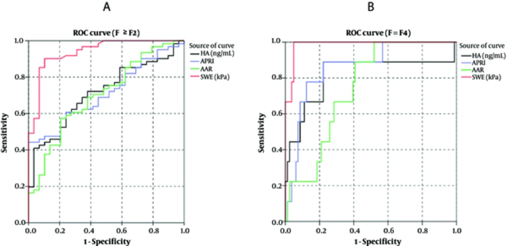
Receiver operating characteristic (ROC) curves of hyaluronic acid (HA), aspartate aminotransferase (AST) to platelet (PLT) ratio index (APRI), AST to alanine aminotransferase (ALT) ratio (AAR), and shear wave elastography (SWE) for the diagnosis of fibrosis stage F ≥ F2 (A) and fibrosis stage F = F4 (B).
| Test | Youden Index | Cut-off value | AUROC | Sensitivity (%) | Specificity (%) | Accuracy (%) | PPV (%) | NPV (%) |
|---|---|---|---|---|---|---|---|---|
| HA | ||||||||
| F ≥ 2 | 0.376 | 352.93 | 0.713 | 40.98 | 96.55 | 58.89 | 96.15 | 43.75 |
| F = 4 | 0.667 | 352.93 | 0.815 | 88.89 | 77.78 | 78.89 | 30.77 | 98.44 |
| APRI | ||||||||
| F ≥ 2 | 0.443 | 0.122 | 0.712 | 44.26 | 100.00 | 62.22 | 100.00 | 46.03 |
| F = 4 | 0.667 | 0.126 | 0.860 | 88.89 | 77.78 | 78.89 | 30.77 | 98.44 |
| AAR | ||||||||
| F ≥ 2 | 0.367 | 0.745 | 0.702 | 57.38 | 79.31 | 64.44 | 85.37 | 46.94 |
| F = 4 | 0.482 | 0.745 | 0.745 | 88.89 | 59.26 | 62.22 | 19.51 | 97.96 |
| SWE | ||||||||
| F ≥ 2 | 0.799 | 6.69 | 0.938 | 90.16 | 89.66 | 90.00 | 94.83 | 81.25 |
| F = 4 | 0.951 | 10.47 | 0.985 | 100.00 | 95.06 | 95.56 | 69.23 | 100.00 |
Abbreviations: AAR, aspartate aminotransferase to alanine aminotransferase ratio; AUROC, the area under the receiver operating characteristic curve; APRI, aspartate aminotransferase to platelet ratio index; HA, hyaluronic acid; NPV, negative predictive value; SWE, shear wave elastography; PPV, positive predictive value.


