
1. Introduction
Hypertension accounts for nearly two-thirds of all deaths worldwide (1). Primary aldosteronism (PA) is a common and curable cause of secondary hypertension and is characterized by renin-independent aldosterone secretion from the adrenal glands (2). It is estimated to account for 5 - 20% of all cases of hypertension, a higher proportion than previously reported (3, 4). Despite these estimates, PA is frequently underdiagnosed, partly because screening is not routinely performed in primary care (2).
Clinical presentations range from resistant hypertension and hypokalemia, sometimes leading to muscle weakness or paralysis, to asymptomatic cases with normal potassium levels (5 - 7).
Importantly, PA is associated with an increased risk of cardiovascular and renal complications. Increased rates of atrial fibrillation, heart failure, stroke, and left ventricular hypertrophy have been documented (8).
Early identification and targeted treatment with mineralocorticoid receptor antagonists or adrenalectomy can significantly improve outcomes (9).
This case underscores the importance of early screening for PA in patients with hypertension to prevent serious complications.
2. Case Presentation
A 50-year-old Gilak woman presented to our clinic with a hypertensive crisis (220/130 mmHg) and quadriparesis. Her hypertension had been diagnosed at age 40, with no family history of high blood pressure. Over the subsequent years, her blood pressure progressively worsened, and she experienced 6 episodes of quadriparesis and 1 episode of paraplegia requiring hospitalization. During these episodes, her serum potassium level ranged from 2.0 to 2.5 mEq/L. She had previously been evaluated by a neurologist and had undergone electromyography and nerve conduction studies (EMG/NCS), which showed nonspecific findings. However, the diagnostic workup was not completed, and the underlying cause of her recurrent episodes remained undetermined.
At presentation, she was taking multiple antihypertensive medications, including amlodipine, valsartan, hydrochlorothiazide, eplerenone, and prazosin, but her blood pressure remained poorly controlled.
On initial examination, her blood pressure was 220/130 mmHg. Other vital signs were as follows: heart rate, 76/min; respiratory rate, 14/min; temperature, 36.7 °C; and arterial oxygen saturation, 96% on room air. She reported no palpitations. She experienced frequent episodes of quadriparesis characterized by sudden weakness and loss of motor function in the limbs, with muscle power of 2/5 and a downgoing Babinski response, but normal sensory and cranial nerve examinations. The patient was alert and had fluent speech. Other physical examination findings were normal.
Cardiac auscultation revealed normal S1 and S2, an additional S4, and a grade 2/6 systolic murmur at the apex and left sternal border. Electrocardiography showed normal sinus rhythm, left posterior hemiblock, left ventricular hypertrophy, and a U wave. Echocardiography showed a left ventricular ejection fraction of 60% and mild mitral and tricuspid regurgitation, with otherwise normal findings.
Laboratory testing revealed marked hypokalemia, with a serum potassium level of 2.3 mEq/L. Other laboratory results were as follows: creatinine, 1.0 mg/dL; fasting blood glucose, 104 mg/dL; triglycerides, 303 mg/dL; magnesium, 1.7 mg/dL; random urine microalbumin, 87.2 mg/dL; intact parathyroid hormone (N-TACT), 55.5 pg/mL; and thyroid-stimulating hormone, 2.91 µIU/mL, measured by chemiluminescence immunoassay. Urinary electrolyte assessment showed increased potassium excretion in a 24-hour urine sample. The patient’s serum sodium, calcium, and thyroid hormone levels were within normal limits.
She was admitted to the hospital for further evaluation and treatment. Her hypertensive crisis was managed by gradually lowering her blood pressure over several hours. Hydrochlorothiazide was immediately discontinued, and potassium chloride was administered intravenously at 15 mEq/hour under continuous electrocardiographic monitoring, with serial serum potassium measurements. The patient was already receiving eplerenone, a mineralocorticoid receptor antagonist with potassium-sparing effects, which was continued. Potassium supplementation was discontinued after the serum potassium level normalized. To evaluate neurologic causes, brain computed tomography (CT) was performed and showed no abnormalities.
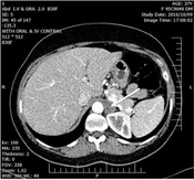
The initial differential diagnosis for acute muscle weakness and severe hypokalemia included primary aldosteronism, familial hypokalemic periodic paralysis, thyrotoxic periodic paralysis, gastrointestinal potassium loss, and neurologic causes of quadriparesis. The combination of resistant hypertension, severe hypokalemia, and muscle weakness raised strong suspicion for PA. The patient was hospitalized for 7 days, during which her serum potassium level normalized to 3.9 mEq/L and her muscle strength was fully restored. She was discharged on antihypertensive therapy and referred for further hormonal evaluation. However, because of poor adherence to the treatment plan, hormonal evaluation was performed 1 month after discharge. At the time of hormonal testing, the patient was receiving antihypertensive medications known to interfere with the renin-aldosterone system; these could not be discontinued because of her uncontrolled blood pressure. Laboratory evaluation revealed an elevated aldosterone concentration of 77.3 ng/dL (reference range, 3.7 - 43.2 ng/dL), a suppressed direct renin concentration of 3.3 µIU/mL (reference range, 5.3 - 99.1 µIU/mL), and an aldosterone-to-renin ratio (ARR) of 24.3, findings consistent with possible PA. Subsequent noncontrast abdominal CT revealed a well-enhancing 2 × 1.5 cm left adrenal mass highly suspicious for an aldosterone-producing adenoma (Figure 1A). The patient’s initial clinical presentation, biochemical evaluation, and abdominal CT findings strengthened the diagnosis of PA. She was subsequently referred for surgical management of the adrenal lesion.
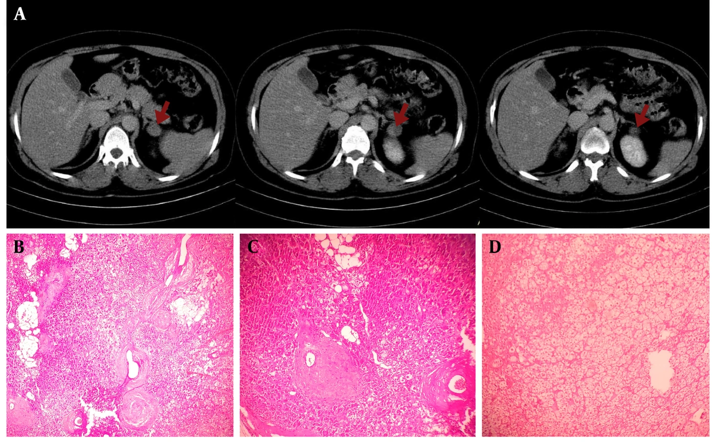
Figure 1.
A, Abdominal CT scan; B-D, pathological findings suggestive of an aldosterone-producing adenoma. A, The red arrows show the 2 × 1.5 cm left adrenal adenoma in the suprarenal region. B, Pale, lipid-rich cells admixed with eosinophilic cells. C, Interface between a hyperplastic zona glomerulosa and adjacent tissue. D, Neoplastic cells with clear to lightly eosinophilic cytoplasm and centrally located round nuclei arranged in nested and trabecular patterns, associated with a rich capillary vasculature. The features are suggestive of an adrenal cortical adenoma.
Because of financial constraints, adrenalectomy was performed 5 months after diagnosis. She underwent laparoscopic unilateral adrenalectomy to remove the adrenal mass. At the time of surgery, her serum potassium level remained within the normal range and her muscle strength was fully preserved; however, she still required antihypertensive medication for blood pressure control.
The definitive diagnosis was established by histopathological examination of the resected tissue, which confirmed an adrenal adenoma (Figure 1B-D).
2.1. Follow-Up
Serial follow-up over 13 months showed progressive improvement. Postoperatively, her blood pressure decreased to 140/90 mmHg, serum potassium remained within the normal range, and neurologic symptoms resolved. During the first 6 months, antihypertensive therapy was gradually tapered to telmisartan 20 mg daily and later discontinued. At the final 13-month follow-up, her blood pressure was 110/70 mmHg without medication. Left ventricular hypertrophy regressed on electrocardiography and echocardiography, and neurologic examination showed full muscle strength (5/5) in all extremities. She experienced no further hypertensive crises or recurrent muscle weakness during follow-up.
Table 1 shows the chronological course of this case.
Table 1.
Chronological Clinical Course
| Time Point | Clinical Events and Management |
|---|---|
| >7 years before presentation | History of poorly controlled hypertension with recurrent episodes of hypokalemia-associated muscle weakness. |
| Initial clinical presentation | Presented to the clinic with severe hypertension, quadriparesis, and severe hypokalemia. |
| Initial evaluation | Laboratory and hormonal investigations raised suspicion for primary aldosteronism (PA). |
| Hospital admission | Referred for inpatient management of severe hypertension and hypokalemia. Hydrochlorothiazide was discontinued, and potassium replacement was initiated with close cardiac and biochemical monitoring. |
| During hospitalization | Additional laboratory investigations, physical examination, neurologic evaluation, and brain imaging were performed. |
| Day 7 of hospitalization | Serum potassium normalized, muscle strength was fully restored, and the patient was discharged on antihypertensive therapy. |
| Post-discharge | One month after discharge, further hormonal tests (renin, aldosterone, and ARR) and abdominal computed tomography were performed. The findings, including an adrenal mass and laboratory results, were consistent with an aldosterone-producing adenoma (APA). The patient was referred for surgical management of the adrenal mass. |
| 5 months after diagnosis | Because of financial constraints, surgery was delayed. The patient subsequently underwent laparoscopic unilateral adrenalectomy to remove the adrenal mass. |
| Histopathological evaluation | Histopathological examination confirmed an adrenal adenoma. |
| Postoperative course | Serum potassium remained normal, and muscle strength was fully preserved. Limited antihypertensive therapy was continued for blood pressure control. |
| 1-year follow-up | The patient experienced no further hypertensive crises or episodes of muscle weakness and was able to discontinue antihypertensive medications. |
3. Discussion
PA is recognized as the most common cause of secondary hypertension. The prevalence of PA is 3.2 - 12.7% in primary care and 1 - 29.8% among patients with hypertension referred for specialist care (10, 11). Aldosterone-producing adenomas are the most frequent etiology (12). When left untreated, PA is associated with significantly worse cardiovascular and renal outcomes. Patients with PA have increased rates of left ventricular hypertrophy, atrial fibrillation, myocardial fibrosis, heart failure, coronary artery disease, and stroke (13).
Despite its relatively high prevalence, PA remains markedly underdiagnosed (14). Hypertension with hypokalemia is a classic presentation that should raise suspicion for PA. Up to 30% of hypertensive patients with unexplained hypokalemia may have PA (2). However, screening is applied inconsistently in practice. A recent survey found that renin and aldosterone measurements were requested by only 7% of general practitioners in Italy and 8% in Germany (15).
Our case illustrates this diagnostic gap. The patient had profound hypokalemia and muscle weakness, yet her underlying PA remained undiagnosed for nearly 10 years. During this period, she was treated with multiple antihypertensive medications, including amlodipine, valsartan, and hydrochlorothiazide, but did not achieve stable blood pressure control. The diagnosis was made only after she developed severe hypertension and repeated episodes of quadriparesis. This finding is consistent with a recent population-level study showing that, even among patients with recurrent or severe hypokalemia and resistant hypertension, the rate of screening for PA remains extremely low, at less than 5% (2). That study also showed that referral to subspecialists, such as cardiologists or endocrinologists, was associated with higher screening rates, as in this case, in which a cardiology evaluation ultimately led to the diagnosis. Conversely, older age and comorbidities such as diabetes were associated with lower screening rates (2).
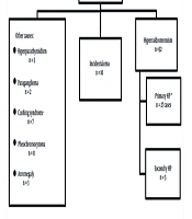
Guidelines from the European Society of Cardiology (ESC) and the European Society of Hypertension (ESH) have increasingly emphasized screening for secondary causes of hypertension. The 2018 guidelines did not provide a clear recommendation specifically for PA screening; however, they recommended screening for secondary hypertension in selected groups, such as younger patients (< 40 years) with grade 2 hypertension, patients with early-onset hypertension, and those with resistant hypertension (5). More recently, the 2024 guideline update moved toward a broader screening approach; with class 2a evidence, it advocates renin and aldosterone measurement in all adults with hypertension ≥ 140/90 mmHg, particularly when suggestive signs or symptoms are present (Figure 2) (6).
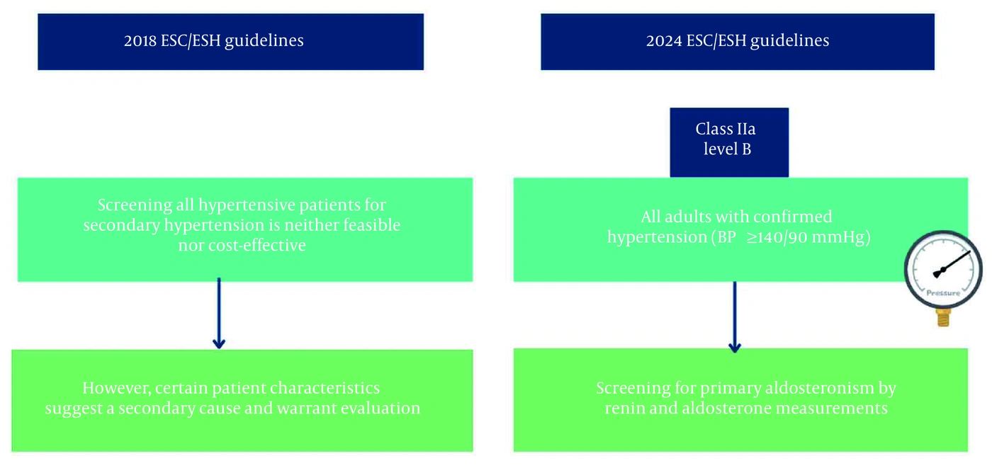
Figure 2.
2024 ESC/ESH guidelines for screening for secondary hypertension. Abbreviations: BP, blood pressure; ESC, European Society of Cardiology; ESH, European Society of Hypertension.
This broader approach may improve detection because hypokalemia is present in only 9 - 37% of patients with primary aldosteronism, with most patients presenting with normokalemia (7). Because general practitioners are the first point of contact for most patients with hypertension, increasing their awareness of PA is essential.
The gold standard for the initial diagnosis of PA is the ARR, often followed by confirmatory testing. To improve ARR sensitivity and specificity, hypokalemia should be corrected, and interfering medications withheld for an appropriate period before testing. Many antihypertensive drugs can distort the ARR, causing false-positive or false-negative results (16). Eplerenone, valsartan, and hydrochlorothiazide can interfere with the renin-aldosterone axis, thereby affecting the ARR and limiting the ability to perform a medication-free assessment (7, 12). In this case, hypokalemia was corrected first; however, discontinuation of antihypertensive therapy was difficult because of severe hypertension. Eplerenone was also clinically useful for controlling potassium loss. Therefore, the ARR had to be interpreted in the context of these potentially interfering factors. Nevertheless, despite treatment with potentially interfering medications, the patient demonstrated a biochemical pattern consistent with primary aldosteronism, with suppressed renin and substantially elevated aldosterone, suggesting renin-independent aldosterone secretion (7). Some antihypertensive medications, such as doxazosin and hydralazine, have minimal effects on ARR and can be used during testing (17). Confirmatory testing was not performed because the patient required urgent treatment during her first admission. In severe clinical presentations, immediate treatment may take priority over further diagnostic testing (7). Furthermore, she was unwilling to undergo further evaluation because of financial constraints at later visits. In this case, the diagnosis of PA was established through a combination of clinical presentation, biochemical findings, adrenal imaging, postoperative clinical improvement, and histopathological confirmation.
Once PA is confirmed, the next step is subtyping to guide therapy. Cross-sectional imaging with CT or magnetic resonance imaging (MRI) of the adrenal glands is the standard first test for subtype classification. For patients with unilateral aldosterone excess, the optimal therapy is unilateral laparoscopic adrenalectomy (18).
In a literature review by Shi et al., 43 cases of PA associated with hypokalemic rhabdomyolysis were identified. Most patients were middle-aged women. All showed good recovery with normalization of potassium, and in many patients, blood pressure also returned to normal, similar to our patient (19).
Other potential differential diagnoses were considered less likely based on the clinical and laboratory findings. Familial hypokalemic periodic paralysis was considered unlikely in the absence of a personal or family history of similar episodes, whereas thyrotoxic periodic paralysis was excluded by normal thyroid function tests. Gastrointestinal potassium loss was unlikely given the absence of vomiting, diarrhea, or other gastrointestinal symptoms. Neurologic causes were also considered less likely given the otherwise unremarkable neurologic examination, normal brain CT, and nonspecific EMG/NCS findings.
This case had several limitations. We were unable to follow the exact protocol for ARR measurement. Immunohistochemistry results, such as CYP11B2 staining, were not obtained. Contrast-enhanced adrenal CT was not performed, which limited detailed characterization of the adrenal mass. In addition, follow-up adrenal imaging was not performed after treatment to assess interval changes in the lesion. Finally, the exact values of some laboratory tests reported as being within the normal range were unavailable.
3.1. Conclusions
This case, together with evolving guideline recommendations, supports recognition of PA as a common, treatable, yet underdiagnosed condition among patients with hypertension. Earlier and broader implementation of systematic screening strategies may enable earlier diagnosis, targeted therapy, and improved cardiovascular and renal outcomes.