
1. Background
Growth disorders and malnutrition are major health problems during childhood and may adversely affect physical growth, development, and long-term health. Growth disorders refer to inadequate or abnormal physical growth and are commonly evaluated using serial measurements of weight, length or height, and, when appropriate, head circumference, interpreted according to standardized age- and sex-specific growth references (1, 2). Early recognition of abnormal growth is important because prolonged growth impairment may be associated with adverse effects on physical development, cognitive function, learning, and overall health (3, 4).
Childhood malnutrition has a multifactorial etiology involving biological, nutritional, social, environmental, and socioeconomic factors (5). Although the epidemiology of malnutrition varies substantially across countries and populations, inadequate dietary intake, impaired nutrient absorption, increased metabolic requirements, and underlying diseases may all contribute to impaired growth. Growth disorders are particularly common during early childhood, when nutritional requirements are high and growth is rapid (1). Failure to thrive (FTT) is generally characterized by inadequate weight gain or failure to maintain an expected pattern of growth over time and is assessed using serial anthropometric measurements, including weight and length or height (1, 6). The causes of FTT are traditionally categorized as organic, non-organic, or mixed. Organic causes include acute or chronic diseases that impair nutrient intake, absorption, or utilization or increase energy requirements, whereas non-organic causes may be associated with inadequate dietary intake and environmental, social, or psychosocial factors. In many children, multiple factors may coexist. Management is directed toward identifying and addressing the underlying cause and providing adequate nutritional support to restore appropriate growth. Prolonged or untreated growth failure may be associated with adverse developmental, behavioral, educational, and psychosocial outcomes (7, 8), with prognosis influenced by the underlying etiology and social and socioeconomic circumstances (9).
Nutritional status and growth may also be associated with changes in the size and morphology of visceral organs. The liver and spleen are particularly relevant because their dimensions vary substantially during childhood and may be affected by age, sex, body size, nutritional status, and underlying disease. Several pediatric conditions, birth-related factors, and medications can alter liver or spleen morphology and dimensions. Accurate assessment of these organs is therefore important when evaluating children with suspected hepatosplenic abnormalities. However, clinical assessment by palpation and percussion may have limited accuracy, particularly for detecting mild enlargement or changes that are not clinically apparent (10).
Ultrasonography is widely used to assess abdominal organs in children because it is noninvasive, does not involve ionizing radiation, is relatively inexpensive, and can be performed repeatedly when clinically indicated. Nevertheless, ultrasonographic organ measurements may vary according to the imaging technique, anatomical landmarks, patient position, equipment, and characteristics of the study population. Differences between ultrasonography and other imaging-based measurements have also been reported, emphasizing the importance of standardized measurement procedures and population-specific reference data (11, 12). In addition, liver dimensions have been reported to correlate with body size, particularly weight, suggesting that the interpretation of organ dimensions without considering age and anthropometric characteristics may be misleading (12).
Previous research has examined liver morphology and hepatic abnormalities in children with severe malnutrition. For example, one study evaluating hepatic steatosis in children with severe malnutrition found no significant association between the degree of steatosis and overall liver size (13). This observation suggests that nutritional status and liver dimensions may not have a simple or uniform relationship and highlights the need to evaluate organ dimensions while accounting for relevant demographic and anthropometric characteristics. Establishing appropriate reference values for liver and spleen dimensions in children is challenging because these organs undergo substantial growth throughout childhood. Published reference values may differ according to age range, population characteristics, anthropometric variables, imaging techniques, and the anatomical landmarks used for measurement (14-15). Consequently, comparisons of organ dimensions between children with different nutritional statuses should account for age and body size rather than relying solely on unadjusted measurements. Despite the clinical importance of these considerations, evidence regarding ultrasonographic liver and spleen dimensions among children with malnutrition remains limited, particularly in populations in resource-limited settings.
2. Objectives
This study compared ultrasonographic liver and spleen dimensions between children with malnutrition and healthy controls. It also examined whether these associations varied by selected demographic and perinatal characteristics, including sex, age, feeding type, birth weight, and gestational age.
3. Methods
3.1. Study Design and Participants
This analytical case-control study compared ultrasonographic liver and spleen dimensions between children with malnutrition and healthy children. Participants were recruited from the Pediatric Gastroenterology Clinics of Ali Ibn Abi Talib and Ali Asghar Hospitals, affiliated with Zahedan University of Medical Sciences, during 2022 - 2023. Children who met the predefined criteria for malnutrition constituted the case group, whereas healthy children with normal anthropometric measurements constituted the control group. Age and sex were selected as matching variables because both are important determinants of childhood growth and normal abdominal organ dimensions. Individual 1:1 matching was performed, with each child in the case group matched to one control of the same sex and similar age. An allowable age difference of ± 6 months was used for children younger than 5 years and ± 12 months for children aged 5 years or older. Controls were recruited from the same clinical setting and during the same study period. When more than one eligible control was available, the control with the closest age to the corresponding case was selected. Because the study was observational, the findings were interpreted as associations rather than causal effects. The study did not assess the effects of nutritional rehabilitation, the diagnostic accuracy of ultrasonography, longitudinal changes in organ dimensions, or the clinical benefits or harms of routine hepatosplenic ultrasonography.
Children were excluded if they had genetic syndromes, metabolic disorders, hereditary short stature, or chronic renal, cardiac, gastrointestinal, or hematologic diseases. Children with incomplete anthropometric measurements or technically inadequate ultrasonographic examinations were excluded from the corresponding analyses.
3.2. Anthropometric Assessment and Definition of Malnutrition
Anthropometric assessment was performed before ultrasonography by trained clinical staff using standardized procedures. World Health Organization (WHO) growth references were used for anthropometric classification. The WHO Child Growth Standards were applied to children younger than 5 years, whereas the WHO 2007 Growth Reference was used for children aged 5 years and older. Weight-for-age (WFA) and height/length-for-age (HFA/LFA) were determined according to the child’s age and sex using the appropriate WHO reference. Children were classified as having malnutrition if both WFA and HFA/LFA were below the fifth percentile of the applicable WHO reference. Children with discordant anthropometric findings, in whom only one of these indicators was below the fifth percentile, were not classified as cases according to the predefined study criteria. Controls were required to have both WFA and HFA/LFA between the fifth and 95th percentiles and no clinical evidence of chronic or systemic disease. Children whose anthropometric measurements did not meet these predefined criteria were not eligible for the control group. Anthropometric classification was based on calculated WHO reference values rather than subjective clinical assessment.
3.3. Measurement of Weight and Length/Height
Weight and length/height were measured before ultrasonography using standardized procedures and calibrated equipment. Children were lightly clothed and measured without shoes. The same measurement procedures were applied to cases and controls throughout the study. For children younger than 2 years, recumbent length was measured using a calibrated infantometer. The child was placed in the supine position, with the head positioned against the fixed headboard, the trunk and legs aligned with the measuring surface, and the legs gently extended. The feet were positioned perpendicular to the measuring surface, and the movable footboard was brought into contact with the soles. Recumbent length was recorded to the nearest 0.1 cm. For children aged 2 years and older, standing height was measured using a calibrated stadiometer. The child stood upright without shoes, with the heels together and the body positioned according to standard height-measurement procedures. The head was maintained in the Frankfurt horizontal plane, and the movable headpiece was positioned firmly on the crown of the head. Standing height was recorded to the nearest 0.1 cm. Weight was measured in all children using a calibrated digital weighing scale. The child was lightly clothed, without shoes, and positioned centrally on the scale. Weight was recorded to the nearest 0.1 kg. If a measurement was technically unsatisfactory or inconsistent with the measurement procedure, it was repeated. Equipment was checked and calibrated regularly throughout the study period. The same standardized procedures and equipment were used for both the case and control groups.
3.4. Sample Size
Because the primary objective was to compare continuous ultrasonographic liver and spleen dimensions between two groups, the initial prevalence-based sample-size calculation using P = 0.50 and a precision of 0.10 was not considered appropriate for the primary outcome. The sample size was therefore determined based on a two-group comparison of continuous outcomes. The calculation considered the expected difference in organ dimensions, the anticipated standard deviation, a two-sided significance level of 0.05, statistical power of 80%, and a 1:1 allocation ratio. Because liver and spleen dimensions were the principal outcomes, the sample size was determined according to the outcome requiring the larger sample under the selected assumptions. An allowance for incomplete data and technically inadequate ultrasonographic examinations was incorporated into the recruitment target. Subgroup analyses according to sex, age, feeding type, birth weight, and prematurity were not considered independently powered analyses and were therefore interpreted as exploratory.
3.5. Data Collection and Study Procedures
After approval by the institutional ethics committee, written informed consent was obtained from the parents or legal guardians of all participants. Data were collected using a structured data-collection form. Recorded variables included age, sex, feeding type, gestational age, birth weight, current weight, length/height, and relevant anthropometric indices. Anthropometric measurements were obtained before ultrasonography. The radiologist performing the ultrasonographic examination was blinded to the participant’s case-control classification.
3.6. Ultrasonography Assessment
Ultrasonographic examinations of the liver and spleen were performed by a senior radiologist using standard pediatric abdominal ultrasonography techniques. Participants were examined in the supine position using an appropriate abdominal transducer according to the child’s age and body habitus. A low-frequency curvilinear transducer was used when appropriate, whereas a higher-frequency transducer was used for smaller children when necessary. Participants underwent routine preparation for abdominal ultrasonography. When clinically feasible, examinations were performed after a period of fasting to reduce bowel gas and facilitate visualization of the abdominal organs. The liver and spleen were systematically examined in longitudinal and transverse planes.
The primary liver outcome was the maximum craniocaudal length of the right hepatic lobe, measured in the longitudinal plane along the midclavicular line. The measurement was obtained from the superior to the inferior margin of the right hepatic lobe at the point at which the maximum longitudinal dimension was visualized, following standard pediatric abdominal ultrasonographic technique. The primary spleen outcome was the maximum longitudinal length of the spleen, measured along its long axis from the superior pole to the inferior pole using the standard abdominal ultrasonographic approach. Liver and spleen dimensions were recorded in centimeters. When technically feasible, three acceptable measurements were obtained for each organ, and the mean of the three measurements was used as the final value for analysis. If repeated technically adequate measurements could not be obtained, the best-quality measurement was recorded. Liver and spleen echogenicity were assessed qualitatively according to standard sonographic criteria. However, the primary outcomes of the study were the quantitative liver and spleen dimensions.
3.7. Ultrasonography Reliability
Because the study was conducted as part of a routine clinical ultrasonographic assessment and repeated examinations by the same observer were not independently performed to assess measurement reliability, formal intraobserver reliability coefficients were not calculated.
3.8. Statistical Analysis
Statistical analyses were performed using IBM SPSS Statistics version 26.0. Continuous variables were summarized as mean ± standard deviation (SD), and categorical variables were summarized as frequencies and percentages. Distributional characteristics of continuous variables were evaluated using graphical methods and the Shapiro-Wilk test. For continuous outcomes, paired-samples t tests were used when the within-pair differences were approximately normally distributed. The Wilcoxon signed-rank test was used when the normality assumption was not satisfied. For each primary outcome, the absolute mean difference between children with malnutrition and control children was reported together with its 95% confidence interval (CI). P values were interpreted together with the magnitude and precision of the estimated effect.
3.9. Ethical Considerations
The study was approved by the Ethics Committee of Zahedan University of Medical Sciences (Approval Code: IR.ZAUMS.REC.1402.120). Participation was voluntary, and written informed consent was obtained from the parents or legal guardians of all participants. Participant information was kept confidential and used exclusively for research purposes.
4. Results
4.1. Participant Characteristics
A total of 194 children were included in the study, comprising 97 children with malnutrition and 97 healthy controls. Results are presented according to the participants’ demographic and clinical characteristics, anthropometric and ultrasonographic findings, and exploratory subgroup analyses of liver and spleen dimensions by sex, age group, feeding type, birth weight, and gestational age. Because age and sex were used as matching variables, their distributions were expected to be comparable between the two groups. The primary outcomes were ultrasonographic liver and spleen dimensions.
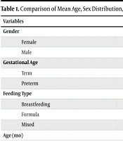
Table 1 presents the demographic and clinical characteristics of the malnutrition and control groups. The mean age was 30.9 ± 27.3 months in the malnutrition group and 35.9 ± 30.1 months in the control group (P = 0.22). The sex distribution was similar between the groups, with males comprising 50.5% of the malnutrition group and 43.3% of the control group (P = 0.31). Gestational age did not differ significantly between the groups (P = 0.16). Among children with malnutrition, 72 (74.2%) were born at term and 25 (25.8%) were preterm, compared with 80 (82.5%) and 17 (17.5%), respectively, among controls. Feeding type differed significantly between the groups (P = 0.001). Breastfeeding was reported in 52 (53.6%) children with malnutrition compared with 67 (69.1%) controls, whereas formula feeding was more frequent among children with malnutrition (33 [34.0%] vs. 11 [11.3%]). Mixed feeding was reported in 12 (12.4%) children in the malnutrition group and 19 (19.6%) children in the control group.
Table 1.
Comparison of Mean Age, Sex Distribution, Feeding Type During the First 6 Months, and Gestational Age Between Case and Control Groups a
| Variables | Case Group | Control Group | Total | P-Value |
|---|---|---|---|---|
| Gender | 0.31 | |||
| Female | 48 (49.5) | 55 (56.7) | 103 (53.1) | |
| Male | 49 (50.5) | 42 (43.3) | 91 (46.9) | |
| Gestational Age | 0.16 | |||
| Term | 72 (74.2) | 80 (82.5) | 152 (78.4) | |
| Preterm | 25 (25.8) | 17 (17.5) | 42 (21.6) | |
| Feeding Type | 0.001 | |||
| Breastfeeding | 52 (53.6) | 67 (69.1) | 119 (61.3) | |
| Formula | 33 (34.0) | 11 (11.3) | 44 (22.7) | |
| Mixed | 12 (12.4) | 19 (19.6) | 31 (16.0) | |
| Age (mo) | 30.9 ± 27.3 | 35.9 ± 30.1 | - | 0.22 |
a Values are expressed as No. (%) or mean ± SD.
4.2. Anthropometric and Ultrasonography Findings
Table 2 compares current weight, height, birth weight, and ultrasonographic liver and spleen dimensions between the malnutrition and control groups. Mean current weight was significantly lower among children with malnutrition than among controls (11,533.9 ± 8,395.3 g vs. 15,515.4 ± 10,621.1 g; P = 0.004). Mean height did not differ significantly between the groups (83.1 ± 26.3 cm vs. 88.8 ± 27.2 cm; P = 0.13). Similarly, mean birth weight did not differ significantly between children with malnutrition and controls (2981.4 ± 298.7 g vs. 3075.3 ± 517.1 g; P = 0.12). Ultrasonography showed statistically significant differences in both liver and spleen dimensions. The mean liver dimension was 90.5 ± 18.4 mm in the malnutrition group and 82.1 ± 24.9 mm in the control group (P = 0.008), indicating a larger mean liver dimension in the malnutrition group in the overall sample. In contrast, the mean spleen dimension was 63.0 ± 21.2 mm in the malnutrition group compared with 69.9 ± 19.9 mm in controls (P = 0.02), indicating a smaller mean spleen dimension among children with malnutrition. Because liver and spleen dimensions may be influenced by age, sex, and body size, these unadjusted differences were further evaluated using multivariable analyses incorporating clinically relevant demographic and growth-related covariates.
Table 2.
Comparison of Weight, Height, Birth Weight, Liver Size, and Spleen Size Between Case and Control Groups at Admission
| Variables and Groups | Mean ± SD | Min | Max | P-Value |
|---|---|---|---|---|
| Current Weight (g) | 0.004 | |||
| Case | 11533.9 ± 8395.3 | 2500 | 35000 | |
| Control | 15515.4 ± 10621.1 | 2500 | 35000 | |
| Current Height (cm) | 0.13 | |||
| Case | 83.06 ± 26.3 | 38 | 135 | |
| Control | 88.8 ± 27.2 | 38 | 135 | |
| Birth Weight (g) | 0.12 | |||
| Case | 2981.4 ± 298.7 | 1700 | 3800 | |
| Control | 3075.3 ± 517.1 | 1700 | 3800 | |
| Liver Size (cm) | 0.008 | |||
| Case | 90.5 ± 18.4 | 50 | 128 | |
| Control | 82.1 ± 24.9 | 50 | 132 | |
| Spleen Size (cm) | 0.02 | |||
| Case | 63.01 ± 21.2 | 27 | 102 | |
| Control | 69.9 ± 19.9 | 27 | 122 |
4.3. Exploratory Subgroup Analyses
Table 3 presents exploratory comparisons of liver and spleen dimensions between children with malnutrition and controls according to sex, age group, feeding type, birth weight, and gestational age. Among boys, the mean liver dimension was 70.6 ± 15.7 mm in the malnutrition group compared with 87.5 ± 14.6 mm among controls (P = 0.001). Similarly, the mean spleen dimension was 53.3 ± 13.9 mm among boys with malnutrition compared with 64.4 ± 17.4 mm among control boys (P = 0.001). Among girls, no statistically significant differences were observed in liver dimension (92.7 ± 20.7 vs. 93.8 ± 27.1 mm; P = 0.800) or spleen dimension (74.1 ± 20.8 vs. 74.9 ± 20.9 mm; P = 0.800). Among children younger than 5 years, the mean liver dimension was lower in the malnutrition group than in controls (74.1 ± 18.6 vs. 83.1 ± 13.5 mm; P = 0.001). The mean spleen dimension was also lower among children with malnutrition (56.8 ± 17.3 vs. 62.5 ± 15.3 mm; P = 0.037). Among children aged 5 years or older, no statistically significant differences were observed in liver dimension (111.8 ± 13.2 vs. 119.6 ± 13.7 mm; P = 0.720) or spleen dimension (91.4 ± 15.7 vs. 91.8 ± 12.4 mm; P = 0.900). Among formula-fed children, the mean liver dimension was lower in the malnutrition group than in controls (69.8 ± 11.4 vs. 92.3 ± 28.0 mm; P = 0.013). The mean spleen dimension was also lower among children with malnutrition (54.5 ± 25.4 vs. 70.2 ± 31.5 mm; P = 0.024). Among breastfed children, no statistically significant differences were observed in liver dimension (87.2 ± 30.5 vs. 90.3 ± 15.5 mm; P = 0.400) or spleen dimension (64.8 ± 24.7 vs. 67.9 ± 19.3 mm; P = 0.400). Similarly, no statistically significant differences were observed among children receiving mixed feeding. Among children with birth weight < 2500 g, both liver and spleen dimensions were lower in the malnutrition group than in controls. The mean liver dimension was 89.2 ± 19.4 mm compared with 104.1 ± 24.2 mm (P = 0.031), while the mean spleen dimension was 64.1 ± 21.2 mm compared with 73.8 ± 15.7 mm (P = 0.025). Among children with birth weight ≥ 2500 g, no statistically significant differences were observed in liver dimension (81.4 ± 23.4 vs. 88.4 ± 16.5 mm; P = 0.160) or spleen dimension (62.9 ± 21.3 vs. 67.8 ± 19.7 mm; P = 0.120). Among term children, no statistically significant difference was observed in liver dimension between the malnutrition and control groups (82.4 ± 21.8 vs. 83.4 ± 22.4 mm; P = 0.800). Among preterm children, however, liver dimension was lower in the malnutrition group (81.6 ± 26.0 vs. 92.2 ± 17.1 mm; P = 0.003). For spleen dimension, no statistically significant difference was observed among term children (64.4 ± 23.0 vs. 70.0 ± 20.7 mm; P = 0.140), whereas among preterm children, spleen dimension was lower in the malnutrition group (57.8 ± 14.3 vs. 69.4 ± 15.9 mm; P = 0.018).
Table 3.
Comparison of Liver and Spleen Sizes by Sex, Age Group, Feeding Type, Birth Weight, and Gestational Age in Children with Malnutrition Versus Controls
| Variables and Group of Participants | Mean ± SD | P-Value |
|---|---|---|
| Gender | ||
| Liver Size (mm) | ||
| Female | 0.800 | |
| Case | 92.7 ± 20.7 | |
| Control | 93.8 ± 27.1 | |
| Male | 0.001 | |
| Case | 70.6 ± 15.7 | |
| Control | 87.5 ± 14.6 | |
| Spleen Size (mm) | ||
| Female | 0.800 | |
| Case | 74.1 ± 20.8 | |
| Control | 74.9 ± 20.9 | |
| Male | 0.001 | |
| Case | 53.3 ± 13.9 | |
| Control | 64.4 ± 17.4 | |
| Age (y) | ||
| Liver Size (mm) | ||
| Under 5 | 0.001 | |
| Case | 74.1 ± 18.6 | |
| Control | 83.08 ± 13.5 | |
| Over 5 | 0.720 | |
| Case | 111.8 ± 13.2 | |
| Control | 119.6 ± 13.7 | |
| Spleen Size (mm) | ||
| Under 5 | 0.037 | |
| Case | 56.8 ± 17.3 | |
| Control | 62.5 ± 15.3 | |
| Over 5 | 0.900 | |
| Case | 91.4 ± 15.7 | |
| Control | 91.8 ± 12.4 | |
| Type of Feeding | ||
| Liver Size (mm) | ||
| Breastfeeding | 0.400 | |
| Case | 87.2 ± 30.5 | |
| Control | 90.3 ± 15.5 | |
| Formula | 0.013 | |
| Case | 69.8 ± 11.4 | |
| Control | 92.3 ± 28.0 | |
| Mixed | 0.100 | |
| Case | 79.1 ± 16.8 | |
| Control | 88.0 ± 15.1 | |
| Spleen Size (mm) | ||
| Breastfeeding | 0.400 | |
| Case | 64.8 ± 24.7 | |
| Control | 67.9 ± 19.3 | |
| Formula | 0.024 | |
| Case | 54.5 ± 25.4 | |
| Control | 70.2 ± 31.5 | |
| Mixed | 0.700 | |
| Case | 63.4 ± 12.9 | |
| Control | 64.5 ± 12.0 | |
| Weight at Birth (g) | ||
| Liver Size (mm) | ||
| < 2500 | 0.031 | |
| Case | 89.2 ± 19.4 | |
| Control | 104.1 ± 24.2 | |
| ≥ 2500 | 0.160 | |
| Case | 81.4 ± 23.4 | |
| Control | 88.4 ± 16.5 | |
| Spleen Size (mm) | ||
| < 2500 | 0.025 | |
| Case | 64.1 ± 21.2 | |
| Control | 73.8 ± 15.7 | |
| ≥ 2500 | 0.120 | |
| Case | 62.9 ± 21.3 | |
| Control | 67.8 ± 19.7 | |
| Gestational Age | ||
| Liver Size (mm) | ||
| Term | 0.800 | |
| Case | 82.4 ± 21.8 | |
| Control | 83.4 ± 22.4 | |
| Preterm | 0.003 | |
| Case | 81.6 ± 26.0 | |
| Control | 92.2 ± 17.1 | |
| Spleen Size (mm) | ||
| Term | 0.140 | |
| Case | 64.4 ± 23.0 | |
| Control | 70.0 ± 20.7 | |
| Preterm | 0.018 | |
| Case | 57.8 ± 14.3 | |
| Control | 69.4 ± 15.9 |
5. Discussion
This case-control study compared ultrasonographic liver and spleen dimensions between children with malnutrition and healthy controls attending pediatric gastroenterology clinics in Zahedan. The groups were generally comparable in terms of age, sex, gestational age, and birth weight; however, current weight was substantially lower among children with malnutrition, as expected based on the anthropometric definition of the study groups. Feeding type during the first 6 months also differed between the groups, with formula feeding being more frequent among children with malnutrition. Overall, the mean spleen dimension was smaller among children with malnutrition, whereas the mean liver dimension was greater in the malnutrition group. Exploratory subgroup analyses indicated that the direction and magnitude of these differences varied by sex, age, feeding type, birth weight, and gestational age. These findings should be interpreted in the context of the observational design and the potential influence of age, body size, and other growth-related characteristics on pediatric organ dimensions.
The difference in feeding patterns between the groups is noteworthy. Formula feeding during the first 6 months was more common among children with malnutrition than among controls, consistent with previous research reporting associations between early feeding practices and childhood nutritional status (16). However, the present case-control design does not allow determination of whether feeding type contributed to subsequent malnutrition or whether other social, nutritional, or clinical factors influenced both feeding practices and nutritional status. Therefore, the observed association should not be interpreted as evidence of a protective causal effect of breastfeeding. Feeding type was considered a potentially relevant covariate in interpreting the organ-dimension findings, and subgroup analyses were performed to explore whether the observed differences varied across feeding categories.
Current weight was substantially lower among children with malnutrition, whereas birth weight did not differ significantly between the groups. This finding is consistent with the study’s anthropometric classification, which was based on current growth status. However, the difference between current and birth weight should not be interpreted as evidence that postnatal factors were the principal causes of malnutrition. A range of postnatal factors, including dietary adequacy, infections, socioeconomic circumstances, and underlying medical conditions, may contribute to childhood malnutrition (15), but these factors were not comprehensively measured or evaluated in the present study.
Previous studies have examined hepatic abnormalities and ultrasonographic findings in children with severe malnutrition (10, 13). Some have reported hepatic steatosis or other biochemical and structural abnormalities in children with malnutrition. However, the presence of hepatic steatosis or biochemical abnormalities does not necessarily imply a consistent reduction in liver dimensions. Therefore, our findings should not be interpreted as demonstrating a specific pathological mechanism underlying the observed differences in liver size. Further studies incorporating biochemical measurements, liver composition, nutritional severity, and longitudinal follow-up are required to clarify these relationships.
The relationship between pediatric organ dimensions and body size is particularly important when interpreting the present findings. Previous research has demonstrated positive associations between liver dimensions and anthropometric measures such as weight (17), whereas other studies have reported differences in liver dimensions among children with growth failure (9). In addition, published reference standards for liver and spleen dimensions vary according to age, sex, height, body weight, population characteristics, imaging equipment, and measurement methodology (14-15). Consequently, a difference in an absolute organ measurement between two groups does not necessarily indicate pathological organ development unless age and body size are appropriately considered.
Reference data from previous pediatric ultrasonography studies further demonstrate that liver and spleen dimensions increase substantially during childhood and that the relationship between organ size and age is not necessarily linear (18). Rather than reproducing the detailed age-specific measurements reported in that external study, our findings should be interpreted in relation to the broader observation that pediatric liver and spleen dimensions are strongly dependent on developmental stage and body size. Differences between our population and previously studied populations may therefore contribute to variation in observed organ dimensions. These considerations support the use of population-appropriate, standardized ultrasonographic measurement protocols when evaluating pediatric organ size.
Our findings are consistent with those of Doğan et al. (19), who prospectively evaluated ultrasonographic solid-organ dimensions in 122 children with primary malnutrition and 68 healthy controls. They reported significantly smaller liver and spleen lengths among children with malnutrition compared with controls (liver: 99.84 ± 19.99 vs. 119.92 ± 26.29 mm, P < 0.001; spleen: 75.58 ± 19.01 vs. 94.72 ± 20.53 mm, P < 0.001). Furthermore, liver and spleen dimensions were positively correlated with anthropometric Z scores, particularly weight and height, suggesting that body size is an important determinant of pediatric organ dimensions. These findings support the biological plausibility of the smaller hepatosplenic dimensions observed in our participants with malnutrition, while also emphasizing the need to account for age and body-size measures when estimating the association between nutritional status and organ dimensions.
The clinical interpretation of ultrasonographic liver and spleen dimensions should therefore be individualized according to age, sex, height, weight, nutritional status, and clinical context. Ultrasonography has important advantages in pediatric abdominal assessment because it is noninvasive and does not involve ionizing radiation. However, our findings do not provide evidence that routine hepatosplenic ultrasonography should be recommended for all children with malnutrition. The present study did not evaluate diagnostic accuracy, clinical outcomes, cost-effectiveness, benefits, or potential harms of routine ultrasonographic screening. Consequently, any clinical use of ultrasonography should be based on the child’s clinical condition and the indication for imaging rather than on the findings of this study alone.
5.1. Study Limitations
This study has several limitations. The case-control design and single-center convenience sampling limit causal inference and generalizability. Residual confounding may persist because socioeconomic, dietary, biochemical, and nutritional-severity variables were not fully assessed. Current weight was closely related to the malnutrition definition and therefore was not treated as an independent confounder. Malnutrition severity and etiologic subtypes were not classified. Ultrasonographic measurements may also be influenced by technical and biological variability. Finally, multiple subgroup analyses may have resulted in chance findings, and the cross-sectional assessment did not allow evaluation of changes following nutritional rehabilitation.
5.2. Conclusions
In this clinic-based case-control study, children with malnutrition differed from healthy controls in ultrasonographic liver and spleen dimensions. In the overall sample, the mean spleen dimension was smaller among children with malnutrition, whereas the mean liver dimension was greater according to the measurements presented in the study data. Differences were also observed across several demographic and perinatal subgroups, although these subgroup findings should be considered exploratory and should not be interpreted as evidence of effect modification or causal relationships without formal interaction analyses.
These findings highlight the importance of considering age, body size, and other growth-related characteristics when interpreting pediatric liver and spleen dimensions. They do not, by themselves, support routine hepatosplenic ultrasonographic screening of all children with malnutrition. Future multicenter prospective studies with standardized ultrasonography protocols, larger and more representative samples, detailed assessment of malnutrition severity and etiology, biochemical measurements, and repeated measurements following nutritional rehabilitation are needed to clarify the longitudinal relationship between nutritional status and pediatric organ dimensions and to establish clinically useful population-specific reference ranges.