
1. Background
Multiple sclerosis (MS) is a chronic inflammatory demyelinating disease of the central nervous system that can also present during childhood or adolescence (1). Pediatric-onset MS (POMS) accounts for approximately 3 - 10% of all MS cases and often follows a relapsing-remitting course characterized by multifocal neurological symptoms, including optic neuritis, motor deficits, ataxia, and sensory disturbances (2, 3). Although children tend to recover more completely after initial attacks, they experience relapses more frequently and accumulate disability at a younger age than adults, underscoring the need for effective long-term disease control (4).
Current disease-modifying therapies used in pediatric MS are largely extrapolated from adult studies (5). Interferon-beta and glatiramer acetate remain first-line therapies because of their safety profiles; however, their limited efficacy and the burden of frequent injections can lead to suboptimal adherence (6, 7). Second-line agents such as fingolimod and natalizumab have demonstrated improved efficacy but raise concerns regarding adverse effects and long-term safety in children (8, 9). Therefore, identifying alternative therapeutic options with stronger and more durable effects remains a major clinical priority.
Accumulating evidence indicates that B cells play a central role in MS pathogenesis, supporting the rationale for B cell–depleting therapies (10, 11). Rituximab (RTX), a monoclonal antibody against CD20-positive B cells (12), has shown promising results in adults with relapsing MS (13) and in some pediatric studies (14-16), suggesting potential benefits in reducing relapse rates and radiological activity.
2. Objectives
This study was designed to evaluate the efficacy and safety of rituximab in preventing clinical relapses in pediatric patients with MS.
3. Methods
3.1. Study Design and Setting
This observational cohort study was conducted at Mofid Children's Hospital, a tertiary referral center in Iran. All consecutive pediatric patients with relapsing POMS who initiated rituximab treatment between April 2018 and March 2024 were identified from hospital medical records. No sampling or convenience selection was performed.
3.2. Participants and Sampling
Patients were eligible for inclusion if they had a diagnosis of relapsing POMS established before 18 years of age, were treated with rituximab at our center, and had a minimum follow-up of 1 year after treatment initiation. Patients were excluded if their final diagnosis was other than multiple sclerosis, including myelin oligodendrocyte glycoprotein (MOG) antibody disease or neuromyelitis optica (NMO) spectrum disorder; accordingly, all patients with a positive serum MOG-IgG result detected using a cell-based assay or a positive NMO antibody result were excluded. Additional exclusion criteria were a primary progressive disease course and incomplete or insufficient medical records.
3.3. Data Collection and Follow-Up
Baseline demographic, clinical, laboratory, and neuroimaging characteristics at the time of rituximab initiation were collected retrospectively from existing medical records. Patients were then followed longitudinally from the date of rituximab initiation until the last available clinical assessment. Follow-up information on clinical relapses and treatment-related adverse events was obtained through medical-record review, supplemented by structured telephone interviews when necessary. The index date for follow-up was the date of the first rituximab infusion. Follow-up continued until the last documented clinical assessment or the end of the study period (March 2024), whichever occurred first.
3.4. Laboratory and Paraclinical Assessments
Cerebrospinal fluid (CSF) analysis was performed to assess the presence of oligoclonal bands using isoelectric focusing followed by immunoblotting. Oligoclonal bands were considered positive when two or more unique bands were detected in the CSF but were absent in the paired serum sample.
The IgG index was calculated as (CSF IgG/serum IgG) ÷ (CSF albumin/serum albumin), with values > 0.7 considered elevated, indicating intrathecal IgG synthesis. IgG index assessment was not performed uniformly in all patients because of variations in laboratory protocols and clinical judgment over the study period.
Testing for MOG antibodies was performed in patients with atypical clinical presentations using cell-based assays. All individuals with positive MOG antibody results were excluded from the study.
3.5. Neuroimaging Protocol
All patients underwent baseline magnetic resonance imaging (MRI) of the brain and spinal cord. Follow-up brain MRI scans were performed in the event of a suspected clinical relapse. Spinal cord imaging was repeated if new spinal cord symptoms emerged.
3.6. Acute-Phase Treatment and Maintenance Therapy
During the acute phase, patients were treated according to standard clinical practice. High-dose intravenous methylprednisolone pulse therapy was administered at a dose of 20 - 30 mg/kg/day (maximum 1 g/day) for 3 - 5 consecutive days. Patients who showed an inadequate clinical response to corticosteroid therapy or presented with severe relapses underwent therapeutic plasma exchange (TPE).
TPE was performed using standard protocols, consisting of 5 - 7 exchange sessions on alternate days, with replacement using albumin and/or fresh frozen plasma as clinically indicated. The decision to initiate TPE was made by the treating neurologist based on relapse severity and response to initial steroid therapy.
Rituximab treatment was initiated after stabilization of the acute attack, typically 2 - 4 weeks after completion of intravenous methylprednisolone and plasma exchange, when performed. Rituximab was administered according to standardized institutional protocols. Induction therapy consisted of 375 mg/m2 weekly for 2 consecutive weeks. Maintenance therapy generally consisted of infusions every 6 - 12 months according to institutional practice.
3.7. Outcome Definition: Clinical Relapse
Clinical relapse was defined as the occurrence of new neurological symptoms or worsening of pre-existing neurological symptoms consistent with central nervous system demyelination, lasting for at least 24 hours, occurring after a period of neurological stability (defined as at least 30 days from the onset of the previous clinical relapse), and in the absence of fever, active infection, or other alternative medical explanations. Each suspected relapse was evaluated and confirmed by a pediatric neurologist based on the clinical history and neurological examination. Pseudo-relapses related to fever, infection, metabolic disturbances, heat exposure, or other transient physiological stressors were excluded through clinical assessment and review of the medical records.
Brain MRI was performed whenever a clinical relapse was suspected to evaluate radiological disease activity. MRI findings were considered supportive but were not required for the diagnosis of a clinical relapse, which remained a clinical diagnosis based on established diagnostic criteria. All suspected relapse events were reviewed by the treating pediatric neurologist. Relapses requiring treatment were managed with intravenous methylprednisolone according to institutional practice.
3.8. Statistical Analysis
Quantitative variables were described using the median and range (minimum-maximum) because of nonnormal distributions. Qualitative variables were presented as frequency and percentage. The Mann-Whitney U test and Fisher exact test were used to compare quantitative and qualitative variables, respectively, between the relapse and non-relapse groups. Multivariable regression analysis was not performed because only 9 patients experienced a clinical relapse during follow-up. The limited number of outcome events would have resulted in unstable adjusted estimates and a substantial risk of model overfitting. Therefore, all analyses were exploratory and based on unadjusted comparisons between the relapse and non-relapse groups.
The annualized relapse rate (ARR) was estimated using negative binomial regression models, with the logarithm of follow-up time included as an offset. A P value < 0.05 was considered statistically significant.
3.9. Ethical Considerations
The study protocol was approved by the Research Ethics Committee of Shahid Beheshti University of Medical Sciences, Tehran, Iran (IR.SBMU.RICH.REC.1403.024). Informed consent was obtained from the parents or legal guardians of all patients before inclusion in the study. The study protocol conformed to the ethical guidelines of the 1975 Declaration of Helsinki, as reflected in prior approval by the institution's human research committee.
4. Results
During the study period, 49 patients treated with rituximab were assessed for eligibility. After application of the predefined inclusion and exclusion criteria, 34 patients were included in the final analysis (Figure 1).
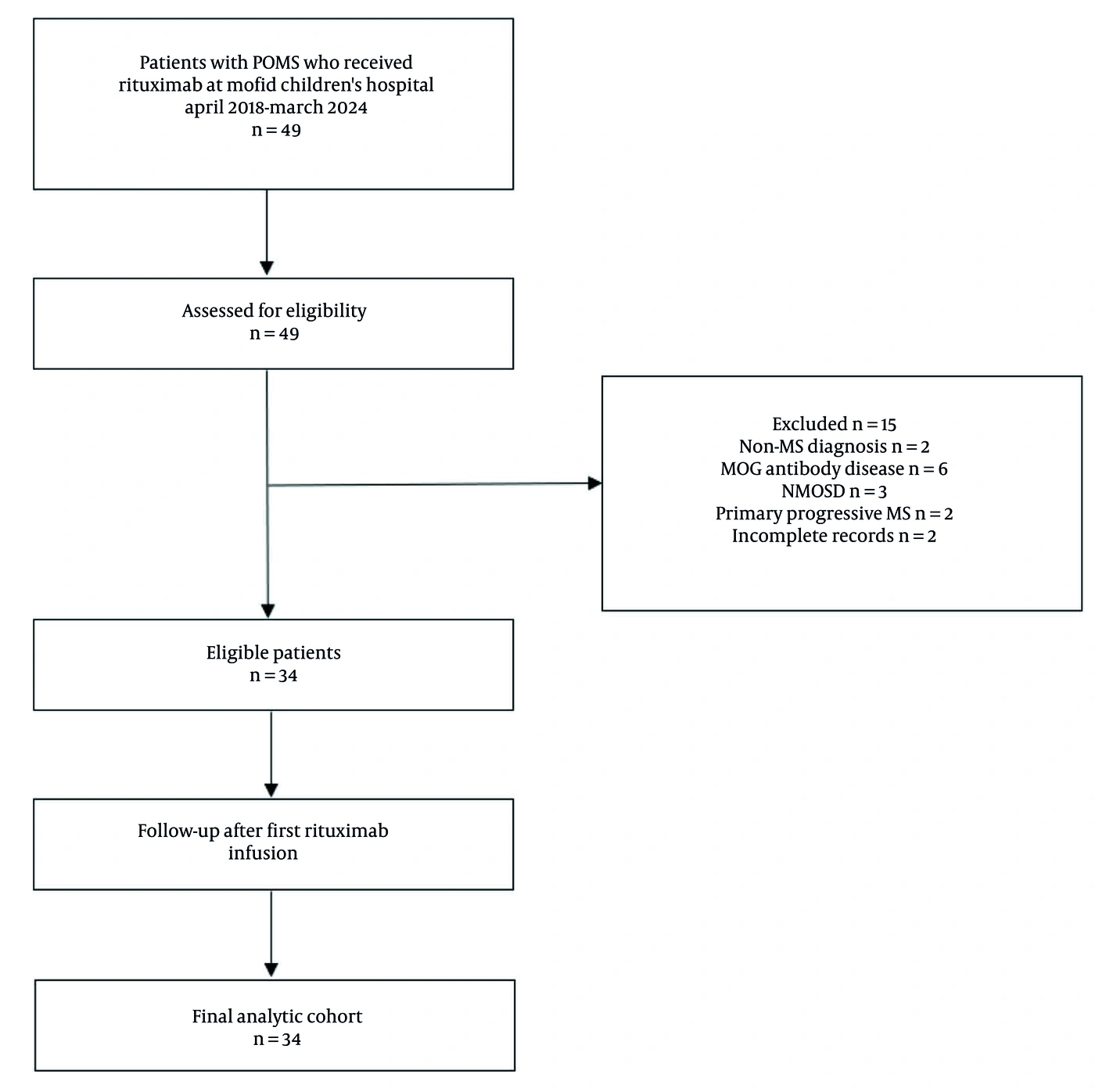
Figure 1.
Flow of patients through screening, eligibility assessment, inclusion, follow-up, and analysis.
Thirty-four patients (17 female and 17 male) with a median age of 15 years (age range: 10 to 18 years) were enrolled. The median age at diagnosis was 11 years (age range: 4 to 17 years).
4.1. Clinical Manifestations and Neurological Examination Findings at Diagnosis
At initial diagnosis, patients presented with diverse neurological manifestations (Table 1). Visual and oculomotor dysfunction was the most common presentation (23 patients, 67.60%), including cranial nerve abnormalities in 8 patients (23.50%) and optic nerve involvement in 15 patients (44.10%). Five patients exhibited overlap of optic and oculomotor nerve dysfunction.
Table 1.
Clinical Manifestations and Neurological Examination Findings of Patients with POMS
| Variables | No. (%) |
|---|---|
| Visual and oculomotor dysfunctions | 23 (67.60) |
| Cranial nerve abnormalities | 8 (23.50) |
| Third nerve palsy | 3 (8.80) |
| Sixth nerve palsy | 2 (5.90) |
| Seventh nerve palsy | 3 (8.80) |
| Optic nerve involvement | 15 (44.10) |
| Decreased visual acuity or vision loss | 9 (26.50) |
| Combined decreased vision with diplopia | 5 (14.70) a |
| Color vision impairment | 2 (5.90) |
| Motor system involvement | 18 (52.90) |
| Muscle tone and strength | |
| Decreased muscle tone | 1 (2.90) |
| Reduced muscle force | 10 (29.40) |
| Pyramidal tract signs | 14 (41.20) |
| Hemiparesthesia | 3 (8.80) |
| Monoparesthesia | 3 (8.80) |
| Monoplegia | 4 (11.80) |
| Hemiplegia with hemiparesthesia | 2 (5.90) |
| Hemiparesthesia with diplegia | 1 (2.90) |
| Quadriplegia | 1 (2.90) |
| Deep tendon reflexes | |
| Hyperreflexia | 5 (14.70) |
| Hyporeflexia | 1 (2.90) |
| Plantar Responses | |
| Extensor plantar response (Babinski sign) | 4 (11.80) |
| Sensory dysfunction | 12 (35.30) |
| Cerebellar dysfunction | 16 (47.10) |
| Ataxia (gait or limb) | 14 (41.20) |
| Dysarthria | 3 (8.80) |
| Nystagmus | 2 (5.90) |
| Brainstem involvement | |
| Dizziness/vertigo | 10 (29.40) |
| Cortical and subcortical manifestations | 9 (26.50) |
| Behavioral disturbances | 8 (23.50) |
| Altered consciousness/drowsiness | 1 (2.90) |
| Sphincter dysfunction | |
| Urinary dysfunction | 2 (5.90) |
a Of the 5 patients with combined decreased vision and diplopia, all had concurrent cranial nerve palsies (3 with third nerve palsy and 2 with sixth nerve palsy), indicating both optic nerve and oculomotor dysfunction. Multiple neurological systems were involved simultaneously in many patients.
Motor system involvement occurred in 18 patients (52.90%), with reduced muscle force in 10 patients (29.40%) and pyramidal tract signs in 14 patients (41.20%). Abnormal deep tendon reflexes (DTR) were noted in 6 patients (17.60%), extensor plantar responses in 4 patients (11.80%), and sensory abnormalities in 12 patients (35.30%).
Cerebellar dysfunction was observed in 16 patients (47.10%), predominantly ataxia in 14 patients (41.20%). Brainstem symptoms, mainly dizziness, occurred in 10 patients (29.40%). Cortical manifestations were present in 9 patients (26.50%), primarily behavioral disturbances. Urinary dysfunction was documented in 2 patients (5.90%). Visual evoked potential (VEP) testing was performed in 23 patients and revealed abnormalities in 10 patients (43.50%), with bilateral involvement in 4 patients (17.40%).
4.2. Baseline Laboratory and Radiological Findings
CSF analysis was performed in 30 of 34 patients (88.2%). Oligoclonal bands were positive in 27 of these 30 patients (90%), supporting the diagnosis of MS through evidence of intrathecal immune activation. The IgG index was assessed in 16 patients and was elevated (> 0.7) in 5 patients (31.3%). The 3 patients with negative oligoclonal bands had typical clinical and radiological features that fulfilled MS diagnostic criteria. Serum MOG antibody testing using cell-based assays was performed in 18 patients (52.9%), and all results were negative.
Brain MRI revealed lesions in the periventricular region in 33 patients (97.10%), the juxtacortical region in 23 patients (67.60%), the brainstem in 8 patients (23.50%), the cerebellum in 8 patients (23.50%), and the corpus callosum in 10 patients (29.40%). T1 hypointense lesions (black holes), indicating more severe tissue damage, were identified in 8 patients (23.50%).
Spinal cord MRI abnormalities were documented in 8 patients (23.50%), with lesions distributed as follows: cervical cord only in 5 patients (14.70%), thoracic cord only in 1 patient (2.90%), lumbar cord only in 1 patient (2.90%), and combined cervical-thoracic involvement in 1 patient (2.90%).
4.3. Follow-Up and Relapse Analysis
Patients were followed from the date of rituximab initiation for a median of 36 months (range, 12 - 60 months). Follow-up duration varied across patients: 12 months for all patients, 18 months for 22 patients, 3 years for 20 patients, and 5 years for 5 patients, reflecting different enrollment times during the study period from April 2018 to March 2024.
During follow-up, 9 patients (26.5%) experienced at least 1 clinical relapse. Of these, 8 experienced a single relapse, whereas 1 patient (PID4) had 2 relapses, at 6 and 12 months after rituximab initiation. The calculated ARR for the entire follow-up period was 0.11 (95% CI, 0.07 - 0.20) relapses per patient-year.
The clinical manifestations of relapse episodes varied among the 9 affected patients. Visual impairment was the most common relapse symptom, occurring in 4 patients (44.4%), followed by gait disturbance in 2 patients (22.2%), ataxia in 1 patient (11.1%), tremor with motor symptoms in 1 patient (11.1%), and urinary dysfunction in 1 patient (11.1%).
All relapse episodes were treated with intravenous methylprednisolone pulse therapy. Brain MRI was performed at the time of relapse in all patients. New or enlarging lesions were documented in 8 of 9 patients (88.9%), whereas 1 patient (PID11) had a clinically confirmed relapse without new MRI lesions. Further demographic and clinical characteristics of patients who experienced relapses are detailed in Table 2.
Table 2.
Demographic and Clinical Characteristics of Patients with Relapse a
| Patient ID | Gender (Age) | Relapse time | Relapse symptoms | Baseline MRI findings | Post-relapse MRI findings |
|---|---|---|---|---|---|
| PID3 | Male (11) | 9 months after initiation of RTX | Visual impairment | Lesions in the periventricular, juxtacortical, the brain stem, and cerebellum/ Lesions in the cervical spinal cord/ Black hole | New lesions in DWM and periventricular areas |
| PID4 | Female (13) | 6 and 12 months after initiation of RTX | Ataxia | Lesions in the periventricular, juxtacortical, the brain stem, cerebellum and CC. | New lesions involving DWM, periventricular areas, and cerebellum |
| PID5 | Female (16) | 3 years after initiation of RTX | Gait disturbance | Lesions in the periventricular and juxtacortical area/ Black hole | New lesions in the periventricular area, DWM, corpus callosum, and a new lesion in the T11 and T12 levels of the spinal cord |
| PID11 | Female (14) | 3 years after initiation of RTX | Visual impairment | Lesions in the periventricular area and the brain stem. | Clinical signs of relapse with no new lesions on MRI |
| PID20 | Male (18) | 6 months after initiation of RTX | Visual impairment | Lesions in the periventricular and juxtacortical area | New lesions in DWM and scattered lesions |
| PID21 | Male (13) | 6 months after initiation of RTX | Tremor and motor symptoms | Lesions in the periventricular area and CC/ Lesions in the cervical spinal cord/ Black hole | New lesion in the right periventricular area |
| PID26 | Male (18) | 12 months after initiation of RTX | Visual impairment | Lesions in the periventricular, juxtacortical, and cerebellum. | Lesion in the corpus callosum with enhancement, new scattered lesions, and involvement of the thoracic spinal cord |
| PID33 | Female (13) | 18 months after initiation of RTX | Urinary dysfunction | Lesions in the periventricular, juxtacortical, and CC. | New scattered lesions |
| PID34 | Female (12) | 9 months after initiation of RTX | Gait disturbance | Lesions in the periventricular, juxtacortical area/ Lesions in the cervical and thoracic spinal cord | New lesion in the occipital and parietal areas with enhancement, spinal atrophy between C5 and C6 without new plaques |
a Abbreviations: RTX, rituximab; DWM, deep white matter; MRI, magnetic resonance imaging; CC, corpus callosum.
Analysis of relapse timing from rituximab initiation showed that 8 of 10 relapse events (80%) occurred within the first 18 months of treatment, whereas no relapses were observed among patients who completed 5 years of follow-up (Figure 2).
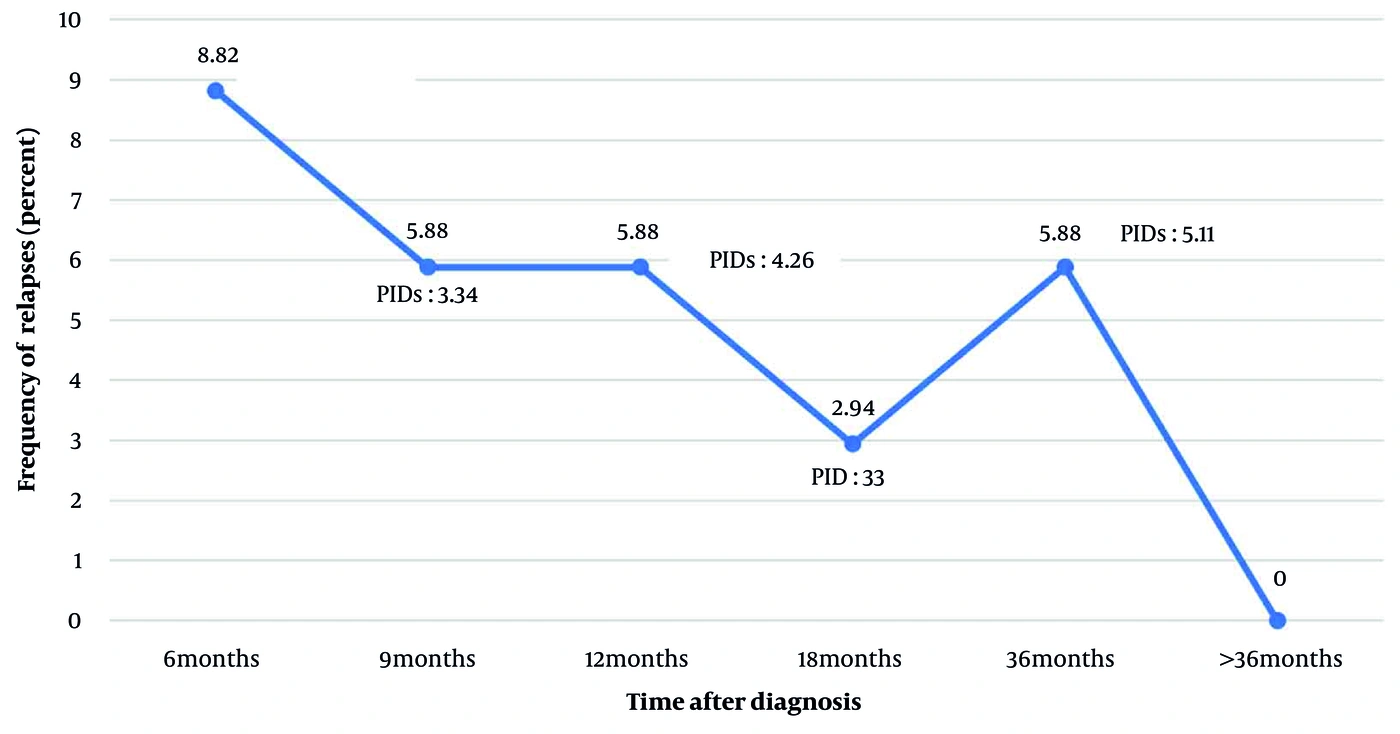
Figure 2.
Temporal distribution of clinical relapse events after initiation of rituximab therapy. Percentages were calculated using the entire study cohort (n = 34) as the denominator. Each labeled point represents an individual patient who experienced a clinical relapse, identified by study ID.
Comparisons between patients who experienced clinical relapse (n = 9) and those who remained relapse-free (n = 25) showed no statistically significant differences in demographic characteristics, including sex (P = 0.50), age at study enrollment (P = 0.39), and age at diagnosis (P = 0.98). Likewise, baseline clinical manifestations, including visual and oculomotor dysfunction, cerebellar dysfunction, brainstem involvement, cortical and subcortical manifestations, and sphincter dysfunction, did not differ significantly between the 2 groups (all P > 0.05).
Similarly, baseline MRI characteristics, including lesion distribution in the periventricular, juxtacortical, brainstem, cerebellar, and corpus callosum regions; the presence of black holes; and spinal cord abnormalities, as well as baseline laboratory findings, including oligoclonal band positivity and elevated IgG index, showed no statistically significant differences between patients with and without relapse during follow-up (all P > 0.05).
In contrast, reduced muscle force, abnormal DTRs, extensor plantar response (Babinski sign), and sensory dysfunction were more frequent among patients who experienced relapse during follow-up than among those who remained relapse-free. Given the limited number of relapse events and the exploratory nature of these unadjusted analyses, these findings should be interpreted as exploratory associations rather than evidence of independent predictors of relapse. Detailed results, including unadjusted odds ratios and 95% confidence intervals where estimable, are presented in Table 3.
Table 3.
Comparison of Demographic, Clinical, Radiological, and Laboratory Characteristics Between Patients with and Without Relapse a
| Variables | Relapse Group (n = 9) | Non-relapse Group (n = 25) | P-Value | OR (95% CI) |
|---|---|---|---|---|
| Sex (male) | 4 (44.40) | 13 (52.00) | 0.50 | 1.35 (0.29 - 6.26) |
| Age (y) | 13 (11 - 18) | 15 (10 - 18) | 0.39 | - |
| Age at diagnosis (y) | 11 (7 - 15) | 11 (4 - 17) | 0.98 | - |
| Visual and oculomotor dysfunctions | 5 (55.60) | 18 (72.00) | 0.30 | 0.48 (0.10 - 2.35) |
| Motor system involvement | 7 (77.80) | 11 (44.00) | 0.08 | 4.45 (0.76 - 25.85) |
| Decreased muscle tone | 1 (11.10) | 0 | 0.26 | Not estimable |
| Reduced muscle force | 6 (66.70) | 4 (16.00) | < 0.001 | 10.50 (1.82 - 60.45) |
| Abnormal pyramidal tract signs | 6 (66.70) | 8 (32.00) | 0.07 | 4.25 (0.84 - 21.49) |
| Abnormal DTR | 5 (55.60) | 1 (4.00) | < 0.001 | 30.00 (2.73 - 328.64) |
| Extensor plantar response (Babinski sign) | 4 (44.40) | 0 | < 0.001 | Not estimable |
| Sensory dysfunction | 6 (66.70) | 6 (24.00) | 0.03 | 6.33 (1.20 - 33.38) |
| Cerebellar dysfunction | 4 (44.40) | 12 (48.00) | 0.58 | 0.86 (0.18 - 4.00) |
| Brainstem involvement | 2 (22.20) | 9 (36.00) | 0.37 | 0.50 (0.08 - 2.98) |
| Cortical and subcortical manifestations | 1 (11.10) | 8 (32.00) | 0.22 | 0.26 (0.02 - 2.50) |
| Sphincter dysfunction | 1 (11.10) | 1 (4.00) | 0.46 | 3.00 (0.16 - 53.71) |
| Abnormal brain MRI findings (baseline) | ||||
| Lesions in the periventricular region | 9 (100.00) | 24 (96.00) | 0.73 | 1.37 (1.11 - 1.69) |
| Lesions in the Juxtacortical region | 7 (77.80) | 16 (64.00) | 0.37 | 1.96 (0.33 - 11.57) |
| Lesions in the brainstem region | 3 (33.30) | 5 (20.00) | 0.35 | 2.00 (0.36 - 10.91) |
| Lesions in the cerebellum region | 2 (22.20) | 6 (24.00) | 0.64 | 0.90 (0.14 - 5.58) |
| Lesions in the corpus callosum region | 3 (33.30) | 7 (28.00) | 0.53 | 1.28 (0.25 - 6.61) |
| Black hole | 4 (44.40) | 4 (16.00) | 0.10 | 4.20 (0.77 - 22.86) |
| Abnormal spinal cord MRI findings (baseline) | 4 (44.40) | 4 (16.00) | 0.10 | 4.20 (0.77 - 22.86) |
| Laboratory findings (baseline) | ||||
| CSF performed | 8 (88.90) | 22 (88.00) | 1.00 | 1.09 (0.09 - 12.06) |
| OCB positive (n = 30) | 8/8 (100.00) | 19/22 (86.40) | 0.64 | 2.52 (0.26 - 24.51) |
| Elevated IgG index (n = 16) | 2/5 (40.00) | 3/11 (27.30) | 0.59 | 2.09 (0.28 - 15.19) |
a Values are expressed as No. (%) or median (min-max) unless otherwise indicated. Odds ratios with 95% confidence intervals were calculated only for binary variables with estimable odds ratios. ORs were not estimated for variables with zero cell counts because sparse data resulted in complete separation and unstable estimates. Abbreviations: DTR, deep tendon reflexes; MRI, magnetic resonance imaging; OCB, oligoclonal bands; CSF, cerebrospinal fluid; OR, odds ratio; CI, confidence interval.
4.4. Safety and Adverse Effects Related to Rituximab Therapy
Fourteen patients (41.2%) experienced at least 1 adverse event related to rituximab therapy. All adverse events were infusion-related and occurred during or within 24 hours after infusion. Most events were mild and transient, including cutaneous reactions (7 patients, 20.6%), nausea and vomiting (3 patients, 8.8%), dizziness (2 patients, 5.8%), and transient increases in blood pressure (2 patients, 5.8%). No delayed or long-term adverse events, serious infections, anaphylaxis, or treatment-related mortality were observed. No patient discontinued rituximab because of adverse effects.
5. Discussion
This study evaluated the efficacy and safety of rituximab in preventing clinical relapses in 34 patients with POMS over a median follow-up of 36 months after rituximab initiation. The observed ARR was 0.11, and 73.5% of patients remained relapse-free during the observation period. These results add to the limited but growing evidence regarding rituximab use in POMS.
To date, data on rituximab use in POMS populations remain limited. Karenfort et al. (19) reported an adolescent with severe MS who achieved complete disease control with rituximab, with no relapses during treatment and no reported adverse effects. Subsequently, Salzer et al. (16) published a case series of 14 pediatric patients with MS treated with rituximab for a median of 23.6 months. None experienced a relapse during rituximab therapy, and 13 of 14 had stable or improved disability scores. More recently, Breu et al. (14) evaluated rituximab in a larger sample of 61 patients, further contributing evidence regarding its efficacy and safety in this population.
Salzer et al. reported no relapses among 14 patients receiving rituximab (16), whereas Breu et al. reported a relapse rate of approximately 10% (14); in our study, relapses occurred in 26.5% of patients. These differences may reflect heterogeneity in patient populations, baseline disease activity, follow-up duration, or rituximab dosing and maintenance strategies. These cross-study comparisons are contextual rather than internally controlled because no study included a concurrent comparator arm; therefore, differences in relapse rates across cohorts cannot be attributed to treatment effect alone.
Despite the occurrence of relapses, the estimated ARR in our study was 0.11, which was numerically lower than the ARR of 0.8 - 1.4 reported in untreated pediatric MS cohorts (20, 21). These comparisons are indirect and should be interpreted as contextual because our study did not include an untreated or active-comparator group.
Furthermore, approximately 80% of relapses occurred within the first 18 months after treatment initiation, and no relapses were observed among patients who completed 5 years of follow-up. This pattern suggests a trend toward progressive disease stabilization over time but should be interpreted cautiously given the limited number of patients with long-term follow-up.
Our findings can also be contextualized using rituximab efficacy data from adult MS populations. Torgauten et al. reported an ARR of 0.03 in adults treated with rituximab, with complete cessation of disease activity in 79% of patients (22). Other systematic reviews and meta-analyses have reported rituximab superiority over placebo and comparability to approved disease-modifying therapies in adult relapsing-remitting MS (13, 23, 24). The overall pattern observed in our study, including a low relapse rate and good tolerability, is broadly consistent with adult observational data. These cross-study and cross-age comparisons are contextual rather than derived from randomized or internally controlled designs and should not be interpreted as head-to-head evidence of comparative efficacy.
One key finding was the temporal clustering of relapses, with 80% occurring within the first 18 months after rituximab initiation, followed by markedly reduced disease activity thereafter. This clustering may reflect residual inflammatory activity before adequate and sustained B-cell depletion was achieved (25, 26). Rituximab selectively targets CD20-positive B cells, but elimination of pathogenic memory B cells, plasmablasts, and their downstream antibody- and cytokine-mediated effector pathways may require time and repeated maintenance dosing (27, 28). Several studies have shown that early after rituximab initiation, autoreactive B-cell clones and pre-existing plasma cells may continue to contribute to inflammatory activity despite peripheral B-cell depletion (29, 30). As B-cell depletion becomes deeper and more stable with subsequent cycles, immunologic control may improve and relapse risk may decline (14, 29).
The observed pattern may also reflect the natural course of early aggressive disease in a subset of patients. Some patients with MS experience more active disease in the initial years after diagnosis (31, 32). The neurological factors identified in our study, including reduced muscle force, abnormal reflexes, extensor plantar responses, and sensory dysfunction, may represent markers of a more aggressive early disease phenotype. Rituximab may eventually control this activity, but complete suppression may require time.
In our study, adverse events were predominantly mild infusion-related reactions, and no serious infections or delayed serious adverse events were observed during follow-up. These findings are broadly consistent with the pediatric literature. Breu et al. reported adverse events in 67% of 60 rituximab-treated pediatric patients with MS, with 65% of events classified as mild to moderate (14). Their cohort had infusion-related reactions in 48% of patients, infectious complications in 20%, hypogammaglobulinemia in 17%, and lymphopenia in 7%. Treatment discontinuation due to adverse events occurred in 7% of patients (4 of 60), including 1 severe case involving an infusion-related toxic reaction and dyspnea. Earlier, smaller series reported more favorable profiles. Salzer et al. documented no acute adverse effects in their 14-patient cohort (16), whereas the single patient reported by Karenfort et al. also tolerated treatment well (19).
The absence of serious infections in our study is reassuring given theoretical concerns regarding B-cell depletion in developing immune systems. The median follow-up of 36 months provided a reasonable period for assessing infection risk, although longer-term safety data remain important. No delayed hypersensitivity reactions or other long-term complications were observed.
5.1. Study Limitations
This study has several important limitations. The relatively small sample size (n = 34) and absence of a control group limit definitive conclusions regarding rituximab efficacy and safety in pediatric MS. The small number of relapse events (n = 9) precluded multivariable analysis, and the identified associations between baseline neurological findings and relapse risk require validation in larger studies. Heterogeneous follow-up duration (12 - 60 months) and individualized rituximab dosing intervals limited uniform assessment of long-term outcomes. Important diagnostic and imaging limitations included the lack of systematic MOG antibody testing in all patients, incomplete CSF analysis in 4 patients, limited IgG index data (16 of 30 CSF samples), and the absence of advanced MRI biomarkers (central vein sign and paramagnetic rim lesions) because standard clinical imaging protocols were used. As a single-center study conducted at a tertiary referral hospital, our study may reflect referral patterns and treatment-selection practices specific to this setting. The decision to treat with rituximab rather than another disease-modifying therapy was individualized rather than protocol-driven, and patients referred to our center may not be representative of the broader POMS population. These factors may limit the generalizability of our findings to other centers and populations.
Despite these limitations, this study provides real-world evidence regarding rituximab use in pediatric MS, and the temporal pattern of relapse clustering warrants further investigation. Future multicenter prospective studies with larger sample sizes, standardized protocols, longer follow-up, and comprehensive immunological and imaging assessments are needed to confirm these findings and establish the optimal role of rituximab in pediatric MS treatment algorithms.
5.2. Conclusions
In this study, rituximab treatment was followed by a low observed ARR, with most relapses clustering within the first 18 months and none observed among patients completing 5 years of follow-up. Adverse events were predominantly mild and transient, with no serious infections or treatment discontinuations. Because this was an uncontrolled observational study without an untreated or active-comparator group, these findings describe the relapse and safety patterns observed in our cohort rather than establishing definitive efficacy or safety relative to other approaches. Specific baseline neurological findings, including reduced muscle force, abnormal reflexes, extensor plantar responses, and sensory dysfunction, were associated with relapse risk and may inform future risk-stratification efforts. Larger multicenter prospective studies with concurrent comparator groups and longer follow-up are warranted to confirm these observations, establish comparative efficacy and safety, and define rituximab's place in pediatric MS treatment algorithms.