


Dear Editor,
A chronic autoimmune disease, rheumatoid arthritis (RA) primarily affects the synovial joints, resulting in pain, swelling, and when left untreated, permanent joint damage and disability (1). Being a systemic disease, RA can affect the lungs, kidneys, heart, and other organ systems (2, 3). To date, only a handful of case series and reports, most notably Heywood & Meyers’ series identifying 7 thoracolumbar RA cases (0.94% of 746 RA patients) and several smaller reports, have documented degenerative thoracolumbar changes in young RA patients without other risk factors (4-6). We are writing to shed light on an important clinical observation regarding degenerative spinal changes in a relatively young patient with a known history of RA who presented with mechanical back pain, aggravated by work and bending, relieved upon rest. The thoracic and lumbar spine X-rays of this 38-year-old female demonstrated multi-level osteophyte formation and end-plate sclerosis as shown in Figure 1. These findings are usually associated with age-related degenerative and spondylotic changes. Osteophytes and end-plate sclerosis are hallmarks of spinal degeneration, frequently due to mechanical stress and advancing age (7). However, the presence of these findings in a young patient with RA raises important considerations about disease progression, coexisting pathology, and treatment strategy. The most recognized site of RA involvement is the cervical spine, particularly at the atlantoaxial joint, while thoracic spine involvement is rare (8). Thoracic involvement in RA is usually attributed to secondary causes such as mechanical stress, inflammatory enthesopathy, or treatment-related bone demineralization rather than primary RA pathology (7, 8).
This case report focuses on the unusual and clinically significant co-existence of inflammatory and mechanical degenerative changes in the thoracic and lumbar spine of a 38-year-old RA patient. The diagnosis of spinal involvement in the present case relied on a combination of clinical presentation, radiographic findings, and exclusion of alternative etiologies. Mechanical back pain, characterized by aggravation on activity and relief at rest, prompted spinal imaging. Plain radiographs of the thoracic and lumbar spine demonstrated multi-level osteophyte formation and end-plate sclerosis, which are consistent with degenerative disc disease. These imaging features were interpreted using standard criteria for spinal degeneration, including Modic-type end-plate changes and osteophytic growth patterns (7, 8). Additionally, no signs of vertebral collapse, syndesmophyte formation, or sacroiliitis were seen, aiding in ruling out other inflammatory spinal disorders including ankylosing spondylitis. Although not performed due to lack of neurological deficits in our case, MRI remains the gold standard for assessment of inflammatory and soft tissue involvement and should be considered in evaluations when required. A thorough rheumatologic review ruled out active systemic flare in our patient, reinforcing the likelihood of mechanical degeneration as a coexisting process in this RA patient.
In the present case, the presence of multi-level osteophytes may be a result of early onset degenerative disc disease and chronic biomechanical stress, potentially accelerated by altered posture and spinal mechanics secondary to joint damage in peripheral joints. Furthermore, the systemic inflammation associated with RA and the use of corticosteroids or immunosuppressants could also lead to abnormal bone remodeling, leading to sclerosis of the vertebral end plates. End-plate sclerosis suggests that intervertebral discs have undergone structural compromise, resulting in increased mechanical load on adjacent vertebrae. In the context of RA, this can be multifactorial, stemming from inflammatory processes, altered mobility, and secondary osteoporosis (8, 9). These findings are significant clinically because they represent a dual pathology: Inflammatory arthritis co-existing with, or predisposing to, early degenerative disc disease, and this overlap complicates diagnosis and management (10). For instance, spinal pain in RA patients may be attributed to systemic inflammation, thereby potentially delaying the recognition of mechanical or degenerative causes which require distinct therapeutic approaches for management. Distinguishing between inflammatory and mechanical spinal pathology is often complex, particularly in young patients with long-standing RA. Early diagnosis is critical, as delayed recognition of degenerative changes can lead to progressive disability, especially when compounded by RA-related joint damage. Standard radiographs, although useful for detecting osteophytes and end-plate sclerosis, may miss early inflammatory or soft tissue changes, underscoring the value of MRI in ambiguous or progressive cases.
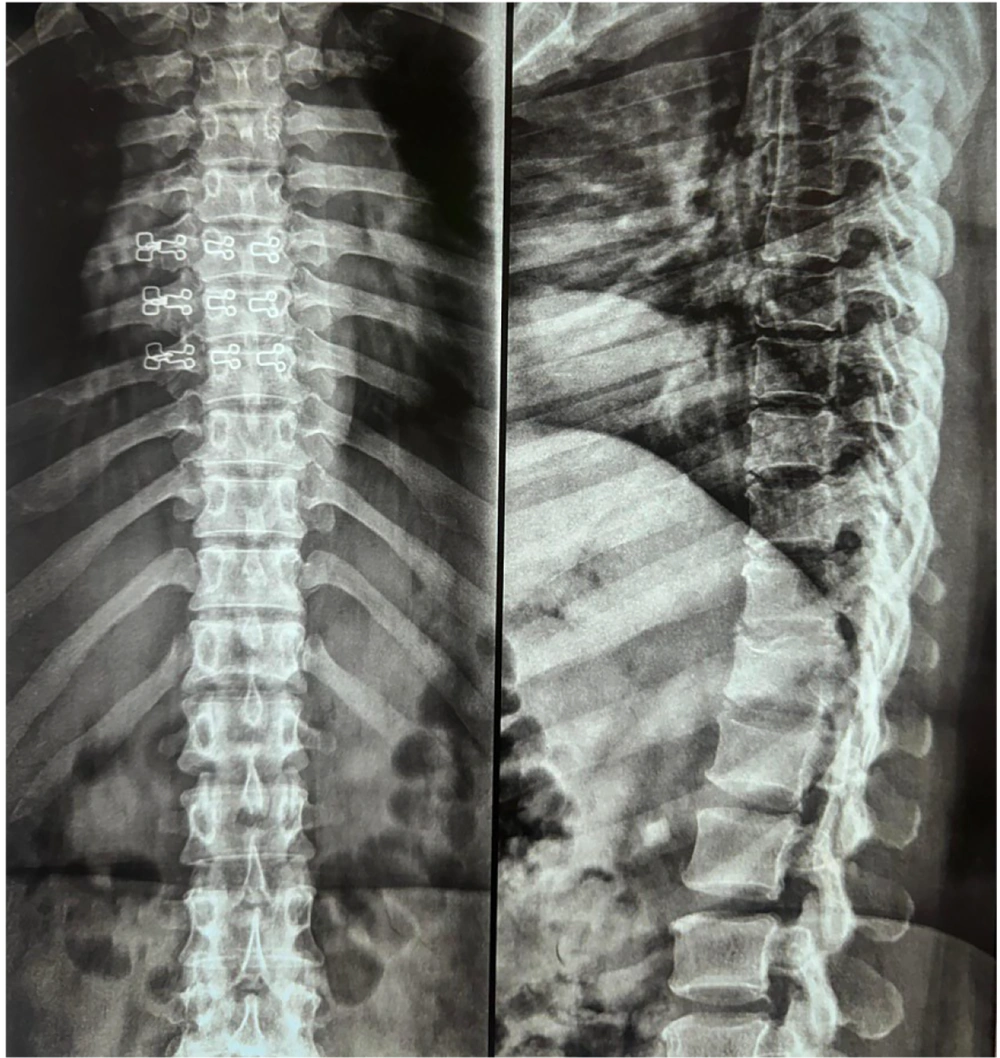
Figure 1.
X-ray of the thoracic and lumbar spine
The management plan in such cases should be multidisciplinary, involving rheumatologists for immunosuppressive therapy, physical therapists for spinal stabilization and postural training, and spine specialists for evaluating surgical or interventional needs in cases of progressive degeneration (11, 12). Disease-modifying anti-rheumatic drugs (DMARDs) are vital to control systemic inflammation in RA, but adjunct strategies including physical therapy, ergonomic correction, and bone-strengthening agents are warranted to improve the associated degenerative changes (13, 14). If neurological symptoms such as pain radiation, sensory loss, or incontinence are present, advanced imaging including MRI should be performed as it provides additional detail about soft tissue involvement, disc pathology, and spinal cord compression (15). With regards to recent advances, nanotechnology offers a novel and promising approach to RA treatment by enabling targeted drug delivery that improves therapeutic outcomes while minimizing side effects (16). Advanced nanoformulations enhance drug solubility, bioavailability, and precision targeting of inflamed tissues (16). These innovations hold significant potential to transform RA management through more effective and personalized therapies.
In conclusion, this case underscores the significance of maintaining a broad differential when interpreting spinal imaging in young patients with RA. Recognizing the co-existence of inflammatory and degenerative processes in the spine is crucial to preventing misdiagnosis and optimizing treatment. The co-existence of RA with degenerative spine disease in the young population, while uncommon, should be kept in mind, especially when clinical symptoms appear disproportionate to typical inflammatory joint activity. Awareness and early recognition of these patterns would aid in diagnosing accurately and tailoring the management plan accordingly. This case underscores the need for heightened clinical vigilance and a coordinated, multidisciplinary treatment plan tailored to the dual pathology in RA patients with spinal involvement.
