J Inflamm Dis

Image Credit:J Inflamm Dis

Challenges of Vancomycin Resistance in Methicillin-Resistant Staphylococcus aureus (MRSA) Among Patients with Complicated Diseases

Author(s):
Hossein AhmadiHossein AhmadiHossein Ahmadi ORCID1, Leila FozouniLeila FozouniLeila Fozouni ORCID1,*, Hamid Reza PordeliHamid Reza PordeliHamid Reza Pordeli ORCID1
1Department of Microbiology, Go.C.,Islamic Azad University, Gorgan, Iran

Journal of Inflammatory Diseases:Vol. 29, issue 3; e165177
Published online:Oct 20, 2025
Article type:Research Article
Received:Aug 06, 2025
Accepted:Oct 13, 2025
How to Cite:Ahmadi H, Fozouni L, Pordeli HR. Challenges of Vancomycin Resistance in Methicillin-Resistant Staphylococcus aureus (MRSA) Among Patients with Complicated Diseases. J Inflamm Dis. 2025;29(3):e165177. doi: https://doi.org/10.69107/jid-165177

Abstract

Background:

Staphylococcal drug resistance is a significant challenge in medicine and has become an emerging threat. Vancomycin-resistant Staphylococcus aureus (VRSA) presents a serious risk to global mortality due to the limited effective management and treatment options available.

Objectives:

This study aimed to investigate the frequency of drug resistance among methicillin-resistant Staphylococcus aureus (MRSA) isolates, focusing on vancomycin resistance in patients with complicated diseases, including immunocompromised, genetic, and chronic metabolic diseases, highlighting these populations as potential emerging foci of drug resistance.

Methods:

Over two years (2023 - 2025), three hundred samples were collected from hospitalized patients with special diseases for staphylococcal analysis. The MRSA and VRSA isolates were identified using both phenotypic methods and polymerase chain reaction (PCR) techniques. Additionally, antibiotic susceptibility to ten classes of antibiotics and the minimum inhibitory concentrations (MICs) of vancomycin were assessed using the Kirby-Bauer method and microdilution broth test, respectively.

Results:

Among 168 MRSA isolates, the prevalence of VRSA isolates was found to be 19.05%. Screening for VRSA indicated that women and individuals aged 36 and older with diabetes were more likely to carry this pathogen. The highest rate of VRSA was detected in wound samples (65.63%), and no cases of VRSA were identified in joint fluid samples (P < 0.05). Additionally, no VRSA isolates were found in individuals with autism, hemophilia, prostate cancer, multiple myeloma, colorectal cancer, hepatitis B, or thalassemia. Gentamicin, an aminoglycoside, had a resistance rate of 53%, making it the least effective agent. In contrast, linezolid demonstrated an effectiveness rate of 98.21%. The MRSA strains isolated from wounds showed a better response to antibiotics.

Conclusions:

The increasing reports of MRSA and VRSA outbreaks highlight the urgent need to revise national treatment guidelines to include effective antimicrobial agents that target resistant staphylococci. It is essential to implement comprehensive surveillance strategies for these agents and to establish robust monitoring systems, particularly for vulnerable patient populations.

1. Background

Antimicrobial resistance (AMR) in clinical Staphylococcus aureus strains has been recognized as a global challenge to public health. It is a significant hospital pathogen that causes mild to severe infections, including skin and soft tissue infections, bacterial endocarditis, pleurisy, and chronic blood infections. This microorganism has a high ability to resist both old and new antibiotics. After the emergence of penicillin-resistant S. aureus, the first strain of methicillin-resistant Staphylococcus aureus (MRSA) was identified in early 1961. For many years, vancomycin was considered the last resort for treating severe MRSA and other resistant gram-positive infections. However, in the late 1980s, vancomycin resistance first appeared in enterococci (VRE), and in the last two decades, it has also emerged in Staphylococcus aureus (VRSA) (1-3). The transfer of the VanA operon from the Tn1546 genetic element by VRE caused the emergence of VRSA (4-6). In 1997, the first clinical isolate of vancomycin-intermediately susceptible Staphylococcus aureus (VISA) was reported, which was not inhibited in vitro at vancomycin concentrations below 4 - 8 μg/mL. In contrast, VRSA was inhibited only at concentrations of 16 μg/mL or higher. The VISA and VRSA strains have evolved from MRSA. The VRSA isolates are usually susceptible to multiple antimicrobial agents. Systemic antimicrobial therapy with effective antibiotics is typically started after a clinical laboratory identifies a VRSA isolate. The uncontrolled use of vancomycin in patients with underlying diseases, such as diabetes, renal failure, cancer, and gangrenous or surgical wounds, facilitates the transfer of vancomycin-resistant plasmids from VRE to VRSA, making the eradication of the bacteria difficult and treatment failure likely (7-9). According to the World Health Organization (WHO), the pathogenicity and antibiotic resistance pattern of S. aureus pose a serious threat to human health worldwide (WHO, 2017) (10). Infections caused by such resistant pathogens are often difficult to treat, and several classes of antibiotics have been used to treat these infections over the past decade, leading to the emergence and spread of multidrug-resistant (MDR) and extensively drug-resistant (XDR) strains (11).

2. Objectives

This study aimed to investigate the frequency of VRSA strains and their resistance to various antibiotics in specific patients with immunodeficiency diseases, genetic disorders, and chronic metabolic conditions. These complicated diseases were analyzed to assess the potential of these patient populations as emerging sources of drug resistance.

3. Methods

3.1. Study Population and Staphylococcal Isolation

In this observational cross-sectional study conducted over two years (2023 - 2025), 300 wound, blood, abscess, and joint fluid samples were examined from hospitalized patients with complicated diseases or immunodeficiencies at hospitals and medical centers in northern Iran. The participants had a mean age of 38.0 years, with an age range of 12 to 78 years. To ensure compliance with research ethics, a questionnaire was created that included variables such as gender, age, sample type, and disease. The inclusion criterion was the presence of signs of infection and a hospital stay of no more than one week. Patients under the age of twelve were excluded from this study.
To identify and confirm bacterial agents, swabs were first placed in thioglycolate transport and transferred to the medical diagnostic laboratory. They were then inoculated onto Chocolate agar, Columbia agar with 5% horse blood, and MacConkey agar (Merck, Germany), and incubated in the presence of 5% CO2 at 37°C for 24 - 48 hours. Staphylococcus aureus strains were identified by examining mannitol-positive colonies through observation of colony morphology, Gram staining, hemolysis, catalase, coagulase (clumping factor test), DNase tests, and finally by the Vitek-2 card system (bioMerieux, France), which is developed for gram-positive bacteria (12).

3.2. Methicillin-Resistant Staphylococcus aureus Confirmation

To identify MRSA isolates, in addition to the phenotypic method using the cefoxitin disk (2), a DNA extraction kit (SinaClone, Iran) was used according to the manufacturer's instructions. Polymerase chain reaction (PCR) was conducted using forward and reverse primers (designed with Oligo 5 software) with the sequences: 5'-AGTTCTGCAGTACCGGATTTGC-3' and 5'-AAAATCGATGGTAAAGGTTGGC-3', which were used to amplify the mecA gene (methicillin resistance gene) (12). Standard strain S. aureus ATCC 33591 and S. aureus ATCC 25923 were used as positive and negative controls, respectively. The presence of 540 bp fragments compared to the marker DNA indicated the presence of MRSA isolates.

3.3. Anti-methicillin-resistant Staphylococcus aureus Susceptibility Testing

In the Kirby-Bauer disk diffusion method, a half McFarland bacterial suspension from overnight cultures of S. aureus was prepared and cultured on Mueller-Hinton agar. The following antibiotic classes were used to assess susceptibility: Aminoglycosides, lipopeptides, ansamycins, fluoroquinolones, glycopeptides, macrolides, anti-beta-lactamases, lincosamides, tetracyclines, and oxazolidinones (product of Padranteb Co., Iran). After placing the disks on the cultures for 16 - 18 hours at 37°C, the inhibition zone around each disk was measured and interpreted according to the CLSI-2021 guidelines (13).

3.4. Vancomycin-Resistant Staphylococcus aureus Screening

The effective and sensitive method for screening VRE is using BHI Agar supplemented with 6 μg/mL of vancomycin (vancomycin screening agar method). Organisms with intermediate resistance to vancomycin demonstrate weak growth on this medium. To identify vancomycin-resistant isolates, the first 10 μL of a 0.5 McFarland suspension of MRSA isolates was cultured on BHI agar containing 6 μg/mL vancomycin (BHI6V), and after 24 hours of incubation at 37°C, they were evaluated in the presence of vancomycin-susceptible Enterococcus faecalis (ATCC 29212) and vancomycin-resistant E. faecalis (ATCC 51299) as negative and positive controls, respectively (13).

3.5. Minimum Inhibitory Concentration Determination

According to recommendations from the Centers for Disease Control and Prevention (CDC), the minimum inhibitory concentration (MIC) was also determined to detect VRSA. Pure cultures were prepared from each resistant isolate grown on BHI6V, and their MICs were determined using the microdilution broth method according to CLSI guidelines, with a focus on vancomycin. For this purpose, an initial concentration of vancomycin equivalent to 1024 µg/mL was inoculated into a 96-well ELISA microplate containing Mueller-Hinton Broth (Merck, Germany). Dilution of drug stocks was continued to a concentration of 2 µg/mL. Subsequently, a suspension of MRSA grown on BHI6V at a concentration of 1.5 × 10⁸ colony-forming units (half McFarland concentration) was added to the wells, and the results were interpreted after 20 - 24 hours of incubation at 37°C. A standard culture of S. aureus ATCC 29213 was used as a positive control culture in antimicrobial susceptibility testing (13, 14).

3.6. Polymerase Chain Reaction Detection of vanA

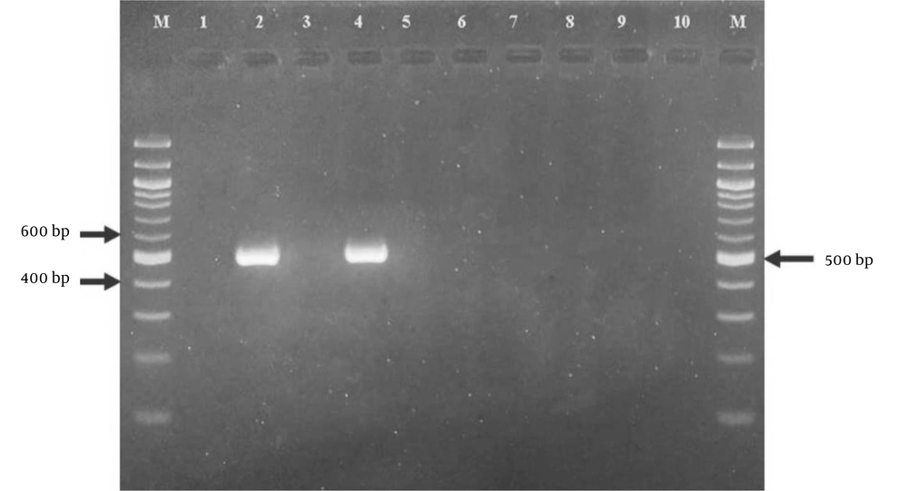
To definitively identify VRSA isolates, after DNA extraction from a 24-hour culture of VRSA isolates in Mueller-Hinton agar, the presence of the vanA gene (the supernatant of the PCR product was measured at an OD260/280 ratio of 1.8 - 2.0 by a nanodrop device) was detected and investigated by PCR using forward and reverse primers (4): (F: 5'—>3'): CATGAATAGAATAAAAGTTGCAAT, (R: 5'—>3'): CCCCTTTAACGCTAATACGATCAA according to the manufacturer's protocol (Cinagene, Iran). The gene was amplified in a program of preincubation at 94°C for 3 minutes, denaturation for 30 seconds, primer annealing at 55°C for 45 seconds, polymerization at 72°C for 1 minute, and finally a final elongation step at 72°C for 10 minutes (15). The presence of the vanA gene was confirmed by electrophoresis on a 2% agarose gel at 150 V in commercial 10× TAE buffer (Fementase) and the observation of a band with a size of 500 bp (Figure 1).
Polymerase chain reaction (PCR) amplification of the <i>vanA</i> gene for methicillin-resistant <i>Staphylococcus aureus</i> (MRSA) isolates
Figure 1.

Polymerase chain reaction (PCR) amplification of the vanA gene for methicillin-resistant Staphylococcus aureus (MRSA) isolates

3.7. Statistical Analysis

Data were analyzed with the SPSS software (ver. 23, IBM Corp., Armonk, NY, USA) using the chi-square test. Graphs were created using Microsoft Excel software (2010). Confidence intervals (CI) were calculated using Stata MP 14 (Stata Corp LP, USA). All analyses were carried out at a significance level of 0.05.

4. Results

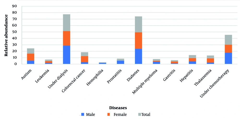
It was found that a significant percentage of individuals undergoing dialysis or with underlying diseases such as diabetes was higher than expected, comprising more than half of the patients evaluated in this study. Conversely, the percentage for hemophilia was the lowest among the conditions assessed. Additionally, men represented the highest frequency of individuals with specific diseases (Figure 2).
Frequency of diseases in hospitalized patients by gender
Figure 2.

Frequency of diseases in hospitalized patients by gender

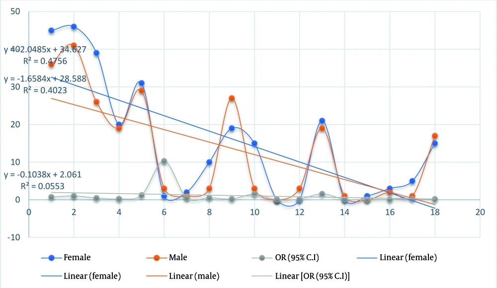
The frequency of individuals over the age of 36 suffering from specific diseases was notably high, reaching 53.38%. A total of 168 samples tested positive for MRSA through both phenotypic and molecular analyses. Most of these samples were collected from wound specimens and patients undergoing dialysis or those with diabetes, depending on the type of condition (P < 0.05). However, no significant difference was found in the frequency of MRSA isolates between women and men across different age groups (Table 1) (Figure 3).
Table 1.Characteristics of the Study Population and Frequency of methicillin-resistant Staphylococcus aureus Isolates a
VariablesGenderP-Value
Female (N = 91)Male (N = 77)
Age (y)0.065
12 - 3545 (49.45)36 (46.75)
≥ 3646 (50.55)41 (53.25)
Specimen0.028 b
Wound39 (42.86)26 (33.77)
Blood20 (21.98)19 (24.67)
Abscess31 (34.06)29 (37.66)
Synovial fluid 1 (1.1)3 (3.9)
Disease0.047 b
Autism2 (2.20)1 (1.3)
Leukemia10 (10.99)3 (3.9)
Under dialysis19 (20.88)27 (35.1)
Colorectal cancer15 (16.48)3 (3.9)
Hemophilia0 (0)0 (0)
Prostate cancer0 (0)3 (3.9)
Diabetes21 (23.08)19 (24.67)
Multiple myeloma0 (0)1 (1.3)
Gastritis1 (1.1)0 (0)
Hepatitis3 (3.3)2 (2.6)
Thalassemia5 (5.49)1 (1.3)
Under chemotherapy 15 (16.48)17 (22.1)

a Values are expressed as No. (%).

b Significant difference (P < 0.05).

Ordered logistic regression model for variables associated with methicillin-resistant <i>Staphylococcus aureus</i> (MRSA) populations
Figure 3.

Ordered logistic regression model for variables associated with methicillin-resistant Staphylococcus aureus (MRSA) populations

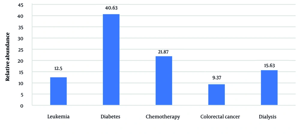
In susceptibility testing, cefoxitin demonstrated a resistance rate of 100%, followed by gentamicin at 53%, making them the least effective antibiotics. In contrast, linezolid exhibited an effectiveness rate of 98.21%, marking it as the most effective antibiotic. Notably, the MRSA isolates from wounds elicited a better response to antibiotics (Table 2). Among the MRSA isolates, MDR strains accounted for 72.2% of the total. The frequencies of VRSA, VISA, and vancomycin-sensitive Staphylococcus aureus (VSSA) were reported to be (32, 19.05%), (28, 16.67%), and (108, 64.28%), respectively. Among the VRSA isolates, 71.88% displayed the presence of the vanA gene, as evidenced by a 500 bp band. The VRSA screening showed that women and individuals aged 36 and older with diabetes were more likely to carry this pathogen. The highest rate of VRSA was found in wounds (65.63%), followed by abscess (25%) and blood samples (9.37%). No VRSA isolates were found in individuals with autism, hemophilia, prostate cancer, multiple myeloma, colorectal cancer, hepatitis B, and thalassemia (Figure 4).
Table 2.Frequency Distribution of the Drug-Resistance Patterns Among Methicillin-Resistant Staphylococcus aureus Isolates Exposed to Various Classes of Antibiotics
Sample TypesAntibiotics
GENKANDRIFCIPCMNORDOTECLZDAZM
Wound (R%)19.088.755.6107.653.015.50011.181.1005.33
Abscess (R%)21.2513.409.851.176.8415.4312.155.6710.753.5006.61
Blood (R%)12.6520.1114.305.3811.1125.6117.4731.2312.594.331.797.11
Antibiotic resistance potential (Total%)
S30.3628.5741.0775.5937.5029.1746.4336.3139.8868.4598.2157.74
I16.6629.1729.1717.8636.9026.7818.4526.7925.6022.62023.21
R52.9842.2629.766.5525.6044.0535.1236.9034.528.931.7919.05

Abbreviations: GEN, gentamicin 10; K, kanamycin 30; AN, amikacin 30. D, daptomycin 30; RIF, rifampicin 10; CIP, ciprofloxacin 5; CM, clindamycin 2; NOR, norfloxacin 5; DO, doxycycline 30; TEC, teicoplanin 30; LZD, linezolid 10; AZM, azithromycin 15; S, sensitivity; I, intermediate; R, resistance.

Prevalence of vancomycin-resistant <i>Staphylococcus aureus</i> (VRSA) isolates in patients based on disease type
Figure 4.

Prevalence of vancomycin-resistant Staphylococcus aureus (VRSA) isolates in patients based on disease type

5. Discussion

Reports indicate approximately 5 million global deaths related to AMR caused by bacteria, significantly affecting Asian and Middle Eastern countries (2, 16). Over the years, S. aureus has developed various mechanisms of drug resistance, making it difficult to treat with conventional antibiotics. Published systematic reviews and meta-analyses have assessed the epidemiology of VRSA globally, showing its prevalence across different years and regions (17). Although the incidence of VRSA in developed countries is relatively low, it can be said that its burden in developing countries, such as Iran, is quite high and significant. According to the findings of the present study, the prevalence of VRSA was estimated at 19.05%, which was consistent with some reports in other parts of the world (especially Africa) (18) and significantly higher compared to some other studies (17). Furthermore, our findings are higher than the systematic reviews and meta-analyses conducted in Iran up to 2012, which reported a prevalence of 2.4% from thirteen studies (19). Of course, this prevalence has increased over the last 12 years and has been addressed in various studies within the country (1, 20, 21).
The high rate of VRSA observed in the patients in this study may be attributed to insufficient surveillance of the types of infections present or the inappropriate use of antibacterial drugs among these patients compared to other groups (22). Additionally, this issue is likely worsened by the irrational use and widespread availability of over-the-counter antibacterial drugs in many developing countries (23). In summary, it can be acknowledged that today, the frequent use of vancomycin as the choice for the treatment of infections caused by MRSA and other MDR gram-positive pathogens has led to the emergence of S. aureus isolates with high resistance to vancomycin or other last-line antibiotics (24, 25).
The results of this study showed different levels of VRSA frequency in different individuals in terms of disease type, age, gender, and type of sample studied, which indicates the existence of changes among the parameters studied. It can be said that a kind of heterogeneity or significant difference (P < 0.05) was observed in some findings of this study, such that VRSA was more common in individuals with diabetes, aged older than 35 to 78 years, women, and wound samples. The possible reason for this heterogeneity could be differences in methodology, study participants, study design, and sample size, all of which affect the prevalence of VRSA. Another part of this study was to evaluate the resistance of S. aureus isolates to different antibiotic groups and determine the resistance pattern for each of the MRSA and VRSA isolates, which was subsequently determined by the type of drug resistance of each isolate. The differences in study results can be attributed to variations in sample size, the selected antibiotic stress, bacterial origin, and methods used for determining drug resistance. Additionally, VRSA frequently exhibits MDR to various antimicrobial drugs; however, in the current study, MDR strains accounted for 91% of the total VRSA isolates.
In the present study, the highest antibiotic resistance observed in MRSA isolates was to cefoxitin (100%), followed by gentamicin at 52.98% and ciprofloxacin at 44.05%. The lowest resistance was noted for linezolid at 1.79%. Studies in other regions assessing the antibiotic resistance patterns of MRSA isolates have yielded both similar and different results compared to the present study. In a study involving ocular samples from Gorgan, 80% of the isolates were identified as MRSA (12). A study conducted in 2024 reported a 40% resistance rate attributed to MRSA. In the current research, all resistant isolates were identified as MRSA; these were found to be sensitive to linezolid, and one isolate was also sensitive to daptomycin. However, all isolates displayed resistance to other antibiotics from various groups. This issue, along with the relative sensitivity of more than 16% of the isolates to vancomycin (VISA), will be of particular importance in analyzing bacterial reactions to antibiotics used in most therapeutic procedures related to microbial infections. In the study in Tabriz (2008), no resistant (VRSA) or intermediate susceptible (VISA) cases were reported to vancomycin (26). In another study in southern India, 16 strains of S. aureus showed MICs of about 4 - 8 μg/mL against vancomycin, which were reported as VISA isolates. The results of these studies do not match the results of the present study, but in the reports, 68.7% of the strains had MICs of 16 μg/mL, indicating an increase in vancomycin resistance in recent years (27).
In this regard, a 2021 systematic review and meta-analysis (with data up to 2020) showed that the global prevalence of VRSA has more than tripled in the past two decades, from 2% before 2006 to 7% between 2015 and 2020. The study also stated that the highest prevalence of VRSA was in Africa (16%) and Asia (5%), followed by North America (4%), South America (3%), and Europe (1%).
The main objective of the present study was to investigate vancomycin resistance and the presence of the vanA gene in MRSA isolates. Current molecular analyses indicated that the vanA resistance gene was detected in 71.88% of VRSA isolates, similar to a meta-analysis study conducted in 2021 (17). Totally, reports indicate that VRSA strains have been identified in several countries, including Pakistan, Iran, and others. The USA, Nigeria, and India (28). Some reports indicate that the prevalence of VRSA isolates is increasing in Iran, while others show a decrease in prevalence over the past five years (3, 29). The observed heterogeneity and discrepancies may be attributed to the shift in research approaches and healthcare priorities beginning in 2020, in response to the SARS-CoV-2 (COVID-19) pandemic. As a result, the number of studies conducted during this time has decreased, which may lead to an underestimation of findings from these periods.
The present study has some limitations. These include the small number of isolates categorized by disease type due to the cross-sectional design, the absence of VISA analysis, and the lack of molecular typing beyond vanA. However, a key strength of this study is the monitoring and measurement of drug resistance in S. aureus isolates from specific patients in the northern region of the country.

5.1. Conclusions

This finding, along with the increasing reports of MRSA and VRSA outbreaks across the country, complicates treatment. It affects incidence rates, mortality, hospital length of stay, and healthcare costs, particularly for vulnerable patients. This situation underscores the urgent need to update and develop national treatment guidelines. These guidelines should include alternative and highly effective antimicrobial agents that specifically target resistant strains of Staphylococcus. Furthermore, it is essential to implement comprehensive surveillance strategies for antimicrobial agents, supported by robust systemic monitoring.
To effectively contain the transmission of VRSA, prioritizing infection control measures is crucial. This includes enforcing contact precautions, conducting thorough screenings, properly sterilizing healthcare equipment, and maintaining a clean and hygienic environment. Due to its strong antibacterial effects, linezolid is a valuable option for treating severe infections caused by drug-resistant S. aureus.

Footnotes

References

  • 1.
    Thompson T. The staggering death toll of drug-resistant bacteria. Nature. 2022. https://doi.org/10.1038/d41586-022-00228-x.
  • 2.
    Hosseini M, Fozouni L. The Glycopeptide-Susceptibility of Multidrug-Resistant/Extensively Drug-Resistant Staphylococcus aureus in Skin Infections. Middle East Journal of Rehabilitation and Health Studies. 2023;11(1). https://doi.org/10.5812/mejrh-138166.
  • 3.
    Cheung GYC, Bae JS, Otto M. Pathogenicity and virulence of Staphylococcus aureus. Virulence. 2021;12(1):547-69. [PubMed ID: 33522395]. [PubMed Central ID: PMC7872022]. https://doi.org/10.1080/21505594.2021.1878688.
  • 4.
    Stogios PJ, Savchenko A. Molecular mechanisms of vancomycin resistance. Protein Sci. 2020;29(3):654-69. [PubMed ID: 31899563]. [PubMed Central ID: PMC7020976]. https://doi.org/10.1002/pro.3819.
  • 5.
    Magiorakos AP, Srinivasan A, Carey RB, Carmeli Y, Falagas ME, Giske CG, et al. Multidrug-resistant, extensively drug-resistant and pandrug-resistant bacteria: an international expert proposal for interim standard definitions for acquired resistance. Clin Microbiol Infect. 2012;18(3):268-81. [PubMed ID: 21793988]. https://doi.org/10.1111/j.1469-0691.2011.03570.x.
  • 6.
    Bamigboye BT, Olowe OA, Taiwo SS. Phenotypic and Molecular Identification of Vancomycin Resistance in Clinical Staphylococcus Aureus Isolates in Osogbo, Nigeria. Eur J Microbiol Immunol (Bp). 2018;8(1):25-30. [PubMed ID: 29760962]. [PubMed Central ID: PMC5944423]. https://doi.org/10.1556/1886.2018.00003.
  • 7.
    Foster TJ. Antibiotic resistance in Staphylococcus aureus. Current status and future prospects. FEMS Microbiol Rev. 2017;41(3):430-49. [PubMed ID: 28419231]. https://doi.org/10.1093/femsre/fux007.
  • 8.
    Fait A, Andersson DI, Ingmer H. Evolutionary history of Staphylococcus aureus influences antibiotic resistance evolution. Curr Biol. 2023;33(16):3389-3397 e5. [PubMed ID: 37494936]. https://doi.org/10.1016/j.cub.2023.06.082.
  • 9.
    Blaskovich MAT, Hansford KA, Butler MS, Jia Z, Mark AE, Cooper MA. Developments in Glycopeptide Antibiotics. ACS Infect Dis. 2018;4(5):715-35. [PubMed ID: 29363950]. [PubMed Central ID: PMC5952257]. https://doi.org/10.1021/acsinfecdis.7b00258.
  • 10.
    List C. WHO MOdel List Of Essential MediCine5. 20th ed. Geneva: WHO; 2017.
  • 11.
    Touaitia R, Mairi A, Ibrahim NA, Basher NS, Idres T, Touati A. Staphylococcus aureus: A Review of the Pathogenesis and Virulence Mechanisms. Antibiotics (Basel). 2025;14(5). [PubMed ID: 40426537]. [PubMed Central ID: PMC12108373]. https://doi.org/10.3390/antibiotics14050470.
  • 12.
    Hosseini M, Fozouni L, Ahani Azari A. [Efficacy of Fluoroquinolones on Methicillin-Resistant Staphylococcus aureus Strains Isolated from External Ocular Infections]. J Gorgan UniV Med Sci. 2023;25(2):75-82. FA.
  • 13.
    CLSI. M100. Performance standards for antimicrobial susceptibility testing: 31st informational supplement. 31th ed. Wayne, PA: Clinical and Laboratory Standards Institute; 2021.
  • 14.
    Hasan R, Acharjee M, Noor R. Prevalence of vancomycin resistant Staphylococcus aureus (VRSA) in methicillin resistant S. aureus (MRSA) strains isolated from burn wound infections. Tzu Chi Medical Journal. 2016;28(2):49-53. [PubMed ID: 28757721]. https://doi.org/10.1016/j.tcmj.2016.03.002.
  • 15.
    Clark NC, Teixeira LM, Facklam RR, Tenover FC. Detection and differentiation of vanC-1, vanC-2, and vanC-3 glycopeptide resistance genes in enterococci. J Clin Microbiol. 1998;36(8):2294-7. [PubMed ID: 9666008]. [PubMed Central ID: PMC105034]. https://doi.org/10.1128/JCM.36.8.2294-2297.1998.
  • 16.
    Antimicrobial Resistance C. Global burden of bacterial antimicrobial resistance in 2019: a systematic analysis. Lancet. 2022;399(10325):629-55. [PubMed ID: 35065702]. [PubMed Central ID: PMC8841637]. https://doi.org/10.1016/S0140-6736(21)02724-0.
  • 17.
    Wu Q, Sabokroo N, Wang Y, Hashemian M, Karamollahi S, Kouhsari E. Systematic review and meta-analysis of the epidemiology of vancomycin-resistance Staphylococcus aureus isolates. Antimicrob Resist Infect Control. 2021;10(1):101. [PubMed ID: 34193295]. [PubMed Central ID: PMC8247230]. https://doi.org/10.1186/s13756-021-00967-y.
  • 18.
    Belete MA, Gedefie A, Alemayehu E, Debash H, Mohammed O, Gebretsadik D, et al. The prevalence of vancomycin-resistant Staphylococcus aureus in Ethiopia: a systematic review and meta-analysis. Antimicrob Resist Infect Control. 2023;12(1):86. [PubMed ID: 37649060]. [PubMed Central ID: PMC10468870]. https://doi.org/10.1186/s13756-023-01291-3.
  • 19.
    Askari E, Zarifian A, Pourmand M, Naderi-Nasab M. High-level vancomycin-resistant Staphylococcus aureus (VRSA) in Iran: A systematic review. Journal of Medical Bacteriology. 2012;1(3-4):53-61.
  • 20.
    Mlynarczyk-Bonikowska B, Kowalewski C, Krolak-Ulinska A, Marusza W. Molecular Mechanisms of Drug Resistance in Staphylococcus aureus. Int J Mol Sci. 2022;23(15). [PubMed ID: 35897667]. [PubMed Central ID: PMC9332259]. https://doi.org/10.3390/ijms23158088.
  • 21.
    Fozouni L, Pishdad Z, Malekpour Kolbadinezhad S. The Potency of Tedizolid, Linezolid, and Vancomycin Against Extensively Drug-Resistant Staphylococcus aureus Clinical Isolates. Zahedan Journal of Research in Medical Sciences. 2022;24(4). https://doi.org/10.5812/zjrms-114238.
  • 22.
    Fraser JL, Mwatondo A, Alimi YH, Varma JK, Vilas V. Healthcare-associated outbreaks of bacterial infections in Africa, 2009-2018: A review. Int J Infect Dis. 2021;103:469-77. [PubMed ID: 33333248]. https://doi.org/10.1016/j.ijid.2020.12.030.
  • 23.
    Unni S, Siddiqui TJ, Bidaisee S. Reduced Susceptibility and Resistance to Vancomycin of Staphylococcus aureus: A Review of Global Incidence Patterns and Related Genetic Mechanisms. Cureus. 2021. https://doi.org/10.7759/cureus.18925.
  • 24.
    Zhang S, Sun X, Chang W, Dai Y, Ma X. Systematic Review and Meta-Analysis of the Epidemiology of Vancomycin-Intermediate and Heterogeneous Vancomycin-Intermediate Staphylococcus aureus Isolates. PLoS One. 2015;10(8). e0136082. [PubMed ID: 26287490]. [PubMed Central ID: PMC4546009]. https://doi.org/10.1371/journal.pone.0136082.
  • 25.
    Helen K, Ashlesha K. Vancomycin-Resistant Staphylococcus aureus: Formidable Threat or Silence before the Storm? Journal of Infectious Diseases and Epidemiology. 2019;5(5). https://doi.org/10.23937/2474-3658/1510093.
  • 26.
    Sh A, Mr N. [Sensitivity of Staphylococcus aureus strains isolated from clinical specimens against vancomycin by using E-test in Tabriz]. Med J Tabriz Univ Med Sci. 2009;30(2):17-23. FA.
  • 27.
    Aligholi M, Emaneini M, Jabalameli F, Shahsavan S, Dabiri H, Sedaght H. Emergence of high-level vancomycin-resistant Staphylococcus aureus in the Imam Khomeini Hospital in Tehran. Med Princ Pract. 2008;17(5):432-4. [PubMed ID: 18685289]. https://doi.org/10.1159/000141513.
  • 28.
    Moravvej Z, Estaji F, Askari E, Solhjou K, Naderi Nasab M, Saadat S. Update on the global number of vancomycin-resistant Staphylococcus aureus (VRSA) strains. Int J Antimicrob Agents. 2013;42(4):370-1. [PubMed ID: 23880172]. https://doi.org/10.1016/j.ijantimicag.2013.06.004.
  • 29.
    Shekarabi M, Hajikhani B, Salimi Chirani A, Fazeli M, Goudarzi M. Molecular characterization of vancomycin-resistant Staphylococcus aureus strains isolated from clinical samples: A three year study in Tehran, Iran. PLoS One. 2017;12(8). e0183607. [PubMed ID: 28854219]. [PubMed Central ID: PMC5576738]. https://doi.org/10.1371/journal.pone.0183607.

Crossmark
Crossmark
Checking
Share on
Cited by
Metrics

Ordering Reprints

Articles are published under the Creative Commons license stated on each article. No permission or royalty fee is required for uses permitted by that license. CCC handles optional bulk and customized reprint orders. Any quotation covers production and delivery services only, not copyright permission. > Request Reprints from CCC 

Search Relations

Author(s):

Related Articles