


1. Background
Mucormycosis is a rapidly progressive and life-threatening invasive fungal infection (1-3). It predominantly affects individuals with impaired immune function, particularly those with uncontrolled diabetes, malignancies, organ transplants, or receiving corticosteroid therapy (4-7). Globally, mucormycosis is among the most commonly reported opportunistic invasive fungal infections after candidiasis and aspergillosis, although its precise ranking varies by region and study population (4, 8, 9).
The global increase in mucormycosis cases, especially during the COVID-19 pandemic, has underscored its clinical significance and the urgent need for more effective treatment strategies (10-13). Current therapeutic approaches involve extensive surgical debridement combined with systemic antifungal drugs, such as amphotericin B (14, 15). However, treatment remains challenging because of delayed diagnosis, high recurrence rates, increasing antifungal resistance, and the nephrotoxic profile of available medications, which may limit prolonged use or preclude treatment entirely in medically complex patients (10, 16, 17).
Given these challenges, alternative or adjunctive therapies have attracted interest as strategies to enhance fungal eradication, reduce drug-related toxicity, and improve patient outcomes (15, 18). Photodynamic therapy (PDT), which uses a photosensitizer activated by a specific light wavelength to generate reactive oxygen species capable of destroying fungal cells, offers several potential advantages, including localized treatment, low systemic toxicity, selective tissue targeting, and repeatability without cumulative adverse effects (19-21). Although PDT has demonstrated efficacy against fungi such as Candida and Aspergillus, its application in Mucorales infections remains insufficiently explored (22, 23).
2. Objectives
Given the paucity of studies evaluating PDT for Mucorales infections, this study aimed to assess the effects of a 660-nm diode laser in combination with two different photosensitizers, phycocyanin (PC) and methylene blue (MB), on Rhizopus oryzae, a member of the order Mucorales. Clarifying the potential role of PDT in difficult-to-treat opportunistic fungal infections may facilitate the development of novel treatment strategies for vulnerable patient populations.
3. Methods
This study was approved under ethical approval code IR.IAU.DENTAL.REC.1401.003.
3.1. Study Design, Setting, and Period
This in vitro experimental study was conducted in the Medical Mycology Research Laboratory, Department of Medical Microbiology, Islamic Azad University of Tehran, between March 2023 and September 2023. All experiments were performed under standardized laboratory conditions in a biosafety level 2 facility.
The study followed a completely randomized experimental design consisting of 22 independent treatment groups (Table 1). Each group represented a unique combination of photosensitizer type, laser power, and irradiation time, in addition to the control groups.
Table 1.The Examined Groups
| Group Number | Experimental Condition | Details |
|---|---|---|
| 1 | Rhizopus with methylene blue | 0.1 mL |
| 2 | Rhizopus with phycocyanin | 2 mg/mL |
| 3 | Rhizopus with laser | 660 nm, 100 mW, 250 seconds |
| 4 | Rhizopus with laser | 660 nm, 100 mW, 500 seconds |
| 5 | Rhizopus with laser | 660 nm, 100 mW, 1000 seconds |
| 6 | Rhizopus with laser | 660 nm, 200 mW, 125 seconds |
| 7 | Rhizopus with laser | 660 nm, 200 mW, 250 seconds |
| 8 | Rhizopus with laser | 660 nm, 200 mW, 500 seconds |
| 9 | Rhizopus with phycocyanin and laser | 2 mg/mL, 660 nm, 100 mW, 250 seconds |
| 10 | Rhizopus with phycocyanin and laser | 2 mg/mL, 660 nm, 100 mW, 500 seconds |
| 11 | Rhizopus with phycocyanin and laser | 2 mg/mL, 660 nm, 100 mW, 1000 seconds |
| 12 | Rhizopus with phycocyanin and laser | 2 mg/mL, 660 nm, 200 mW, 125 seconds |
| 13 | Rhizopus with phycocyanin and laser | 2 mg/mL, 660 nm, 200 mW, 250 seconds |
| 14 | Rhizopus with phycocyanin and laser | 2 mg/mL, 660 nm, 200 mW, 500 seconds |
| 15 | Rhizopus with methylene blue and laser | 0.1 mL, 660 nm, 100 mW, 250 seconds |
| 16 | Rhizopus with methylene blue and laser | 0.1 mL, 660 nm, 100 mW, 500 seconds |
| 17 | Rhizopus with methylene blue and laser | 0.1 mL, 660 nm, 100 mW, 1000 seconds |
| 18 | Rhizopus with methylene blue and laser | 0.1 mL, 660 nm, 200 mW, 125 seconds |
| 19 | Rhizopus with methylene blue and laser | 0.1 mL, 660 nm, 200 mW, 250 seconds |
| 20 | Rhizopus with methylene blue and laser | 0.1 mL, 660 nm, 200 mW, 500 seconds |
| 21 | Rhizopus alone | Negative control in culture medium |
| 22 | Rhizopus with amphotericin B | Positive control, 32 µg/mL |
3.2. Study Population
In this study, the standard strain of Rhizopus oryzae (ATCC-56536), procured in lyophilized form from an industrial microorganism repository, was analyzed (24). The strain was rehydrated and cultured on Sabouraud dextrose agar (SDA), followed by incubation at 25°C for 2 - 3 days. Subsequently, a suspension was prepared to match the 0.5 McFarland standard (0.5 × 10⁶ colony-forming units per milliliter [CFU/mL]) using sterile distilled water.
3.3. Sample Size
The sample-size calculation indicated that six biologically independent samples per group were required. The sample size for each group was calculated using an effect size of 0.44, a significance level of α = 0.05, and a power (β) of 0.2. Accordingly, a total of 132 independent cultures (6 wells × 22 groups) were prepared and analyzed.
3.4. Photosensitizer and Laser Application
PC (Ghoghnoos, Iran) at 2 mg/mL and MB (Merck, Germany) at 0.1 mL, equivalent to a final concentration of 0.05 mg/mL, were prepared and diluted using 0.01% physiological serum. A 100-μL aliquot of the prepared Rhizopus oryzae suspension was added to each well of a 96-well microplate, followed by the addition of 100 μL of the photosensitizer.
A 660-nm laser device (Hammers, Iran) was used. The power settings (100 mW and 200 mW) and exposure times (125, 250, 500, and 1000 seconds) varied. The laser, with a cross-sectional area of 0.5 cm2, was applied at a distance of 1 cm from the suspension surface in each well.
The delivered energy was calculated as follows:
Energy (J) = Power (W) × Exposure time (seconds)
The fluence was calculated as follows:
Fluence (J/cm2) = Energy (J)/beam area (0.5 cm2) (Table 2).
Table 2.Optimal Dose Calculation in Each Experimental Group
| Power | Time (s) | Energy (J) | Fluence (J/cm2) |
|---|---|---|---|
| 100 mW (0.1 W) | 125 | 12.5 | 25 |
| 100 mW | 250 | 25 | 50 |
| 100 mW | 500 | 50 | 100 |
| 100 mW | 1000 | 100 | 200 |
| 200 mW (0.2 W) | 125 | 25 | 50 |
| 200 mW | 250 | 50 | 100 |
| 200 mW | 500 | 100 | 200 |
| 200 mW | 1000 | 200 | 400 |
The selected exposure times (125 - 1000 seconds) and power outputs (100 and 200 mW) were based on pilot experiments. Device power limits and thermal safety considerations also guided parameter selection.
The laser output was calibrated before each experimental session using a calibrated power meter to ensure accurate power delivery. The beam was aligned perpendicular to the sample surface. During irradiation, the sample temperature was monitored with a digital infrared thermometer to avoid thermal effects exceeding ± 1°C. The laboratory temperature was maintained at 25 ± 1°C, with relative humidity between 45% and 55%, because both parameters influence fungal growth.
To ensure precise exposure, black paper with a hole matching the well size was used to cover the remainder of the plate.
3.5. Rhizopus Oryzae Colony Count Measurement
After laser exposure, approximately 100 µL of the irradiated Rhizopus oryzae suspension (0.5 × 10⁶ CFU/mL) was plated onto SDA plates. The suspension was evenly spread across the plates using a glass spreader. The plates were then incubated at 25°C for 2 - 3 days. The resulting colonies on each plate were counted. This procedure was performed eight times, referring to repeated technical measurements of plated dilutions to improve counting accuracy. These repeated counts were averaged to generate a single CFU/mL value for each experimental unit.
Each culture was freshly prepared from independent inocula to ensure the biological independence of the replicates.
The experimental unit for statistical analysis was a single well containing an independently prepared Rhizopus oryzae culture exposed to a specific treatment condition.
3.6. Statistical Analysis
Treatment effectiveness was assessed by counting CFU/mL. All data were transferred to SPSS version 24. Data normality was assessed using the Shapiro-Wilk test. One-way analysis of variance (ANOVA) was conducted to compare mean colony counts across the different treatment groups, and Tukey's honestly significant difference (HSD) test was used for post hoc pairwise comparisons. Although the design contained factorial elements, the primary aim was to compare treatment combinations as clinically relevant protocols rather than to estimate interaction effects.
4. Results
Figure 1 presents the mean CFU/mL values for each treatment group subjected to PDT, stratified by the photosensitizer used. Table 3 provides the complete numerical data for all 22 groups, including sample size (n = 6), mean CFU/mL ± standard deviation (SD), minimum and maximum values, and corresponding P values (Figure 1 and Table 3).
Table 3.Complete Numerical Outcomes of All 22 Experimental Groups
| Main Groups and Experimental Condition | No. | Mean CFU/mL ± SD | Minimum | Maximum | P Value |
|---|---|---|---|---|---|
| No photosensitizer | |||||
| 250-second 100 mW laser | 6 | 0.75 ± 0.10 | 0.6 | 0.9 | 0.00 |
| 500-second 100 mW laser | 6 | 0.72 ± 0.12 | 0.6 | 0.9 | 0.00 |
| 1000-second 100 mW laser | 6 | 0.67 ± 0.08 | 0.6 | 0.8 | 0.00 |
| 125-second 200 mW laser | 6 | 0.68 ± 0.12 | 0.5 | 0.8 | 0.00 |
| 250-second 200 mW laser | 6 | 0.60 ± 0.06 | 0.5 | 0.7 | 0.00 |
| 500-second 200 mW laser | 6 | 0.62 ± 0.13 | 0.5 | 0.8 | 0.00 |
| Methylene blue | |||||
| Without laser | 6 | 1.37 ± 0.08 | 1.3 | 1.5 | 0.01 |
| 250-second 100 mW laser | 6 | 1.22 ± 0.08 | 1.1 | 1.3 | 0.00 |
| 500-second 100 mW laser | 6 | 1.22 ± 0.08 | 1.1 | 1.3 | 0.00 |
| 1000-second 100 mW laser | 6 | 1.32 ± 0.10 | 1.2 | 1.4 | 0.006 |
| 125-second 200 mW laser | 6 | 1.13 ± 0.08 | 1.1 | 1.3 | 0.00 |
| 250-second 200 mW laser | 6 | 1.15 ± 0.06 | 1.1 | 1.2 | 0.00 |
| 500-second 200 mW laser | 6 | 1.13 ± 0.08 | 1.0 | 1.2 | 0.00 |
| Phycocyanin | |||||
| Without laser | 6 | 1.28 ± 0.08 | 1.2 | 1.4 | 0.001 |
| 250-second 100 mW laser | 6 | 0.30 ± 0.09 | 0.2 | 0.4 | 0.00 |
| 500-second 100 mW laser | 6 | 0.27 ± 0.05 | 0.2 | 0.3 | 0.00 |
| 1000-second 100 mW laser | 6 | 0.28 ± 0.08 | 0.2 | 0.4 | 0.00 |
| 125-second 200 mW laser | 6 | 0.28 ± 0.08 | 0.2 | 0.4 | 0.00 |
| 250-second 200 mW laser | 6 | 0.15 ± 0.05 | 0.1 | 0.2 | 0.00 |
| 500-second 200 mW laser | 6 | 0.25 ± 0.05 | 0.2 | 0.3 | 0.00 |
| Control groups | |||||
| Positive control: Amphotericin B | 6 | 0.10 ± 0.00 | 0.1 | 0.1 | 0.00 |
| Negative control | 6 | 1.50 ± 0.00 | 1.5 | 1.5 | 0.00 |
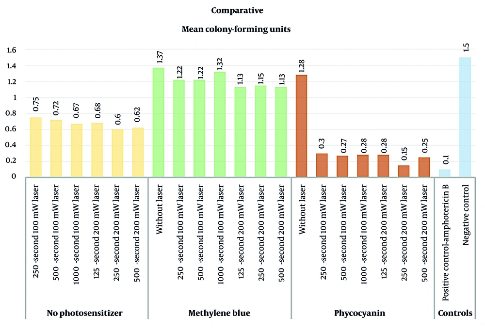
Figure 1.
The illustration of mean CFU/mL of each experimental group categorized according to the utilized photosensitizer.
The lowest mean colony count was observed in the Rhizopus group treated with 32 µg/mL amphotericin B (positive control) (0.10 ± 0.00), whereas the highest was observed in the negative control group (1.50 ± 0.00).
PC and MB demonstrated comparable antimicrobial effects when applied independently without laser irradiation (1.28 ± 0.08 and 1.37 ± 0.08 CFU/mL, respectively). However, under laser irradiation, the PC-treated groups showed a markedly greater reduction in colony counts than the MB-treated groups, with CFU/mL values ranging from 0.15 to 0.28 in the PC groups versus 1.13 to 1.32 in the MB groups.
The data showed a normal distribution (P = 0.14); therefore, parametric tests were used for data analysis. One-way ANOVA revealed a significant difference in mean CFU/mL across the treatment groups (P < 0.001). Subsequent post hoc pairwise comparisons using Tukey's HSD test indicated that the Rhizopus oryzae group treated with amphotericin B showed significantly better performance than the other groups (P > 0.05). Only the difference between this positive control group and the group treated with 2 mg/mL PC combined with 125 seconds of laser irradiation was not statistically significant.
The groups treated with PC in combination with laser irradiation showed significantly lower mean colony counts than the other laser-irradiated groups (P < 0.001). Notably, laser irradiation alone also produced a significantly greater reduction in Rhizopus colonies than MB (P < 0.001).
Regarding variations in laser irradiation power and duration within groups, no significant differences were observed (P > 0.05).
5. Discussion
The present study investigated the comparative photodynamic effects of PC and MB on the fungal pathogen Rhizopus oryzae, using amphotericin B as the positive control. The findings revealed significant differences in antifungal efficacy among the treatments, with amphotericin B demonstrating the most pronounced reduction in CFU/mL. This finding is consistent with its status as the standard antifungal therapy for mucormycosis; however, its nephrotoxicity, infusion reactions, and limitations in patients with systemic comorbidities underscore the ongoing need for complementary or alternative antifungal strategies (25, 26).
Notably, the group treated with PC combined with 125 seconds of laser irradiation exhibited CFU reductions comparable to those of amphotericin B, suggesting that PC-assisted PDT may provide clinically relevant antifungal activity under certain optimized conditions.
The observed superior performance of PC over MB supports the hypothesis that photosensitizer selection is a critical determinant of PDT efficacy. PC’s natural origin, favorable tolerability, and advantageous photophysical properties, including high absorption at approximately 620 - 660 nm and the generation of reactive oxygen species, may contribute to its enhanced antifungal effect. Moreover, the reduction in fungal colonies following laser application alone, although less pronounced, suggests that irradiation may exert inherent antifungal or photothermal effects, highlighting the importance of protocol optimization (27-29).
These findings are consistent with previous research on PDT applications in other fungal infections, including Candida and Aspergillus. However, studies specifically examining PDT for Mucorales remain limited. Liu et al. (11) demonstrated MB-PDT-mediated reductions in minimal inhibitory concentrations, and differences in light sources, methodology, and evaluation metrics may explain discrepancies relative to our results. The potential for PC-based PDT to act synergistically with, or reduce dependence on, conventional antifungals warrants further investigation.
Although the outcomes of this in vitro study are encouraging, they cannot be directly extrapolated to clinical practice because of biological, anatomical, and host-related complexities that are absent under laboratory conditions. Light penetration depth, interaction with necrotic tissues, oxygen availability, and local immune responses are variables that require validation in animal models and controlled clinical trials.
Before the clinical translation of PDT protocols, several additional preclinical steps are required to ensure both efficacy and safety. These include validation in more complex in vitro models, such as multispecies biofilms and tissue-mimicking substrates, to better replicate the clinical environment. Furthermore, optimization of treatment parameters, including photosensitizer concentration, pre-irradiation time, laser wavelength, power density, and exposure time, is necessary to define standardized and reproducible protocols. In vivo studies using appropriate animal models are also essential to evaluate tissue penetration, host response, and potential cytotoxicity to surrounding healthy tissues. Finally, controlled clinical trials are required to confirm antimicrobial efficacy, assess safety in human oral tissues, and determine long-term outcomes compared with conventional antimicrobial approaches. Future studies can also explore dose-response relationships, repeated PDT sessions, and combination regimens with systemic antifungals.
5.1. Conclusions
This in vitro study demonstrated that amphotericin B exhibited the highest antifungal efficacy against Rhizopus oryzae. PDT, particularly when mediated by PC, also produced measurable antifungal effects against Rhizopus oryzae in a controlled laboratory environment. The present study did not evaluate tissue penetration, host-pathogen interactions, toxicity, animal infection models, or combination therapeutic approaches. Therefore, PC-assisted PDT should be considered hypothesis-generating rather than clinically actionable at this stage, and the results cannot be extrapolated to clinical practice or patient management. Further preclinical investigations are required before any potential clinical translation.
