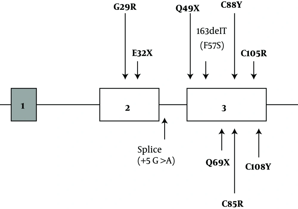
CCH is a rare kind of congenital hypothyroidism with genetic causes, mostly in the TSHβ subunit gene. TSH is a 28 kDa glycoprotein hormone released by the anterior pituitary gland. Similar to other glycoprotein hormones, TSH is a heterotrimeric protein consisting of a common α-subunit (92 aa) and a specific β-subunit (118 aa). The TSHβ gene with 4.9 kb in size contains 3 exons and 2 introns. The first exon contains 37 bp and includes the 5' untranslated region of the gene. The coding region of the gene includes the second exon (163 bp) and the third exon (326 bp), which are separated by a 450 bp intron (
15).
For the TSHβ gene, more than ten different mutations have been identified so far, which all are considered as probable causes of TSH deficiency worldwide. Inheritance of CCH is autosomal recessive (
17). Because in most screening programs, attention is only paid to high TSH levels, and fT4 is often ignored, the diagnosis rate of CCH is often low. Therefore, diagnosis and initiating thyroid hormone replacement may be delayed in these children, resulting in neurodevelopmental retardation and growth failure (
18). Like other CCH pediatric patients with late diagnosis, our patients were also diagnosed between the ages of 3.0 - 6.5 years (Mean ± SD = 4.42).
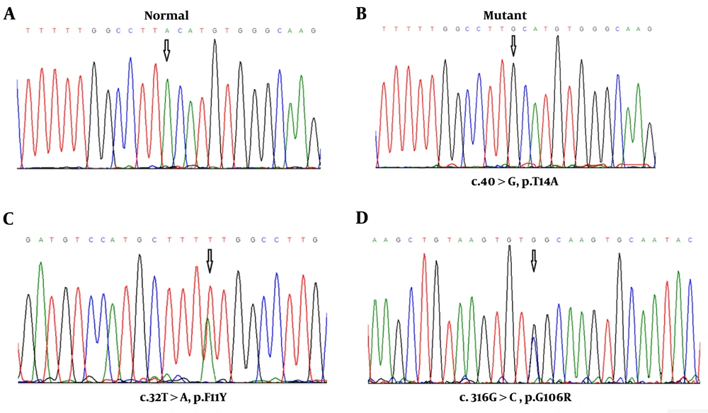
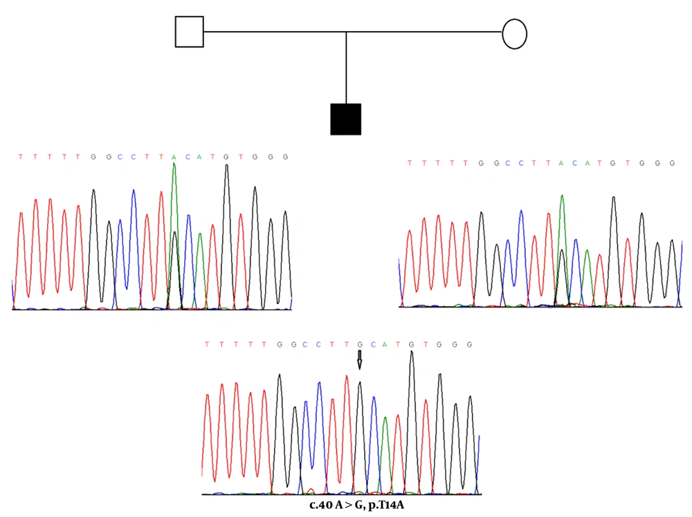
In the present paper, using a PCR-based direct sequencing strategy, we described three different missense mutations in the TSHβ -subunit gene in five (out of seven) Iranian unrelated children patients affected by congenital TSH-deficient hypothyroidism. Two heterozygous missense mutations (c.32T>A: p. F11Y and c.316G>C: p. G106R) are novel and so far has not been detected in any patients with low TSH hypothyroidism. Also, three affected children were homozygous for A-to-G substitution in codon 40 of the TSHβ subunit that leads to the conversion of threonine to an alanine (p.T14A), a common missense variant in the TSHβ gene.
The F11Y mutation in the conserved SMLFGL region of exon 2 of the gene was identified in a six-year-old boy. This new mutation constitutes a T to A transversion (c.32T>A) and results in a change from nonpolar phenylalanine to polar tyrosine in the mutant peptide. The heterozygous (F11Y) and homozygous (T14A) missense mutations found in exon 2 of the TSHß gene, causing amino acid substitutions in the signal peptide of the TSHß-subunit. The observation of these mutations possibly indicates the presence of new mutational hot spots of the TSH ß gene. TSHß-subunit-like other secretory proteins are synthesized as precursors extended at the NH2 terminus by a sequence of 20 amino acids, termed the leader or signal sequence, which is required for translation by ribosomes and protein transport from the membrane. The sequences of signal peptides are heterogeneous, but mostly, the signal peptides have consisted of three conserved domains: a hydrophilic NH2 terminal region of five to eight positively charged basic amino acids, which is essential for translocation, a central hydrophobic core of 7 to 15 amino acids, which is vital for recognition and transferring precursor protein to the ER membrane, and a polar COOH terminal region of approximately six amino acids, which affects the fidelity and efficiency of signal peptidase cleavage as posttranslational processing (
19).
In the present study, the c.32T>A mutation of the TSHß gene was located in the hydrophobic core of the signal peptide, causing an F11Y amino acid substitution. According to the in-silico prediction results, this missense mutation changes the hydropathy determinant of the core region (hydropathy score alters from 2.800 for nonpolar and hydrophobic phenylalanine to -1.300 for polar and hydrophilic tyrosine). Thus, it probably leads to impairment of the recognition and transformation of precursor protein into the endoplasmic reticulum. Also, amino acid sequence alterations near the cleavage site in the COOH terminal region of the signal peptide can disrupt the efficiency of signal peptidase cleavage. The c.40A>G missense variation (T14A) is located in the polar COOH terminal region and changes the hydropathy determinant of this region (hydropathy score alters from -0.700 for polar threonine to 1.800 for non-polar alanine), so it can influence the hydrophobic interactions.
Furthermore, the G106R mutation in exon 3 of the gene was identified in a 3.5-year-old affected girl. In previous studies, exactly a nucleotide before G106R mutation, C105V mutation has been identified in related and unrelated cases from different populations such as Brazilian (
20), German (
21,
22), and Belgium (
23) patients. Due to the high frequency of this mutation in different populations, it’s argued that this region is critical for maintaining the biological and structural integrity of the TSH molecule and represents a mutational hot spot (
11,
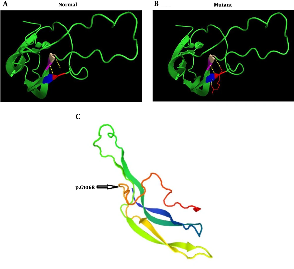
24). Furthermore, the G106R mutation changes the amino acid sequence in the seat belt region of the TSH heterodimer, the important region for the dimerization with the ɑ-subunit and essential for the accurate secretion of the mature TSH. In-silico analysis for novel mutation c. 316G>C revealed the pathogenic nature of this alteration (PolyPhen2: 0.568, the score of SIFT: 0, and Mutation Taster prediction: disease-causing). The prediction model of the tertiary structure of TSHβ protein also indicated that this mutation is part of a coil structure in the protein. This model shows that in the position of 106, glycine amino acid (a small and without side-chain amino acid) connects with valine and cysteine at positions of 71, and 72, respectively. These bonds are conserved and located in important positions of this protein. In the mutant protein, the amino acid arginine has a side chain with three carbons and changes the number of bands in this position, as it binds to the two amino acids of cysteine and valine at positions 105, 72, and 71, respectively. The findings of the prediction model of the tertiary structure of mutant TSHβ protein suggest that the conformation of the TSHβ subunit is probably altered with this mutation.
The other confirmatory evidence that this mutation is a pathogen cause of CCH in our patient roots in the fact that this mutation was not identified in 60 ethnically matched healthy subjects. The interpretation of measurable TSH in parents of this case was difficult. Particularly because both parents did not show any mutation in this region. Nevertheless, early clinical data of this patient with poor growth indicated low serum FT4 and TSH hypothyroidism as well as normal basal values of cortisol and prolactin. Besides, no response to TRH was observed, which in total revealed the necessity of molecular analysis. However, in all affected patients, central congenital hypothyroidism was not distinguished by neonatal screening when only the TSH was considered so that the exact diagnosis of CCH due to TSH deficiency was delayed. Because severe mental retardation has been reported in some children with CCH, using early molecular diagnosis in-time thyroid hormone replacement therapy can be provided.
The current study had limitations, including the necessity of evaluating the frequency of the mutations in a study with a larger sample size. Secondly, inaccessibility of information regarding the family history of the CCH for all participants. Moreover, applied studies are needed to investigate the contribution of these novel variants to the CCH phenotype.
In conclusion, three missense mutations (two novels and one previous reported) in exons 2 and 3 of the TSH ß-subunit gene in 5 sporadic cases of CCH are reported. The findings of the prediction model demonstrated that these mutations may affect the structure of TSH, resulting in impairments in the biological functions of the protein. According to the best knowledge of the authors, this study is the first of its kind in Iran, and our observations suggested that these variations may be associated with non-familial CCHs phenotypes in Iranian pediatric patients. Nevertheless, since the sample size was small, such an association cannot be confirmed. Also, the findings showed that these missense mutations may be more common than previously thought in CCH patients. However, additional studies are required to identify the mechanisms by which these novel mutations lead to genomic instability and contribute to the risk of sporadic CCH in the Iranian population.