


1. Background
2. Objectives
3. Methods
3.1. Cell Culture
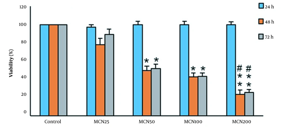
The viability percentage of HT-29 cells in different concentrations and incubation times. Each assay has been done six times (4 replicates per assay), and the mean ± standard deviations have been indicated. * P < 0.05, ** P < 0.01, # P < 0.05; *, # and $ symbols indicate comparison to the control, and MCN50 groups.
3.2. Cell Viability Assay
3.3. Annexin V / PI Apoptosis Assay
3.4. Real-Time PCR
3.5. Western Blotting Analysis
3.6. Statistical Analysis
4. Result
4.1. Cell Viability
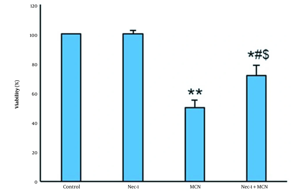
The viability percentage of HT-29 cells (mean ± SD). Each assay was done six times. * P < 0.05, ** P < 0.01, # P < 0.05, $ P < 0.05; *, # and $ indicate comparison to the control, Nec-1, and MCN groups, respectively.
4.2. Annexin V-FITC/ PI Assay
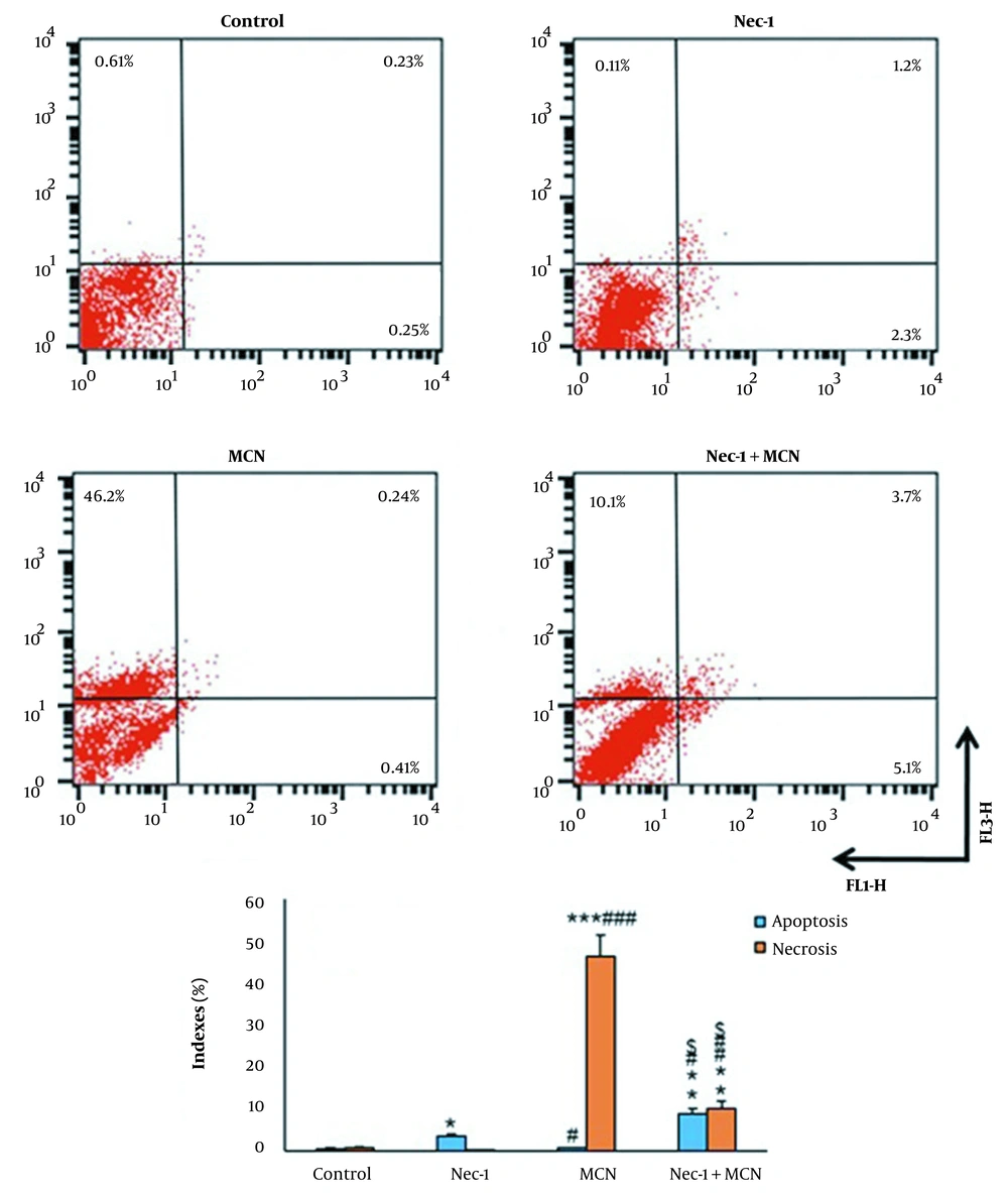
Flow cytometry results of Annexin V/PI staining (mean ± SD, n = 3). Lower left quadrant: Live cells, lower right quadrant: Early apoptosis, upper right quadrant: Late apoptosis, upper left quadrant: Necrotic cells. * P < 0.05, ** P < 0.01, *** P < 0.001, # P < 0.05, ## P < 0.01, ### P < 0.001, $ P < 0.01.
4.3. Real-Time PCR Assay
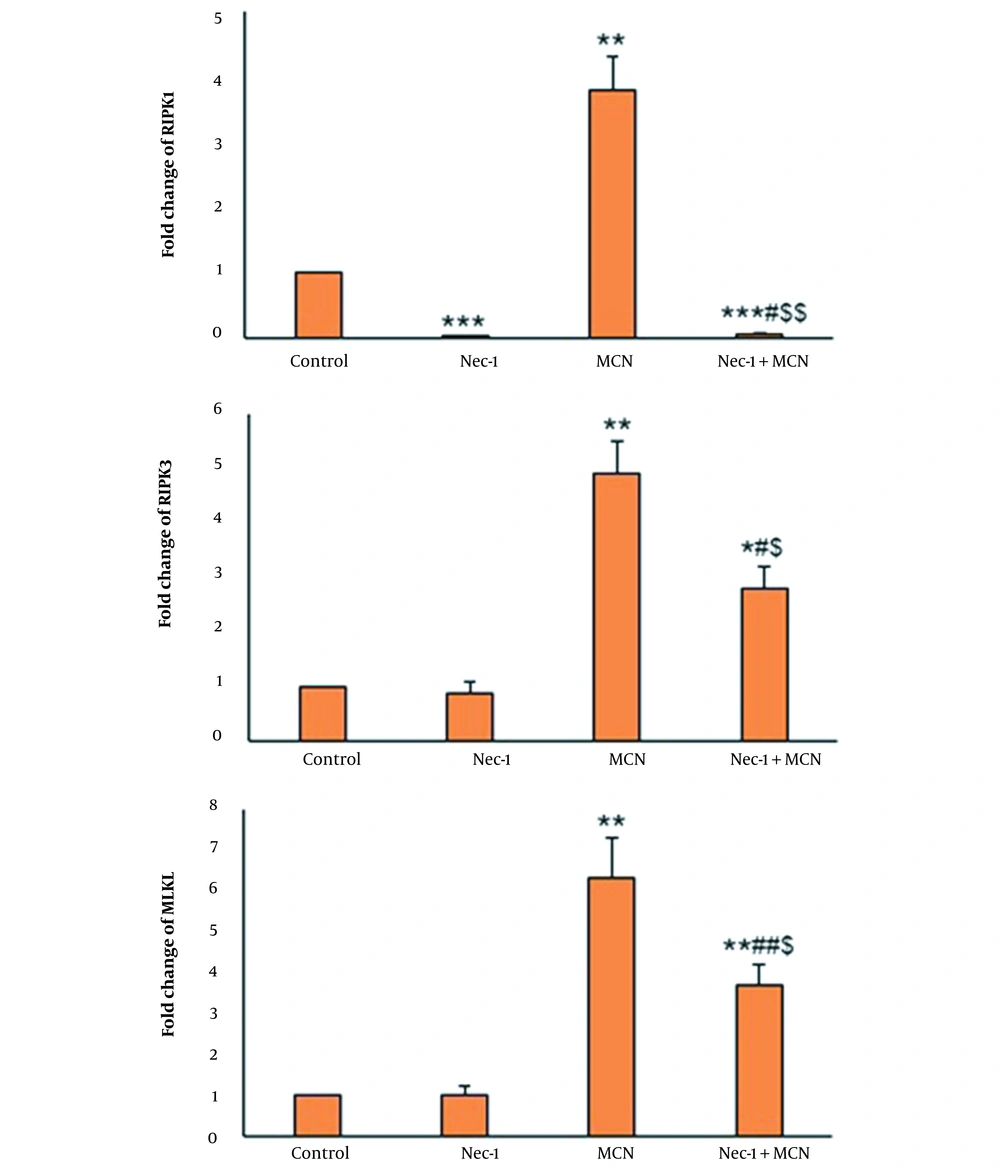
mRNA expression of the different groups (mean ± SD, n = 5). *P < 0.05, ** P < 0.01, *** P < 0.001, # P < 0.05, ## P < 0.01, $ P < 0.05, $$ P < 0.001.
4.4. Western Blotting Analysis
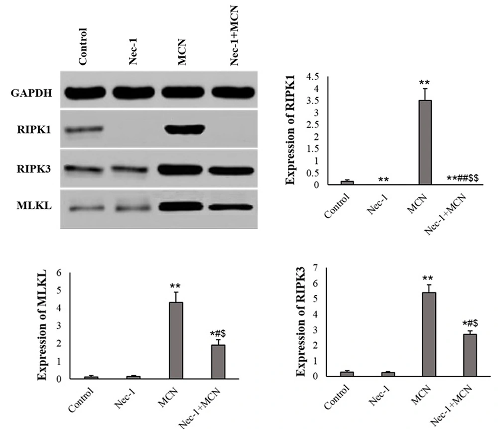
Protein expression of HT-29 cells in different groups (mean ± SD, n = 3). Image J software was used to quantify the protein levels, and the cells normalized to β-actin band intensity. * P < 0.01, ** P < 0.001, # P < 0.01, ## P < 0.001, $ P < 0.01, $$ P < 0.001.
