
Jentashapir J Cell Mol Biol

Image Credit:Jentashapir J Cell Mol Biol

Cytotoxic Effects of Escitalopram on MCF7 Breast Cancer Cells and Normal Hek293 Cells

Author(s):
Atefeh DehghaniAtefeh Dehghani1, Sanaz PashapourSanaz PashapourSanaz Pashapour ORCID2, Keivan SheikhiKeivan Sheikhi1, Abbas ZabihiAbbas ZabihiAbbas Zabihi ORCID3,*
1Department of Biology, Faculty of Medical Sciences, Sanandaj Branch, Islamic Azad University, Sanandaj, Iran
2Department of Forensic Toxicology, Legal Medicine Research Center, Legal Medicine Organization, Tehran, Iran
3Department of Biology, Faculty of Basic Sciences, Rasht Branch, Islamic Azad University, Rasht, Iran

Jentashapir Journal of Cellular and Molecular Biology:Vol. 16, issue 3; e160110
Published online:Sep 02, 2025
Article type:Research Article
Received:Feb 01, 2025
Accepted:Aug 18, 2025
How to Cite:Dehghani A, Pashapour S, Sheikhi K, Zabihi A. Cytotoxic Effects of Escitalopram on MCF7 Breast Cancer Cells and Normal Hek293 Cells. Jentashapir J Cell Mol Biol. 2025;16(3):e160110. doi: https://doi.org/10.5812/jjcmb-160110

Abstract

Background:

The present study primarily aimed to investigate the cytotoxic effects of the antidepressant escitalopram on both cancerous human breast cancer (MCF7) cells and normal human embryonic kidney (HEK293) cells.

Methods:

MCF7 and HEK293 cell lines were randomly divided into control groups and treatment groups exposed to varying concentrations of escitalopram, ranging from 15.625, 31.25, 62.5, 125, 250, to 500 µM. Cell viability was assessed using the MTT assay, and gene expression analysis in MCF7 cells was performed through RT-PCR. To compare the data across the groups, one-way ANOVA followed by Tukey’s post hoc test was conducted.

Results:

A significant reduction in MCF7 cell viability was observed at escitalopram concentrations of 62.5, 125, 250, and 500 µM compared to the control group (P < 0.001). Similarly, significant cytotoxicity was detected in HEK293 cells at 125, 250, and 500 µM (P < 0.001). Moreover, there was a significant increase in BAX gene expression in MCF7 cells (P < 0.01).

Conclusions:

Escitalopram exhibits dose-dependent cytotoxic effects on breast cancer cells, possibly via upregulation of the pro-apoptotic BAX gene. Additionally, off-target effects on normal cells must be considered.

1. Background

Breast cancer is a leading cause of cancer-related mortality in women worldwide (1). By 2030, the global incidence is expected to reach 3.2 million new cases annually. In Iran, the increasing prevalence of breast cancer is linked to lifestyle, diet, physical inactivity, and genetic predisposition. Surgical intervention remains primary, yet metastasis limits its use. Therefore, alternative strategies are required (2-5). Escitalopram, a selective serotonin reuptake inhibitor (SSRI), is commonly used to treat depression and anxiety. Beyond neurological roles, SSRIs may have cytotoxic effects on various cells, including cancerous ones (6-9). Escitalopram has shown potential in reducing MCF7 cell viability through BAX upregulation and BCL2 downregulation. Yet, few studies have explored the apoptotic mechanisms in depth (10, 11).

2. Objectives

The present study evaluates the cytotoxic effects of escitalopram on MCF7 and HEK293 cells, focusing on cell viability, gene expression, and possible apoptotic mechanisms.

3. Methods

Escitalopram (research grade) was obtained and dissolved in DMSO. Working concentrations were prepared at 15.625, 31.25, 62.5, 125, 250, and 500 µM. MCF7 and HEK293 cell lines were acquired from the Pasteur Institute of Iran and maintained in DMEM with 10% FBS and 1% penicillin-streptomycin at 37°C in a 5% CO2 incubator.

3.1. MTT Assay

MCF7 and HEK293 cells were seeded at 5 × 10⁴ cells/well and treated with escitalopram concentrations for 48 hours. Post-treatment, MTT reagent (0.5 mg/mL) was added for 3 hours. Formazan crystals were dissolved in DMSO and absorbance measured at 570 nm using an ELISA reader. All experiments were performed in triplicate (12-19).

3.2. Gene Expression Analysis

Cells were treated with selected concentrations for 48 hours. Total RNA was extracted and cDNA synthesized using commercial kits. RT-PCR was performed using gene-specific primers with conditions: 95°C for 15, 60°C for 30, 72°C for 15 minutes. Relative gene expression was analyzed using the 2-ΔΔCt method (20-25). β-ACTIN was used as the housekeeping gene (Table 1).
Table 1.Primer Sequences of BCL2, and BAX Genes
GenesSequences (5’-3’)Accession Number
β-ACTINNM_001101.5
ForwardGGCACCCAGCACAATGAAG
ReverseCCGATCCACACGGAGTACTT
BAXNM_138761.4
ForwardCGGCAACTTCAACTGGGG
ReverseTTCAGCCCAACAGCCG
BCL-2NM_000633.3
ForwardGGTGCCGGTTCAGGTACTCA
ReverseTTGTGGCCCTTCTTTGAGTTCG

3.3. Statistical Analysis

IC50 values were calculated using nonlinear regression. Data were analyzed by one-way ANOVA with Tukey’s post hoc test. P-values < 0.05 were considered significant. Results are presented as mean ± SE.

4. Results

4.1. Effect of Escitalopram on MCF7 Viability

Escitalopram at 62.5, 125, 250, and 500 µM significantly reduced MCF7 viability compared to control (P < 0.001). No significant changes were observed at 15.625 and 31.25 µM (Figure 1).
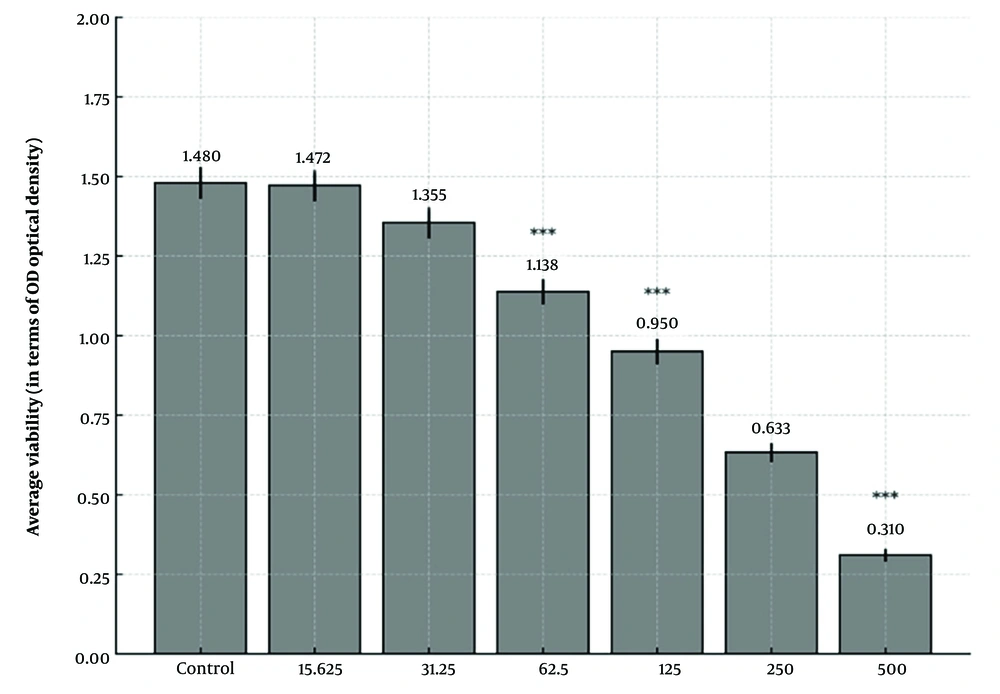
Comparison of the effects of various concentrations of escitalopram on the viability of MCF7 cells. *** indicates a statistically significant difference compared to the control group (P &lt; 0.001).
Figure 1.

Comparison of the effects of various concentrations of escitalopram on the viability of MCF7 cells. *** indicates a statistically significant difference compared to the control group (P < 0.001).

4.2. Effect on HEK293 Cells

HEK293 cell viability significantly decreased at 125, 250, and 500 µM escitalopram (P < 0.001), and moderately at 62.5 µM (P < 0.01) (Figure 2).
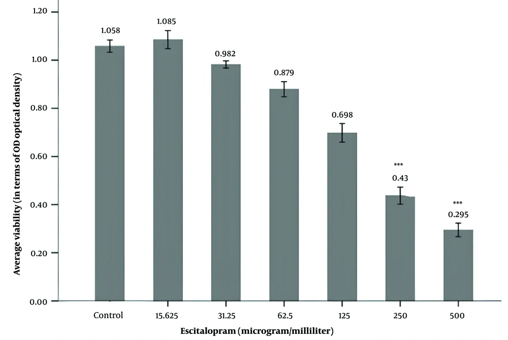
Comparison of the effects of various concentrations of escitalopram on the viability of HEK293 cells. *** indicates a statistically significant difference compared to the control group (P &lt; 0.001).
Figure 2.

Comparison of the effects of various concentrations of escitalopram on the viability of HEK293 cells. *** indicates a statistically significant difference compared to the control group (P < 0.001).

4.3. Gene Expression Analysis in MCF7

A significant increase in BAX mRNA levels was found in treated MCF7 cells (** P < 0.01). Similarly, BCL2 expression increased significantly (** P < 0.01), contrary to expectations. Relative quantification (RQ) values are presented in (Figures 3 and 4).
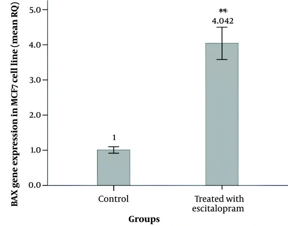
Comparison of the effects of escitalopram on the expression level of the BAX gene and in MCF7 cells. *Indicates significance compared to the control group. ** P &lt; 0.01. (Abbreviation: RQ, relative quantification).
Figure 3.

Comparison of the effects of escitalopram on the expression level of the BAX gene and in MCF7 cells. *Indicates significance compared to the control group. ** P < 0.01. (Abbreviation: RQ, relative quantification).
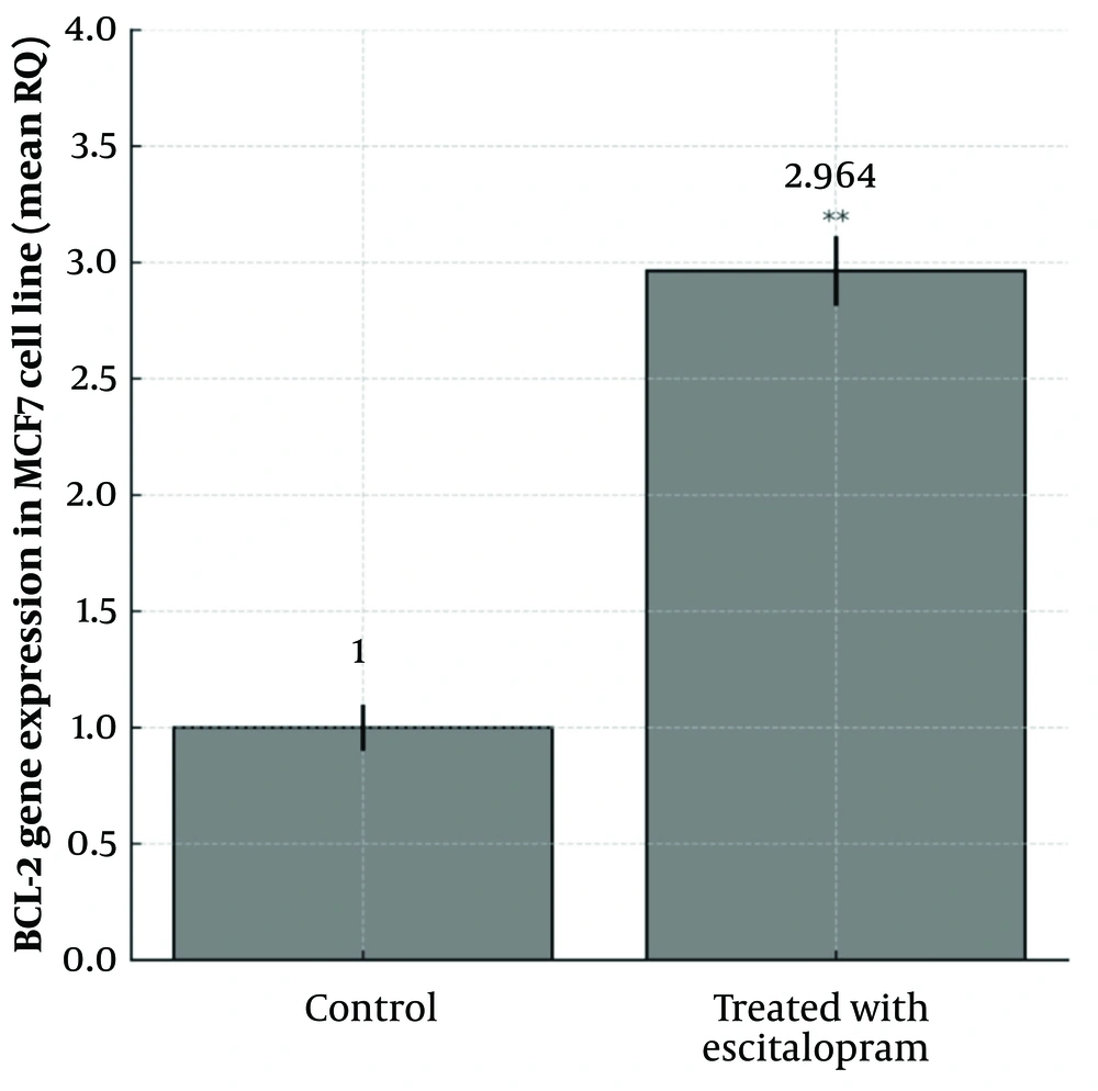
Comparison of the effects of escitalopram on the expression level of the BCL2 gene and in MCF7 cells. *Indicates significance compared to the control group. ** P &lt; 0.01. (Abbreviation: RQ, relative quantification).
Figure 4.

Comparison of the effects of escitalopram on the expression level of the BCL2 gene and in MCF7 cells. *Indicates significance compared to the control group. ** P < 0.01. (Abbreviation: RQ, relative quantification).

5. Discussion

Given the ability of antidepressant drugs to inhibit cancer cells (1, 6, 7, 26), the impact of escitalopram on reducing the proliferation of breast cancer cells remains a highly debated topic. This study aimed to investigate the effects of escitalopram on breast cancer and normal cells using MTT assay and RT-PCR methods, to elucidate the cellular and molecular mechanisms by which escitalopram may eliminate breast cancer cells. The results of this study indicate that escitalopram exerts cytotoxic effects on MCF7 and HEK293 cell lines in a dose-dependent manner. In MCF7 cells, the cytotoxic effect may be linked to escitalopram’s ability to induce oxidative stress, mitochondrial dysfunction, or alterations in signaling pathways involved in cell survival (10, 11, 26-28). Previous studies have demonstrated that SSRIs, including escitalopram, can modulate calcium homeostasis, disrupt mitochondrial membrane potential, and activate apoptotic pathways, which could contribute to anticancer effects in breast cancer cells. The selective toxicity towards cancerous cells, if confirmed, could support further exploration of escitalopram as an adjuvant therapy in breast cancer treatment.
On the other hand, the cytotoxicity observed in HEK293 cells raises concerns regarding potential off-target effects in non-cancerous cells. HEK293 cells are widely used as a model for normal human cells, and significant cytotoxicity in these cells may indicate possible side effects of escitalopram beyond its neurological targets. This finding suggests that while escitalopram may have potential anticancer effects, its safety and specificity toward cancer cells must be carefully considered before clinical application.
Several studies support these findings, showing that SSRI antidepressants significantly reduce the viability of MCF7 cancer cells by increasing BAX gene expression and decreasing BCL2 gene expression (1, 10). Interestingly, despite previous studies indicating a downregulation of BCL2 by SSRIs, our findings showed a significant upregulation. This discrepancy may be due to differences in cell type, experimental conditions, or compensatory feedback mechanisms. Further investigation is warranted. Many studies have demonstrated that serotonin reuptake inhibitors can effectively inhibit breast cancer cells (27-30). Several studies indicated that escitalopram could reduce the viability of breast cancer cells (11, 28). Although further research is needed to comprehensively examine the effects of escitalopram on both cancerous and normal breast cells, this study focuses on the cytotoxic effects of escitalopram on breast cancer cells in a cell culture environment. The findings of this research may contribute to a better understanding of the potential and limitations of using escitalopram in breast cancer treatment and aid in developing more effective innovative therapeutic methods.
Future studies should focus on elucidating the precise molecular mechanisms underlying escitalopram-induced cytotoxicity, including its effects on apoptosis-related proteins, reactive oxygen species (ROS) generation, and mitochondrial integrity. Additionally, in vivo studies and clinical investigations are necessary to determine the therapeutic relevance of escitalopram in cancer treatment and its potential risks to normal tissues (31).

5.1. Conclusions

Escitalopram exerts dose-dependent cytotoxic effects on MCF7 breast cancer cells through BAX gene upregulation. However, observed cytotoxicity in HEK293 cells necessitates further safety evaluation.

Footnotes

References

  • 1.
    Amini Khodashahri F, Lashani M, Saki S. The effects of sertraline on breast cancer (MDAMB-231) cells viability in vitro. Journal of Biological Studies. 2022;5(1):93-102. https://doi.org/10.62400/jbs.v5i1.6383.
  • 2.
    Zabihi A, Pashapour S, Malakijoo N. Evaluation of cytotoxicity of rubiadine on MCf7 and AGO cell lines. Toxicology Communications. 2022;4(2). https://doi.org/10.53388/2022020209.
  • 3.
    Shoorabeh FF, Goodarzi E, Shafeai F, Pordanjani SR, Abbasi M. Pattern of burden cancer breast and relationshipin to human development index in Iran 2009 to 2019: an observational study based on the Global Burden of Diseases. BMC Womens Health. 2024;24(1):540. [PubMed ID: 39334063]. [PubMed Central ID: PMC11428874]. https://doi.org/10.1186/s12905-024-03378-4.
  • 4.
    Soleimani A, Adeli S. Incidence of Cancer from 2015 to 2019 in Northwestern of Iran: A Population-Based on Cancer Registry Profile of Maragheh County. Jundishapur Journal of Health Sciences. 2024;16(2). https://doi.org/10.5812/jjhs-144589.
  • 5.
    Liao L. Inequality in breast cancer: Global statistics from 2022 to 2050. Breast. 2025;79:103851. [PubMed ID: 39580931]. [PubMed Central ID: PMC11625356]. https://doi.org/10.1016/j.breast.2024.103851.
  • 6.
    Gul SO, Korkut A, Aydemir E. Combined Effect of Sertraline and Capecitabine on Breast Cancer Cell Lines In Vitro and In Silico Evidence for Synergistic Interaction. Scientia Pharmaceutica. 2024;92(3). https://doi.org/10.3390/scipharm92030038.
  • 7.
    Baldissera AB, Boia-Ferreira M, Basilio ABC, Resende JSS, Castro MAA, Chaim OM, et al. Sertraline as a potential cancer therapeutic approach: Biological relevance of TCTP in breast cancer cell lines and tumors. Adv Med Sci. 2023;68(2):227-37. [PubMed ID: 37379765]. https://doi.org/10.1016/j.advms.2023.06.001.
  • 8.
    Kazi KJ, English CD, Ivantsova E, Souders Ii CL, Martyniuk CJ. Transcriptome networks and physiology related to cardiac function and motor activity are perturbed in larval zebrafish (Danio rerio) following exposure to the antidepressant citalopram. Environ Pollut. 2024;361:124767. [PubMed ID: 39168440]. https://doi.org/10.1016/j.envpol.2024.124767.
  • 9.
    Mohanthi S, Sutha J, Gayathri M, Ramesh M. Evaluation of the citalopram toxicity on early development of zebrafish: Morphological, physiological and biochemical responses. Environ Pollut. 2024;357:124399. [PubMed ID: 38906410]. https://doi.org/10.1016/j.envpol.2024.124399.
  • 10.
    Cho YW, Kim EJ, Nyiramana MM, Shin EJ, Jin H, Ryu JH, et al. Paroxetine Induces Apoptosis of Human Breast Cancer MCF-7 Cells through Ca(2+)-and p38 MAP Kinase-Dependent ROS Generation. Cancers (Basel). 2019;11(1). [PubMed ID: 30634506]. [PubMed Central ID: PMC6356564]. https://doi.org/10.3390/cancers11010064.
  • 11.
    Patel R. Anti-proliferative effects of selected antidepressant agents on human metastatic breast cancer cell line, MDA-MB-231. Long Island University, The Brooklyn Center; 2013.
  • 12.
    Pashapour S, Heshmati M, Mousavi Z, Esmaeili S. The Cytotoxicity of the Chloroform and Petroleum Ether Fractional Extracts of Galium verum L. in HepG2 and HT29 Cell Lines. Journal of Kermanshah University of Medical Sciences. 2020;24(2). https://doi.org/10.5812/jkums.101079.
  • 13.
    Wdowiak A, Farahmandlou N, Tajik A, Pashapour S, Ahmadi R. The Cytotoxic Effect of Estradiol Valerate, Progesterone, and Testosterone on Brain Glioblastoma (A172) , Colorectal Cancer (HT29) and Human Embryonic Kidney (HEK293) Cells and the Expression Levels of Bax, Bcl-2, and KAI-1/CD82 in A172 and HT29 cells. Journal of Biological Studies. 2021;4(3):106-19. https://doi.org/10.62400/jbs.v4i3.6153.
  • 14.
    Norouzi S, Ahmadi R, Pashapour S. [The cytotoxic effects of Tolmetin on evaluation of Bax and Bcl2 genes expression level in cervical cancer cells (Hela)]. Feyz Med Sci J. 2020;24(1):31-7. FA.
  • 15.
    Hojatipour T, Pashapour S, Almasirad A, Mousavi Z. The cytotoxic activity evaluation of an arylhydrazone derivative of mefenamic acid on HEPG2 liver cancer cells and normal gingival HGF cells. Gene Reports. 2023;33. https://doi.org/10.1016/j.genrep.2023.101810.
  • 16.
    Ghazazani Z, Heshmati M, Asgarpanah J, Pashapour S. Evaluation of Molecular Docking and Cytotoxicity by Pycnocycla bashagardiana Fruit Essential Oil in Colon Cancer Cell Lines. Journal of Kermanshah University of Medical Sciences. 2023;26(4). https://doi.org/10.5812/jkums-131043.
  • 17.
    Zabihi A, Pashapour S, Mahmoodi M. Cell Therapy and Investigation of the Angiogenesis of Fibroblasts with Collagen Hydrogel on the Healing of Diabetic Wounds. Turk J Pharm Sci. 2023;20(5):302-9. [PubMed ID: 37933815]. [PubMed Central ID: PMC10631366]. https://doi.org/10.4274/tjps.galenos.2022.62679.
  • 18.
    Lavasani RSM, Pashapour S, Almasirad A, Mousavi Z. Effects of an aryl hydrazone derivative of naproxen on HepG2 liver cancer and HGF gingival fibroblasts cell lines. The Nucleus. 2023;66(2):183-93. https://doi.org/10.1007/s13237-023-00428-4.
  • 19.
    Pashapour S, Zabihi A, Behrouzi R. Investigating the cytotoxic effect of ibuprofen concentration in liver cancer cells (HepG2) and normal fibroblast (AGO). Toxicology Advances. 2022;4(4). https://doi.org/10.53388/20220202015.
  • 20.
    Pashapour S, Heshmati M, Mousavi Z, Esmaeili S. The effects of methanolic extract of the aerial parts of Galium verum on HT29 and AGO cell lines. Nucleus J. 2022:1-10. https://doi.org/10.1007/s13237-021-00380-1.
  • 21.
    Pashapour S, Heshmati M, Mousavi Z, Esmaeili S. The Apoptotic Effect of Methanolic Extract of Galium verum on HT29 Cell Line. Journal of Biological Studies. 2022;4(4):210-20. https://doi.org/10.62400/jbs.v4i4.6347.
  • 22.
    Heshmati M, Hasani-Reza Abad N, Pashapour S. Evaluating the Effects of Silymarin on Expressing SBDSP1 and CASC11 Genes in HCT116 Colon Cancer Cells. Journal of Kermanshah University of Medical Sciences. 2022;26(2). https://doi.org/10.5812/jkums-122802.
  • 23.
    Hosseini SHR, Pashapour S, Farhadi M, Zabihi A. Human papillomavirus infection and its relationship with common polymorphism of HLA gene by PCR method. Gene Reports. 2023;31. https://doi.org/10.1016/j.genrep.2023.101767.
  • 24.
    Farzaneh S, Bandad S, Shaban F, Heshmati M, Barikrow N, Pashapour S. The Expression of miR-34c-5p Induces G0/G1 Cell Cycle Arrest and Apoptosis in SW480 Colon Cancer Cell. Iran J Pharm Res. 2023;22(1). e135501. [PubMed ID: 38116556]. [PubMed Central ID: PMC10728859]. https://doi.org/10.5812/ijpr-135501.
  • 25.
    Pashapour S, Heshmati M, Mousavi Z, Esmaeili S. Effect of whole methanolic extract of Galium verum on AGO cell line. Toxicology Communications. 2022;4(2). https://doi.org/10.53388/20220202010.
  • 26.
    Dong F, He K, Zhang S, Song K, Jiang L, Hu LP, et al. SSRI antidepressant citalopram reverses the Warburg effect to inhibit hepatocellular carcinoma by directly targeting GLUT1. Cell Rep. 2024;43(10):114818. [PubMed ID: 39388353]. https://doi.org/10.1016/j.celrep.2024.114818.
  • 27.
    Hanbashi A. Anti-Proliferative Mechanism of Selective Serotonin Re-Uptake Inhibitor-Sertraline-on Human Metastatic Breast Cancer Cell Line MCF7. Long Island University, The Brooklyn Center; 2014.
  • 28.
    Salama M, Elamin A, Youssif M, Mattar NA. Induction of DNA damage and growth arrest by citalopram in breast cancer cells mediated via activation of Gadd45a and apoptotic genes. Ultrastruct Pathol. 2025;49(2):158-69. [PubMed ID: 39825580]. https://doi.org/10.1080/01913123.2025.2454691.
  • 29.
    Duarte D, Falcao SI, El Mehdi I, Vilas-Boas M, Vale N. Honeybee Venom Synergistically Enhances the Cytotoxic Effect of CNS Drugs in HT-29 Colon and MCF-7 Breast Cancer Cell Lines. Pharmaceutics. 2022;14(3). [PubMed ID: 35335887]. [PubMed Central ID: PMC8952811]. https://doi.org/10.3390/pharmaceutics14030511.
  • 30.
    Warchal SJ, Dawson JC, Shepherd E, Munro AF, Hughes RE, Makda A, et al. High content phenotypic screening identifies serotonin receptor modulators with selective activity upon breast cancer cell cycle and cytokine signaling pathways. Bioorg Med Chem. 2020;28(1):115209. [PubMed ID: 31757681]. [PubMed Central ID: PMC6961118]. https://doi.org/10.1016/j.bmc.2019.115209.
  • 31.
    Zabihi A. The role of biological macromolecules in the regulation of angiogenesis in glioblastoma: Focus on vascular growth factors, integrins, and extracellular matrix proteins. Int J Biol Macromol. 2025;311(Pt 1):143838. [PubMed ID: 40319984]. https://doi.org/10.1016/j.ijbiomac.2025.143838.

Crossmark
Crossmark
Checking
Share on
Cited by
Metrics

Purchasing Reprints

  • Copyright Clearance Center (CCC) handles bulk orders for article reprints for Brieflands. To place an order for reprints, please click here (   https://www.copyright.com/landing/reprintsinquiryform/ ). Clicking this link will bring you to a CCC request form where you can provide the details of your order. Once complete, please click the ‘Submit Request’ button and CCC’s Reprints Services team will generate a quote for your review.
Search Relations

Author(s):

Related Articles