
1. Background
Stuve-Wiedemann syndrome (SWS; #OMIM 601559) is a rare genetic disorder inherited in an autosomal recessive manner. It is characterized by a combination of osteoarticular symptoms and a dysautonomic presentation (1). Dysautonomia, a condition affecting the nervous system, causes disruptions in the body's autonomic functions, such as blood pressure and heart rate (2). While initially perceived as a fatal condition in infancy, researchers have since identified several patients exhibiting symptoms of SWS (3, 4). Given the rarity of this disease, a more comprehensive and precise delineation of the clinical symptoms is crucial to enhance the care and ensure the long-term survival of affected individuals.
Initially reported by Stuve and Wiedemann in 1971, SWS was later acknowledged as a distinct disorder in 2000. Various individuals have reported an association between SWS and a lethal variant of Schwartz-Jampel syndrome (SJS) referred to as "SJS Type 2". The SJS type 2, also known as SWS, is a rare, usually lethal, form of SJS characterized by myotonia (muscle stiffness), skeletal dysplasia, contractures, and early death. It is distinct from SJS type 1, which is caused by mutations in a different gene (1, 5).
The molecular and genetic underpinnings of the disease were established in 2004 by Dagoneau et al., who identified several mutations in the leukemia inhibitory factor receptor (LIFR) gene (#OMIM 601559), located on chromosome 5p13 (6). The SWS is identified by a blend of symptoms associated with bone participation and dysautonomia; therefore, the condition is categorized under both skeletal dysplasias (a subtype of curved bone dysplasias) and ciliary neurotrophic factor (CNTF) pathway-related disorders (7).
Over recent years, there have been documented cases of individuals exhibiting the symptoms of SWS despite not having LIFR mutations. Conversely, there have also been instances of patients with LIFR mutations but with an incomplete SWS phenotype (like dysautonomic features without long bone participation), suggesting a variance in both the physical and genetic characteristics of this syndrome (3, 8).
With the development of genomic sequencing technologies in the early 2000s, there has been a significant enhancement in the diagnostic methods and rapid detection of genetic disorders. Notably, techniques such as whole-exome sequencing (WES) or whole-genome sequencing (WGS) have the capability to identify over 5,000 different phenotypic and genetic conditions in a single test. The diagnostic precision of WES and WGS is estimated to be around 35 - 40% (9-12). Genome sequencing increases the ability to identify previously unknown and rare genetic disorders (13, 14). Approximately 6,000 to 8,000 rare diseases have been discovered, 80% of which have identifiable genetic underpinnings (15, 16).
2. Objectives
In this study, by using this technique, we will investigate potentially pathogenic variants of the LIFR gene in the rare disease SWS, which follows a Mendelian pattern.
3. Methods
The patients studied included three families from Khuzestan province who had been referred to the Noorgene Ahvaz Medical Genetics Laboratory between 1400 and 1402. Inclusion criteria included having symptoms of SWS syndrome, including skeletal disorders and respiratory distress, and confirmation of these symptoms by a specialist physician.
The patient's consent to participate in the study was also obtained. After collecting the samples and obtaining consent from the families, genetic counseling and finally drawing a family tree were performed. In the next step, 5 cc of peripheral blood from the patients and their parents was prepared and poured into a Falcon tube containing 0.5% ethylenediamine-tetraacetate (EDTA). After DNA purification using the salting out method, the quality and quantity of the extracted genetic material were evaluated using agarose gel loading and the Nanodrop device, respectively.
After sequencing using the WES method, bioinformatic analysis was performed. Raw sequencing data (FASTQ files) were first assessed for quality using FastQC (v0.11.9). Adapter trimming and low-quality read filtering were carried out using Trimmomatic (v0.39). Clean reads were then aligned to the human reference genome GRCh37/hg19 using BWA-MEM (v0.7.17). Post-alignment processing, including sorting and marking duplicate reads, was performed using SAMtools (v1.10) and Picard (v2.23.8). Variant calling was conducted with GATK HaplotypeCaller (v4.1.8.1) following best practice workflows. Variants were annotated using ANNOVAR (April 2018 version). Identified variants were further analyzed and visualized using IGV (Integrative Genomics Viewer, v2.8) to confirm read alignment and zygosity. The Illumina HiSeq 2500 platform was used to determine the genome sequence.
After sequencing the data using the WES method, the results were analyzed. The sequences of the primers used are shown in Table 1.
Table 1.
Leukemia Inhibitory Factor Receptor Gene Primer Sequences
| Name | Primer Sequencing |
|---|---|
| LIFR-Ex9-F | 5’ TTACGAATTTGCCCGTTTTT ’3 |
| LIFR-Ex9-R | 5’TTCAGAAATCAAAAATTATCAGAAAGA’3 |
Abbreviation: LIFR, leukemia inhibitory factor receptor.
To perform PCR, 10 μL of Red Mix solution was added to 0.5 μL of primers F and R. The PCR protocol begins with an initial denaturation step at 94°C for 5 minutes to ensure complete separation of DNA strands. This is followed by 30 cycles of denaturation at 94°C for 30 seconds, primer annealing at 60°C for 30 seconds, and extension at 72°C for 30 seconds. After the cycling steps, a final extension is carried out at 72°C for 5 minutes to complete the synthesis of all DNA fragments. The PCR products were loaded onto an electrophoresis gel (Figure 1).
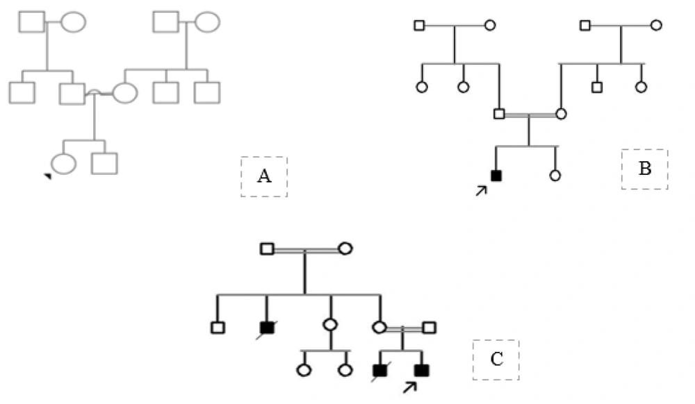
Figure 1.
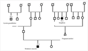
Pedigree of the first family (A), second family (B), and third family (C)
To confirm the variant obtained in the patient and parents, sequence reading was performed using an ABI3130XL sequencer and then analyzed using Chromas software and the NCBI and ENSEMBL databases.
4. Results
In this study, three families from Khuzestan province were examined. The first family had a 2-year-old girl with various symptoms, including skeletal dystrophy, muscular dystrophy, and growth retardation. The other family members were normal for this disease (Figure 1A). In the second family, the patient was a 2-year-old boy from a consanguineous marriage with bone abnormalities, short stature, and respiratory and pulmonary disorders. There was no previous similar patient in their family (Figure 1B). The third family also had a 1-month-old sick boy, the result of a family marriage, with bone abnormalities, respiratory distress, and hyperthermia. There was a similar 3-month-old infant in their family who had died. In the second generation of this family tree, there was a sick uncle with similar symptoms who had died at 8 months of age (Figure 1C).
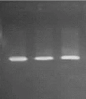
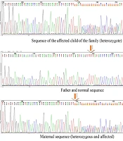
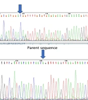
After analyzing the exome data with a 100X read depth, different variants were obtained for all three patients. According to their clinical symptoms, more focus was placed on 42 genes associated with muscular and skeletal dystrophies. The results for the first two patients were normal regarding the LIFR gene, and further analysis is needed to find the cause of the disease. However, the third patient showed a new variant, NM_001127671.1 c.1267A>G (p.Ile423Val). This variant is a VUS according to in silico studies. In the next step, this variant was confirmed as heterozygous in both parents using trench sequencing (Figure 2). The PCR products were loaded on a 1.7% agarose gel. The resulting product has 481 bp and was run alongside a 100 bp ladder (Figure 3).
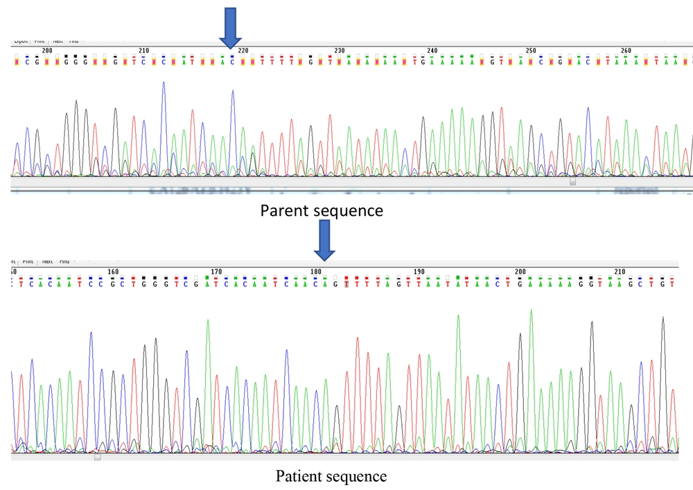
Figure 2.
Patient sequence: Sequencing of exon 9 of the leukemia inhibitory factor receptor (LIFR) gene in the patient shows that he is homozygous for the c.1267A>G: p.I423V mutation. Sequencing of the patient's parents shows that they are carriers for the c.1267A>G: p.I423V mutation.
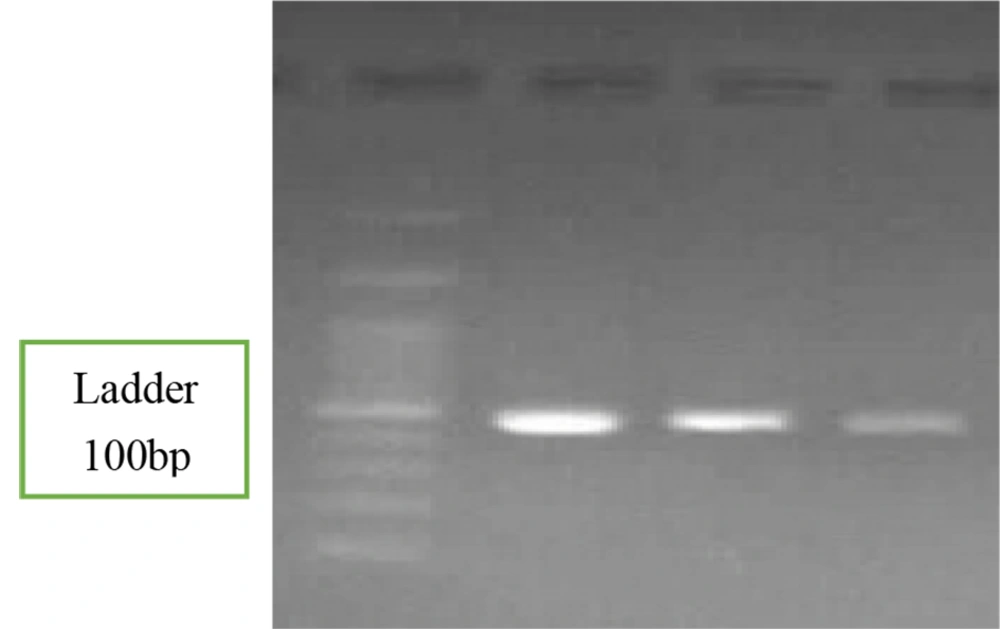
Figure 3.
PCR products loaded on agarose gel resulting from amplification of exon 9 of the leukemia inhibitory factor receptor (LIFR) gene
5. Discussion
The SWS (MIM 601559) is a genetic disorder inherited in an autosomal recessive pattern, distinguished by traits such as a mask-like facial expression, skeletal anomalies, and limited fetal growth. The syndrome was initially documented by Stuve and Wiedemann in 1971 in two siblings who experienced early fatal outcomes (17). Due to their similar clinical and radiological characteristics, SWS and SJS type 2 are often considered to be a single disorder. This syndrome is identified by traits such as short stature, curvature of the limbs more prominent in the lower limbs, camptodactyly, breathing difficulties, and episodes of elevated body temperature typically linked to feeding or swallowing challenges. Other clinical manifestations include a compressed mouth, underdevelopment of the middle of the face, congenital joint contractures, low muscle tone, and occasional anomalies. The SWS is connected to a notable rise in neonatal mortality, primarily attributed to respiratory complications and malignant hyperthermia that does not respond well to treatment (17).
The exact incidence and prevalence of SWS in the world are not known, as the number of patients is very small. Studies suggest that SWS is caused by mutations in the LIFR gene on chromosome 5p13. Several mutations in this gene were identified in 19 families with the syndrome. In families from the United Arab Emirates (UAE), an insertion and frameshift (653 654T) were identified, indicating a founder effect in this region (18).
Bhalla et al. studied that in SWS, respiratory distress and hyperthermic events are the main causes of premature neonatal death, and most patients are not expected to survive their infancy. This study investigated the survival and clinical course of a 5-year-old boy with SWS. This case report will increase awareness of the syndrome and help facilitate early diagnosis, intervention, and genetic counseling for families (19). The results of this research are consistent with the findings of the present study.
During another study, Chen et al. examined the factor responsible for SWS. The IL6ST gene is responsible for encoding GP130, a shared signal transducer for a group of 10 cytokines in the IL-6 cytokine family. Previous studies have identified factors involved in the IL6ST cytokine that maintain leukemia inhibitory factor (LIF) signaling. The researchers detailed the cases of three separate families in which a minimum of five individuals displayed symptoms resembling the fatal Stuve-Wiedemann-like syndrome. This syndrome is defined by skeletal abnormalities, neonatal lung issues, and accompanied by other characteristics like congenital thrombocytopenia, eczematoid dermatitis, kidney irregularities, and impaired immediate response.
Through their investigation, the team pinpointed various mutations that lead to a loss of function, including a homozygous mutation and an intronic splice variant that triggers exon skipping. Cellular responses to GP130-dependent cytokines, such as IL-6, IL-11, IL-27, oncostatin M (OSM), and LIF, are not observed in functional assays. Patients' cells were genetically altered via lentiviral transduction, which successfully restored the signaling deficiency. This research reveals a new genetic syndrome in humans caused by the absence of signaling from a group of GP130-dependent cytokines and underscores the significance of the LIF signaling pathway in prenatal and perinatal growth (3).
This study, like the present study, emphasizes the importance of the LIFR gene in the embryonic development pathway. In this study, WES testing was performed on three patients from different families. The results showed the presence of the c.1267A>G: p.I423V variant in exon 9 of the LIFR gene in one of the patients. This variant was confirmed as heterozygous in the patient's parents, and the parents were advised to perform molecular screening focusing on this gene before attempting to conceive again.
5.1. Conclusions
Overall, the results of this study emphasize the importance of WES in identifying potential variants associated with SWS syndrome. Notably, the c.1267A>G (p.Ile423Val) variant was identified for the first time in the LfIFR gene. The presence of this variant in the parents, in a heterozygous state, was confirmed using the standard Sanger sequencing method. However, while the conclusion highlights the value of WES, it does not address its limitations, such as reduced sensitivity in detecting structural variants, intronic or regulatory region mutations, and low coverage areas that may lead to missed variants. These constraints should be acknowledged when interpreting WES findings, especially in genetically heterogeneous disorders.