
1. Background
Muscular dystrophies are a diverse set of disorders, both clinically and genetically, that impact skeletal muscle. These disorders exhibit a progressive clinical course and entail the deterioration of muscle fibers (1). Muscular dystrophy is a rare condition, with a prevalence of approximately 1 in 5,000 to 10,000 individuals globally (2).
The age at which symptoms develop can vary greatly, ranging from birth in conditions like congenital muscular dystrophy, to childhood in cases such as Duchenne muscular dystrophy, and to adulthood in instances of facioscapulohumeral muscular dystrophy (3). Most muscular dystrophies share common characteristics, such as progressive muscle weakness (involving primarily proximal muscles) and elevated levels of serum creatine kinase (CPK). However, the severity and progression of the disease can differ among various types. Many muscular dystrophies are also associated with additional symptoms, including cardiomyopathy and respiratory muscle weakness, especially in the later stages of the condition (4).
The term "muscular dystrophy" includes a range of inherited conditions that lead to a gradual and widespread deterioration of muscle tissue, stemming from a lack of adequate glycoproteins in the muscle cell membrane (5). This non-communicable disorder manifests in various forms, each characterized by distinct inheritance patterns, age of onset, and severity of muscle degeneration. Different mutations in specific genes give rise to the diverse manifestations seen in this disease (6).
Muscular dystrophy can impact both skeletal and cardiac muscles. The advancement of the condition differs based on the type and seriousness of the disorder but typically involves a progressive weakening of muscles, reduced mobility, and the possibility of respiratory and heart-related issues (7). Multiple gene mutations can cause muscular dystrophy, and the disease can be passed down through different inheritance patterns, such as X-linked, autosomal dominant, or autosomal recessive (8). Mutations in the DMD gene on the X chromosome, responsible for encoding dystrophin, are the primary cause of muscular dystrophy (9). As a result of having only one X chromosome, hemizygous males exhibit this phenotype, highlighting why it is prevalent in this population (10). Additionally, it is worth mentioning that mutations in the dystrophin gene lead to allelic heterogeneity. For instance, variations in the DMD gene can give rise to either Duchenne or Becker muscular dystrophy, depending on the extent of protein deficiency (11).
Tibial muscular dystrophy (TMD; identified in the Online Mendelian Inheritance in Man database as MIM 600334), is a muscle disease that affects the lower legs. It's a late-onset condition, typically showing up in adulthood, and it's passed down through families in a dominant pattern, meaning you only need one copy of the faulty gene to develop it. This condition was first identified in Finnish patients by Lillback et al. The TMD primarily affects the front part of the lower leg, specifically the tibialis anterior muscle. Symptoms generally start to appear between the ages of 35 and 45, or later. Unlike some other types of muscular dystrophy, individuals with TMD do not develop heart muscle disease (cardiomyopathy) and it does not affect facial muscles (12).
The primary candidate gene associated with the TMD locus is the TTN gene, responsible for producing the massive muscle protein titin, also known as "connectin" (13). The titin gene comprises 363 exons encompassing 38,138 amino acid residues (4,200 kDa) in its coding region (14). Developed in the last ten years, next-generation sequencing (NGS), also known as massively parallel sequencing, enables the concurrent sequencing of numerous DNA fragments without prior sequence information (15). The NGS shows promise as a valuable diagnostic tool for identifying muscular dystrophies. Whole exome sequencing (WES) has demonstrated diagnostic yields ranging from 25% to 73% in pediatric patients with neuromuscular disorders (16). However, the success rate may vary depending on the specific patient group and the filtering methods employed. The use of WES allows for genetic diagnosis in individuals with unusual symptoms or exceptionally rare disorders (17).
2. Objectives
In this study, an attempt will be made to investigate muscular dystrophy using the WES technique.
3. Methods
A cross-sectional investigation was conducted on a family exhibiting mild muscle weakness, who were referred to the Noorgene Medical Genetics Laboratory in Ahvaz for diagnostic evaluation between the years 1401 and 1402. Inclusion criteria for the study involved experiencing symptoms of muscle weakness disorders, such as a sensation of weakness in the muscles, which were confirmed by a specialist physician. Additionally, consent from the patients to participate in the research was obtained.
3.1. Case Presentation
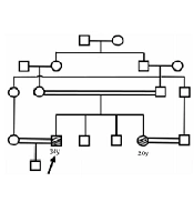
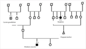
The patient, a 24-year-old male, presented with mild muscle weakness. After examining other family members, it was found that he had a healthy brother and two affected siblings with similar muscle weakness symptoms. Examination of the parents revealed that the mother of the family also had muscle weakness (Figure 1).
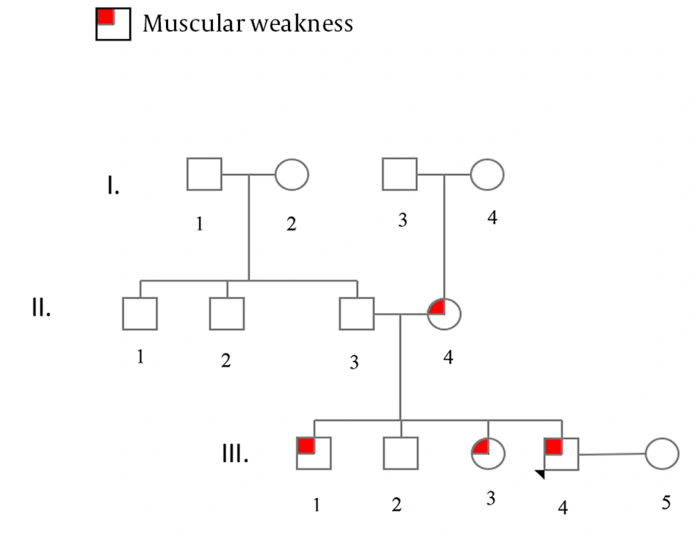
Figure 1.
Family trees related to the TTN gene
3.2. Participant Recruitment and Genetic Analysis
Participants were selected for the study based on the presence of clinically confirmed symptoms and supported by imaging results, as assessed by a specialist physician. Before their participation, all individuals provided informed consent.
3.3. Sample Collection and DNA Extraction
Genomic DNA was isolated from 5 mL peripheral blood samples using a salting-out extraction method. The quality and quantity of the extracted DNA were subsequently evaluated via Nanodrop spectrophotometry and agarose gel electrophoresis.
3.4. Whole-Exome Sequencing and Variant Analysis
The WES was performed on the Illumina HiSeq2500 platform, and the resulting sequencing data were analyzed using standard bioinformatic pipelines to extract relevant information. Following this, criteria for quality score and minimum read depth were set at 30 and 10x, respectively, with a focus on identifying rare variants by maintaining a minor allele frequency (MAF) under 0.01. For this study, a minimum read depth of 32 and a coverage of 35 were deemed acceptable. Sanger sequencing was then employed to confirm the identified genetic variations, utilizing specific primers detailed in Table 1.
Table 1.
Sequence of Primers Used for Polymerase Chain Reaction of TTN Gene
| Variables | Sequence (5' -> 3') | Length | Tm | GC% | Self-complementarity | Self-3' complementarity |
|---|---|---|---|---|---|---|
| TTN-Ex46-F | GGCAGCAGAATCTGAGACCT | 20 | 59.46 | 55.00 | 3.00 | 0.00 |
| TTN-Ex46-R | GGGGTTTGGTCCTCCAGTAG | 20 | 59.38 | 60.00 | 3.00 | 2.00 |
3.5. Polymerase Chain Reaction and Sequencing Confirmation
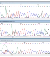
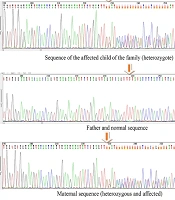
Polymerase chain reaction (PCR) amplification was conducted in a 20 μL reaction volume containing a Red Mix solution, both forward and reverse primers, double-distilled water, and the extracted DNA template. The amplified products were then analyzed by electrophoresis (Figure 2) and subsequently validated using an ABI 3130XI sequencer, with Chromas software employed for sequence verification (Figure 3).
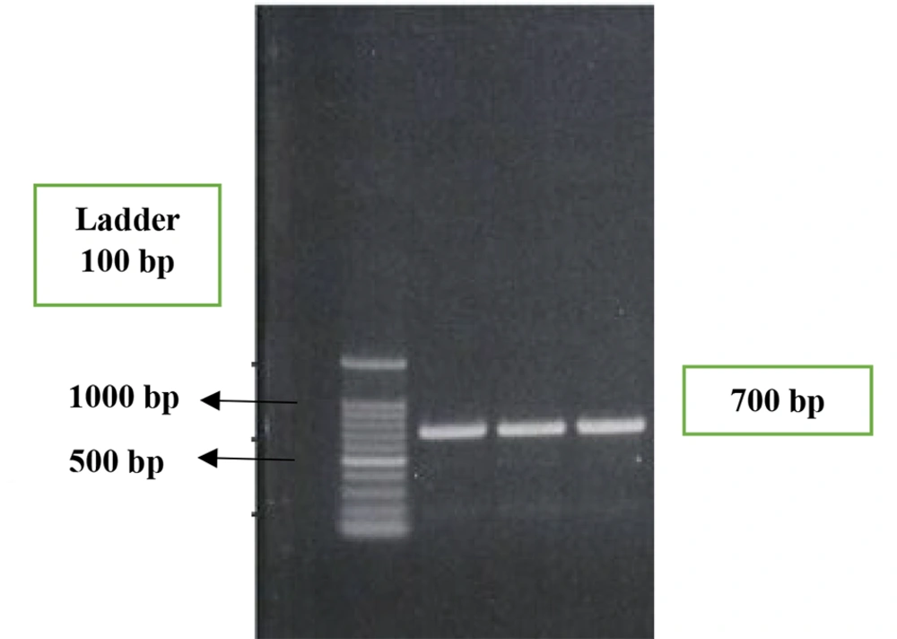
Figure 2.
Electrophoresis of polymerase chain reaction (PCR) products
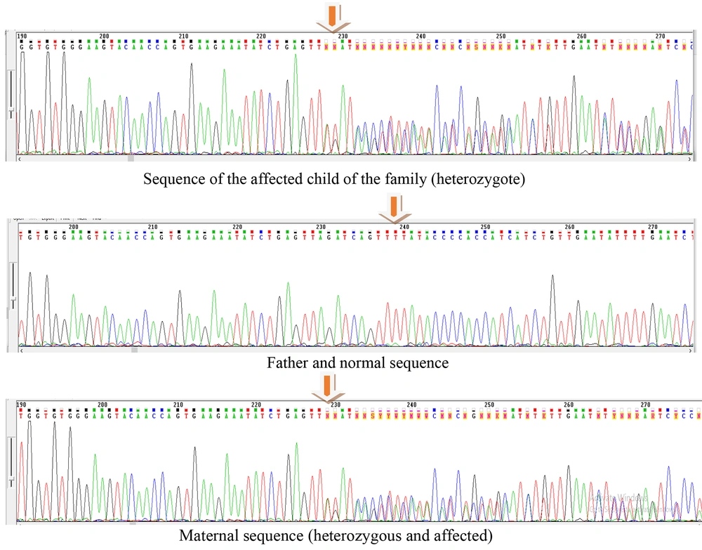
Figure 3.
Sequencing results of the parents and the affected child: The affected child exhibits a heterozygous genotype for the identified TTN mutation (c.14952_14960del; p.Leu4984_Gln4986del). The father shows a normal (wild-type) sequence, whereas the mother is heterozygous for the same deletion, confirming maternal inheritance. Sequence alignment reveals that all samples are identical in the surrounding region and codon numbering, with the only difference being the presence or absence of the deleted nucleotides, which determines the heterozygous or homozygous status of each individual.
3.6. Variant Annotation and Pathogenicity Prediction
The identified genetic variants were compared against the ENSEMBL and NCBI BLAST databases. Furthermore, in silico analyses were performed using Polyphen2, MutationTaster, SIFT, and Predict SNP software to assess the potential pathogenicity of the identified mutation.
4. Results
Exome sequencing was performed using the Illumina HiSeq 4000 (200x) instrument and according to the instructions of the Agilent SureSelect V6 kit. The sensitivity and specificity of this kit in detecting point mutations and small deletions and additions are estimated to be more than 95%. The results of the exome analysis identified more than 235,621 variants in the patient, and after applying the filter, the variant NM_1333793:c.14952_14960del (p.Leu4984_Gln4986del) was finally detected in the patient (Figure 4).

Figure 4.
All stages of variant calling, annotation, filtering, and the number of variants in each step
Sequencing in the patient confirmed the exome results, and the mutation NM_1333793:c.14952_14960del (p.Leu4984_Gln4986del) was confirmed as heterozygous in the patient, which is consistent with the autosomal dominant pattern of the patient's family tree. This mutation was confirmed as heterozygous in the patient's mother. The patient's father was also sequenced, and the result was normal.
Based on information retrieved from databases like OMIM, the identified mutation is responsible for genetic muscle disorders. The sequence findings from various samples show consistency in the specific region and codon number. The variation in individuals being heterozygous or homozygous is solely due to differences in the nucleotide types deleted. This deletion is located within the A-band region of titin, which contains numerous fibronectin type III (FN3) and immunoglobulin (Ig)-like domains. These domains play essential roles in maintaining sarcomere integrity and passive elasticity of muscle fibers. Loss of three consecutive residues in this region may disrupt the folding or stability of these structural motifs, potentially impairing titin’s function in muscle contraction and elasticity. After selecting the candidate variant, primers were designed for the relevant region and amplified using the conventional technique with the Amplicon (Denmark) kit and the recommended temperature conditions in the kit for the desired genomic region, and then confirmed by sequencing.
5. Discussion
Tardive TMD is an inherited myopathy characterized by late-onset symptoms (12). Weakness and muscle atrophy typically affect the front part of the leg, specifically the tibialis anterior muscle. Symptoms typically manifest between the ages of 35 and 45, although onset can occur at a later age (18). The disease advances gradually, with severe disability being uncommon in the majority of patients, who can maintain their ability to walk (19). In more severe cases, older patients may require walking aids. Conversely, some individuals experience mild symptoms and may not even be aware of their condition (20). Hence, although the disease follows a dominant inheritance pattern, patients reach reproductive age and transmit the faulty gene to their offspring. In this study, after confirming the disease in the proband, it was determined by examining the family tree that the mother of the family was affected and had transmitted the disease to three of her four children.
Due to its positioning on 2q31, the gene responsible for the massive skeletal muscle protein titin (188840) emerged as a compelling candidate, both locally and functionally, for potentially harboring the causal mutations of TMD (14). In a study by Hackman et al., it was uncovered that an 11-base pair deletion/insertion in the final exon (exon 363) of the TTN gene (188840.0004) served as the underlying cause of TMD in 81 Finnish patients from 12 distinct families (21). The patients were initially examined in the 1980s. During the early identification of the disease, categorizing the disorder was challenging due to the diverse range of characteristics present among family members and extended relatives. Among the numerous individuals affected by TMD, only a small number within that family exhibited severe limb-girdle muscular dystrophy (LGMD). Before this, Markesbery et al. (1974) had reported on a single family with a comparable autosomal dominant late-onset distal myopathy (LODM). Outside Finland, Hofmann et al. (2008) reported TTN mutations associated with hereditary myopathy with early respiratory failure (HMERF), a disorder overlapping clinically and genetically with TMD. Their study identified mutations in the FN3 domains within the A-band region, further supporting the crucial role of titin structural domains in maintaining skeletal muscle integrity (22). Moreover, Ohlsson et al. identified mutations in exon 344 leading to HMERF in European patients, indicating that TTN mutations in the distal A-band region consistently underlie late-onset myopathies (23).
In this study, genomic DNA was extracted from a peripheral blood sample and subjected to WES. A gene filtering strategy focusing on neuromuscular disorders was then implemented to identify potential disease-causing mutations. Additionally, targeted sequencing was performed to validate the identified variant. This revealed a novel missense variant (c.41529G > C; p.Arg13843Ser) in the TTN gene of a patient presenting with lower limb weakness, occasional tongue fasciculations, and mild scoliosis. This variant leads to an arginine-to-serine substitution within the titin protein, potentially inducing structural alterations relevant to the pathophysiology of TMD.
5.1. Conclusions
In conclusion, the application of NGS techniques facilitates the investigation of novel and previously uncharacterized variants associated with TMD, thereby contributing to a more comprehensive genetic diagnostic panel for this disease. In this study, the variant NM_1333793:c.14952_14960del (p.Leu4984_Gln4986del) was classified as pathogenic, a finding supported by both bioinformatics analyses and congruence with the familial pedigree patterns observed in the studied families.