Anomalous Muscle Bundles Causing Subpulmonic Stenosis

Author(s):
Soheila ChamanianSoheila Chamanian1, Majid JalalyazdiMajid Jalalyazdi1, Reza Jafarzadeh EsfehaniReza Jafarzadeh Esfehani2,*, Nima NakhaiiNima Nakhaii3
1Atherosclerosis Prevention Research Center, Imam Reza Hospital, Faculty of Medicine, Mashhad University of Medical Sciences, Mashhad, IR Iran
2Student Research Center, Sabzevar University of Medical Sciences, Sabzevar, IR Iran
3Faculty of Medicine, Mashhad University of Medical Sciences, Mashhad, IR Iran
*Corresponding Author: Corresponding author: Reza Jafarzadeh Esfehani, Atherosclerosis Prevention Research Center, Imam Reza Hospital, Faculty of Medicine, Mashhad University of Medical Sciences, Mashhad, IR Iran. Tel: +98-9354851877, Fax: +98-5144264462, E-mail: Email: [email protected]

Jentashapir Journal of Cellular and Molecular Biology:Vol. 6, issue 6; e29630
Published online:Dec 14, 2015
Article type:Case Report
Received:Apr 30, 2015
Accepted:Jun 15, 2015
How to Cite:Chamanian S, Jalalyazdi M, Jafarzadeh Esfehani R, Nakhaii N. Anomalous Muscle Bundles Causing Subpulmonic Stenosis. Jentashapir J Cell Mol Biol. 2015;6(6):e29630. doi: https://doi.org/10.17795/jjhr-29630

Abstract

Introduction:

Muscular subpulmonic stenosis causing right ventricular outflow tract obstruction is an uncommon cardiac disease, especially when first diagnosed in adults. Diagnosis can be challenging when it is minimally symptomatic.

Case Presentation:

We report a 40-year-old man who was referred to cardiology clinic for work-up of dyspnea and a heart murmur found on his physical examination. A transesophageal echocardiogram (TEE) revealed a muscular subpulmonic stenosis and a small perimembranous ventricular septal defect (VSD) with left-to-right shunt and severe right ventricular outflow tract obstruction (RVOT). Right heart catheterization showed an estimated gradient of 75 mmHg across the two chambers due to subpulmonary stenosis. The pressure in the proximal right ventricular chamber was 100/25 mmHg and 25/17 mmHg in the distal chamber. Surgical correction was successfully performed. A postoperative TEE showed no residual gradients across the RVOT.

Conclusions:

Full echocardiographic examination of the right ventricle, including RVOT is essential for the diagnosis of this disease. Surgical correction appears to be a successful strategy for management of this rare condition in adults.

1. Introduction

Anomalous right ventricular muscle bundle (ARVMB) is a form of congenital subvalvular pulmonic obstruction (1). The muscle bundle can be single or multiple (1). This bundle can divide the right ventricle into two chambers and make double chamber right ventricle (DCRV) (2). The hypertrophied muscle band can make two chambers with a pressure gradient across (3). Transesophageal echocardiography (TEE) is a good diagnostic tool as it estimates resultant systolic pressure gradient within the ventricle (2). While DCRV causing right ventricular outflow obstruction is a rare finding in adults, it is usually diagnosed in childhood and is commonly associated with ventricular septal defect (VSD) (1). When ARVMB and VSD present simultaneously, it may be diagnosed as Tetralogy of Fallot or VSD with infundibular stenosis (1). Sometimes the space between anomalous muscles can be confused surgically with VSD (4). Moderator and anomalous muscle bundle is attached parietaly to the right ventricle anterior wall; however, the moderator band does not usually present as obstructive factor (4). Obstructive muscle should be resected in symptomatic adults as well as repairing any associated condition and symptomatic patients with gradient over than 40 mmHg should undergo surgery as well (5). In this report, we will discuss a case of anomalous muscle bundles causing subpulmonic stenosis and its surgical management.

2. Case Presentation

A 40-year-old man with a history of mild dyspnea on exertion since 10 years ago was referred to our cardiology clinic. His symptoms became worsen since last year. His physical examination revealed a loud murmur and referred to cardiologist for further work-up. On physical examination a grade IV/VII harsh systolic ejection murmur most prominent at the left lower sternal border, and a right ventricular heave were found. A 12 lead electrocardiogram showed normal sinus rhythm, right ventricular hypertrophy, and right axis deviation.
Transthoracic echocardiogram (TTE) showed normal left ventricular function and dimensions, a small perimembranous VSD with left-to-right shunt, moderate right ventricular hypertrophy (RVH), moderate right atrial enlargement (RAE), and mild tricuspid regurgitation (TR). The patient also underwent transesophageal echocardiography (TEE) that showed severe RAE, severe RVH, severe right ventricular outflow tract (RVOT) obstruction with excessive hypertrophy of the infundibulum, and right ventricular muscle bundles. There was a small restrictive perimembranous VSD measuring 3 mm and mild TR. There was also mild mitral valve and aortic valve regurgitation. A patent foramen ovale was also noted. Right cardiac catheterization demonstrated high right ventricular pressures of 100/25 mmHg compared to aortic pressure of 123/88 mmHg. On pullback across the RVOT, the pressure was 100 mmHg in the proximal chamber and 25 mmHg in the distal chamber with a 75 mmHg pressure gradient (Figures 1 and 2). There was no pressure gradient between the RVOT and the main pulmonary artery. The Qp:Qs ratio was 1.3. Right ventriculogram demonstrated RV subvalvular/infundibular stenosis secondary to RVH. Left cardiac catheterization was unremarkable.
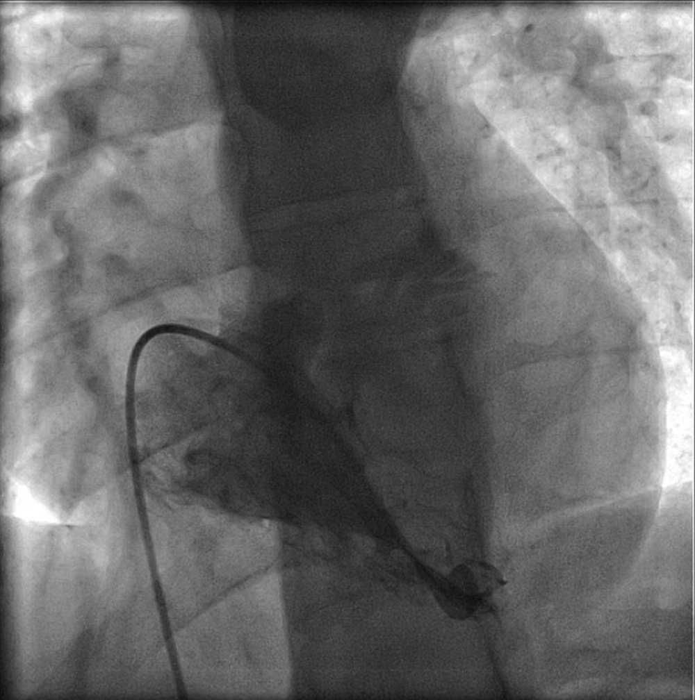
Ventriculography of Right Ventricle, Anteroposterior View
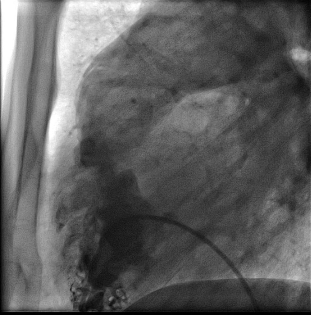
Figure 1.

Ventriculography of Right Ventricle, Anteroposterior View

Ventriculography of Right Ventricle, Lateral View
Figure 2.

Ventriculography of Right Ventricle, Lateral View

The patient underwent repair of the subpulmonic stenosis. Substantial RV muscle bundle resection was done through a right atrial approach with closure of the VSD and patch augmentation of the RVOT. The patent foramen ovale (PFO) was also closed. There were no operative or postoperative complications. A postoperative TEE demonstrated moderate RAE, moderate RVH, no RVOT obstruction, no VSD, and normal biventricular systolic function.

3. Discussion

DCRV is a congenital heart defect which is seen in 0.5% - 2% of congenital heart disease cases (2). The anomalous muscle bundle divides the ventricle into a low pressure outlet and high pressure inlet portion (2). There are various etiologies reported for the obstructive form of this disease. Increased blood flow among VSD causing supraventricular hypertrophy, hypertrophy of superior displaced moderator band, and septoparietal trabeculations in combination with acquired factors are 3 causal bases (5). While DCRV is mostly presented between 4 months to 20 years of age, our case was a 40-year-old adult (2). DCRV can be asymptomatic or as our case, presented with exertional dyspnea as the most prevalent presentation (2). Other presenting symptoms include angina, dizziness, and even syncope (6). These patients might have normal chest x-ray or even normal electrocardiograph if there is no significant ventricular shunt or significant obstruction (1). TTE and TEE are two simple diagnostic techniques. TEE is more capable than TTE in defining the details of DCRV, including the pressure gradient (2). Cardiac computed tomography, magnetic resonance imaging, and catheterization may also be required for complete characterization of this congenital heart anomaly in adults (7). While VSD is a common coexisted lesion, pulmonary valve stenosis, subaortic stenosis, Tetralogy of Fallot, double outlet right ventricle, and complete or corrected transposition of great vessels are other coexisting lesions (5). In addition to a restrictive perimembranous VSD, the reported case had RAE, mild TR, severe RAE, severe RVH, severe RVOT obstruction with excessive hypertrophy of the infundibulum, and right ventricular muscle bundles. Khoueiry et al. reported a 45-year-old female with persistent dyspnea on exertion for 2 years (3). Her TTE showed small VSD and colour Doppler TEE showed turbulence with high velocity jet in proximal portion of right ventricle as well as an abnormal muscle bundle separating in right ventricle (3). In magnetic resonance imaging, they confirmed a VSD lesion without significant shunt and double chamber right ventricle (3). Park et al. reported a case of asymptomatic isolated DCRV in adulthood (8). A turbulent flow jet with mosaic pattern was present and moderate tricuspid regurgitation, right ventricular dilatation and hypertrophy were found (8). TEE revealed an anomalous muscle bundle dividing right ventricle to two chambers (8). They did not find any shunt between right and left parts of the heart and confirmed their diagnosis with MRI (8).
DCRV has good survival and low long-term morbidity (9). As the obstruction in DCRV is progressive and can lead to heart failure, surgical treatment should be generally considered (5). Anomalous muscle resection can be made through a right ventriculotomy, right atriotomy or combined transatrial-transpulmonary approach (5). Ventriculotomy is rarely used and might be necessary in patients with severe right ventricular obstruction (5, 10). Long-term prognosis of intracardiac repair of DCVR is excellent (10). In conclusion, muscular subpulmonic stenosis is a rare condition in adult patients. Full echocardiographic examination of the right ventricle, including RVOT is essential for the diagnosis. Invasive diagnostic studies and CMR can further define the anatomy and pressure gradients to confirm diagnosis and guide therapeutic decisions. Surgical correction appears to be a successful strategy for management of this rare condition in adults.

Footnotes

  • Authors’ Contributions:Study concept and design: Soheila Chamanian, Majid Jalalyazdi, Nima Nakhaii, and Reza Jafarzadeh Esfehani; Acquisition of data: Soheila Chamanian, Majid Jalalyazdi, and Nima Nakhaii; Drafting of the manuscript: Reza Jafarzadeh Esfehani; Critical revision of the manuscript for important intellectual content: Majid Jalalyazdi; Study supervision: Majid jalalyazdi.

References

  • 1.
    Danilowicz D, Ishmael R. Anomalous right ventricular muscle bundle: clinical pitfalls and extracardiac anomalies. Clin Cardiol. 1981;4(3):146-50. [PubMed ID: 7261489].
  • 2.
    Hoffman P, Wojcik AW, Rozanski J, Siudalska H, Jakubowska E, Wlodarska EK, et al. The role of echocardiography in diagnosing double chambered right ventricle in adults. Heart. 2004;90(7):789-93. [PubMed ID: 15201250]. https://doi.org/10.1136/hrt.2003.017137.
  • 3.
    Khoueiry G, Bhat T, Tantray M, Meghani M, Abi Rafeh N, Abdallah M, et al. A rare case of double-chambered right ventricle associated with ventricular septal defect and congenital absence of the pulmonary valve. Clin Med Insights Circ Respir Pulm Med. 2014;8:1-3. [PubMed ID: 24683307]. https://doi.org/10.4137/CCRPM.S11174.
  • 4.
    Lucas RJ, Varco RL, Lillehei CW, Adams PJ, Anderson RC, Edwards JE. Anomalous muscle bundle of the right ventricle. Hemodynamic consequences and surgical considerations. Circulation. 1962;25:443-55. [PubMed ID: 14467101].
  • 5.
    Darwazah AK, Eida M, Bader V, Khalil M. Surgical management of double-chambered right ventricle in adults. Cardiovasc. Dis. 2011;38(3):301.
  • 6.
    McElhinney DB, Chatterjee KM, Reddy VM. Double-chambered right ventricle presenting in adulthood. Ann Thorac Surg. 2000;70(1):124-7.
  • 7.
    Kha LC, Cassano-Bailey A, Cleverley K, Sud M, Strzelczyk J, Jassal DS. Multimodality cardiac imaging of a double chambered right ventricle with intrapulmonary shunting: a case report. BMC Res Notes. 2012;5:516. [PubMed ID: 22999111]. https://doi.org/10.1186/1756-0500-5-516.
  • 8.
    Park JG, Ryu HJ, Jung YS, Kim KJ, Lee BR, Jung BC, et al. Isolated double-chambered right ventricle in a young adult. Korean Circ J. 2011;41(5):272-5. [PubMed ID: 21731569]. https://doi.org/10.4070/kcj.2011.41.5.272.
  • 9.
    Kahr PC, Alonso-Gonzalez R, Kempny A, Orwat S, Uebing A, Dimopoulos K, et al. Long-term natural history and postoperative outcome of double-chambered right ventricle--experience from two tertiary adult congenital heart centres and review of the literature. Int J Cardiol. 2014;174(3):662-8. [PubMed ID: 24836684]. https://doi.org/10.1016/j.ijcard.2014.04.177.
  • 10.
    Hachiro Y, Takagi N, Koyanagi T, Morikawa M, Abe T. Repair of double-chambered right ventricle: surgical results and long-term follow-up. Ann Thorac Surg. 2001;72(5):1520-2. [PubMed ID: 11722036].

Copyright

Copyright © 2015, Ahvaz Jundishapur University of Medical Sciences. This is an open-access article distributed under the terms of the Creative Commons Attribution-NonCommercial 4.0 International License (http://creativecommons.org/licenses/by-nc/4.0/) which permits copy and redistribute the material just in noncommercial usages, provided the original work is properly cited.

Similar Articles

5
May
2012

Simultaneous Transcatheter Closure of Ventricular Septal Defect and Pulmonary Valvuloplasty

Mostafa Behjati-Ardakani,
Mahdi Haddadzadeh,
Shahrokh Rajaei

Behjati-Ardakani M, Haddadzadeh M, Rajaei S. Simultaneous Transcatheter Closure of Ventricular Septal Defect and Pulmonary Valvuloplasty. Inn J Pediatr. 2012;22(4):. doi:

24
Jan
2017

Double Orifice Fissured Subaortic Membrane in the Adult

Giordano Zampi,
marzia cottini,
Amedeo Pergolini,
Vincenzo Polizzi,
Francesco Musumeci

Zampi G, cottini M, Pergolini A, Polizzi V, Musumeci F. Double Orifice Fissured Subaortic Membrane in the Adult. Int J Cardiovasc Pract. 2017;2(1):e130058. doi: https://doi.org/10.21859/ijcp-020107

12
Nov
2022
Int J Cardiovasc Pract

Prevalence and Hemodynamic Consequences of Mitral Valve Abnormalities in Atrial Septal Defect

Maryam Shojaeifard,
Sheida Keshavari,
Saideh Jamshidi,
Hooman Bakhshandeh,
Mohammad Ali Ghaznavai,
Hossein Dehghani Mohammad Abadi
,et al.

Shojaeifard M, Keshavari S, Jamshidi S, Bakhshandeh H, Ghaznavai MA, et al. Prevalence and Hemodynamic Consequences of Mitral Valve Abnormalities in Atrial Septal Defect. Int J Cardiovasc Pract. 2021;6(2):e131707. doi: https://doi.org/10.5812/intjcardiovascpract-131707

1
Sep
2020

Transcatheter Device Closure of Complex Paravalvular Leak after Bioprosthetic and Mechanical Pulmonary Valve replacement in Two Adult Patients

Zahra Khajali,
Ata Firouzi,
Maryam Keshavarz Hedayati,
Fateme Jorfi,
Mohamad Sadra Nazari

Khajali Z, Firouzi A, Keshavarz Hedayati M, Jorfi F, Nazari MS. Transcatheter Device Closure of Complex Paravalvular Leak after Bioprosthetic and Mechanical Pulmonary Valve replacement in Two Adult Patients. Int Cardiovasc Res J. 2020;14(3):e102248. doi:

30
Jun
2021

Percutaneous intervention of pulmonary supravalvular stenosis in two patients.

Zahra Khajali,
Ata Firouzi,
Mohamad Sadra Nazari,
Fateme Jorfi,
Maryam Keshavarz Hedayati

Khajali Z, Firouzi A, Nazari MS, Jorfi F, Keshavarz Hedayati M. Percutaneous intervention of pulmonary supravalvular stenosis in two patients.. Int Cardiovasc Res J. 2021;15(2):e107798. doi:

Download PDF401.92 KB

Crossmark

Crossmark

Checking

Share on
Cited by
Metrics

Ordering Reprints

Articles are published under the Creative Commons license stated on each article. No permission or royalty fee is required for uses permitted by that license. CCC handles optional bulk and customized reprint orders. Any quotation covers production and delivery services only, not copyright permission. > Request Reprints from CCC 

Search Relations

Author(s):

Related Articles