
1. Background
Oral squamous cell carcinoma (OSCC) is the most common head and neck malignancy, and squamous cell carcinoma (SCC) accounts for more than 90% of oral and oropharyngeal malignancies, mainly affecting the tongue and the floor of the mouth. OSCC can spread rapidly, expand locally, and metastasize to other body areas through the lymphatic system. Despite various management strategies, including surgery, radiation, and chemotherapy, fewer than half of affected individuals survive for more than 5 years (1, 2). Given the poor prognosis of OSCC, understanding its etiology and implementing appropriate measures to prevent its development or control it at early or precancerous stages are essential. Risk factors for OSCC include cigarette or pipe smoking, use of tobacco products through sniffing or chewing, excessive alcohol consumption, sun exposure, a family history of cancer, inadequate nutrition, and immunosuppression (3). Given the role of poor oral hygiene, which can lead to periodontitis, in the development of oral tumors, studies investigating potentially involved pathogens have identified many bacterial species in the oral cavity.
Among these bacteria, the anaerobes Fusobacterium nucleatum and F. periodonticum have been implicated in the development of OSCC (4), most likely through mechanisms such as promoting cell proliferation and angiogenesis, disrupting normal apoptosis, facilitating invasion and metastasis, and contributing to chronic inflammation that leads to carcinogenesis (5). In addition to the enrichment of certain bacteria in OSCC samples compared with controls (6), the abundance of some bacteria, such as F. periodonticum, Parvimonas micra, Streptococcus constellatus, Haemophilus influenzae, and Filifactor alocis, has been associated with disease stage, with a higher prevalence in advanced stages (stage V vs stage I) (7). In addition to environmental factors, molecular changes in epithelial cells, including the inactivation of cancer suppressor genes and the overexpression of proto-oncogenes, have been shown to result in tumor cell proliferation and distant metastasis (8). Accordingly, research has focused on bacterial products and metabolites that can induce genetic changes in oral epithelial cells. Porphyromonas gingivalis stimulates the production of inflammatory cytokines, promotes cell proliferation, invasion, and migration, and ultimately leads to apoptotic cell death (9).
2. Objectives
Given the association between microorganisms and the development and staging of OSCC, as well as the limited number of studies examining the role of P. gingivalis in OSCC, the present study aimed to investigate the expression of carcinogenesis-related genes, including c-Myc and p53, in DPS-7 human dental pulp stem cells exposed to P. gingivalis as an in vitro oral cell model.
3. Methods
3.1. Cultivation of P. gingivalis and Identification of the Microorganism by Polymerase Chain Reaction
The standard strain of P. gingivalis (ATCC 33277) was cultured on Brucella agar base (code 111291; Merck, Germany) supplemented with 5% defibrinated sheep blood. To enrich the medium, 5% horse serum, 5 mg/mL hemin, and 5 mg/mL vitamin K were added. Colistin was added to render the culture medium selective for P. gingivalis. The plates were then incubated in an anaerobic jar containing a type A gas pack, catalyst, and indicator. The standard Escherichia coli strain (ATCC 25922) was incubated in LB broth (DNAbiotech Co., Iran) at 37°C. After 28 - 74 hours of incubation at 37°C, a polymerase chain reaction (PCR) assay was performed for accurate identification. First, bacterial genomic DNA was extracted using a Takara extraction kit (TaKaRa, Japan) according to the manufacturer's instructions. Two pairs of specific primers (Table 1) were used to amplify 404- and 141-base pair fragments of the 16S rRNA gene for P. gingivalis and E. coli, respectively (10).
Table 1.
Specific Primers for Molecular Detection of Porphyromonas gingivalis and Escherichia coli
| 16S rRNA | Fragment | Primer 1 | Primer 2 |
|---|---|---|---|
| Porphyromonas gingivalis | 404 bp | AGGCAGCTTGCCATACTGCG | ACTGTTAGCAACTACCGATGT |
| Escherichia coli | 141 bp | AAAGGAGACTGCCAGTGATA | AGGTCGCTTCTCTTTGTATG |
3.2. Cell Culture
Human dental pulp stem cells (DPS-7) were obtained from the Cell Bank of the Iranian Biological Resource Center (IBRC C10371) (characteristics are listed in Table 2). DPS-7 is a nonmalignant dental pulp stem cell line established from a human dental pulp biopsy and was used in this study as an in vitro oral-derived cell model to evaluate the molecular effects of P. gingivalis exposure on carcinogenesis-related pathways. Cells were cultured in RPMI-1640 medium supplemented with 10% fetal bovine serum (FBS; Atlas Biologicals, Fort Collins, CO, USA), 2 mM glutamine, 100 μg/mL penicillin, 0.1 mM nonessential amino acids, and 1 mM sodium pyruvate. DPS-7 cells were maintained in a humidified incubator with 5% CO2 at 37°C.
Table 2.
Characteristics of Dental Pulp Stem Cells (DPS-7)
| Characteristic | Description |
|---|---|
| Cell line name | DPS-7 |
| Cell line type | Human dental pulp stem cells |
| Accession cell no. | IBRC C10371 |
| Organism | Homo sapiens |
| Breed | Unknown |
| Summary | Established from a biopsy of the dental pulp of a 25-year-old male |
| Morphology | Fibroblast-like |
| Medium | Alpha-MEM medium + 20% fetal bovine serum + 2 mM L-glutamine |
| Subculture | Split subconfluent cultures (70% - 80%) 1:2 to 1:3, ie, seeding at 2 - 4 × 10,000 cells/cm2 using 0.25% trypsin and 0.02% EDTA. |
| Incubation | At 37°C with 5% CO2 |
| Storage | 90% FBS + 10% DMSO at about 1 × 10⁶ cells/vial |
| Mycoplasma | Negative |
| Bacteria/fungi | Negative |
| Viruses | Negative for HBV and EBV by PCR and HIV and HCV by RT-PCR |
| Interspecies identification | Confirmed as human by PCR method |
| Release condition | General |
3.3. Treatment of DPS-7 Cells With P. Gingivalis or E. Coli
Cells were divided into three groups: 1) DPS-7 cells, 2) DPS-7 cells exposed to P. gingivalis (P.DPS-7), and 3) DPS-7 cells exposed to E. coli (E.DPS-7). Bacterial suspensions were adjusted to approximately the 0.5 McFarland standard before inoculation, corresponding to an estimated concentration of approximately 1 × 10⁸ CFU/mL. DPS-7 cells were exposed to bacteria at an approximate cell-to-bacteria ratio of 1:100 for a single continuous 42-hour incubation period at 37°C. After incubation, cells were washed with phosphate-buffered saline (PBS) to reduce nonadherent extracellular bacteria and then cultured in fresh antibiotic-containing medium. The same washing procedures, medium replacement, and antibiotic exposure conditions were applied to all experimental groups, including controls.
3.4. Nucleic Acid Extraction and cDNA Reverse Transcription
Total RNA was extracted from DPS-7 cells using a commercial Biosol kit (Fregene Poyesh, Tehran, Iran) according to the manufacturer's instructions. RNA was separated from DNA and cellular proteins by adding 200 µL chloroform, followed by centrifugation at 120000 rpm at 4°C for 15 minutes; the aqueous phase containing RNA was collected. Finally, 30 µL DEPC-treated water was added to dissolve the RNA precipitate, and the quantity and purity of the extracted RNA were confirmed using a NanoDrop device (ND-1000; BioRad, USA) at a wavelength of 260 nm. RNA quality was assessed by electrophoresis on a 1% agarose gel. On gel examination, the presence of 18S and 28S rRNA bands confirmed RNA from DPS-7 cells, whereas the presence of 16S and 23S bands indicated bacterial RNA. Reverse transcription was performed using a cDNA synthesis kit (TAKARA Co.). For further analysis, DNA and synthesized cDNA were stored at -20°C.
3.5. Quantitative PCR for Oncogenic and Tumor Suppressor Genes
To investigate the effect of P. gingivalis on the carcinogenicity of infected cells, the c-Myc and p53 genes were quantitatively evaluated as an oncogene and a tumor suppressor, respectively. Real-time PCR was performed using SYBR Green PCR (TaKaRa, Japan) on an ABI 7500 real-time PCR instrument (Applied Biosystems, Carlsbad, CA, USA). Primers were designed using Allele ID software (version 7.5), and their specificities were evaluated using the EMBL-EBI and NCBI BLAST databases. Primer sequences are presented in Table 3. Each 12.5-µL reaction contained 3 µL of cDNA template, 0.5 µM of the respective forward and reverse primers, 5.25 µL Power SYBR Green PCR Master Mix (Bioneer, Korea), and 2.5 µL nuclease-free water. Quantitative PCR was performed with an initial incubation at 95°C for 1 minute, followed by 40 cycles of denaturation at 95°C for 20 seconds and annealing/extension at 60°C for 35 seconds. All assays for each sample were performed in triplicate in a single batch, and the mean results of the quantitative PCR analyses were used for statistical assessment. The negative control in all analyses contained all components of the reaction mixture, excluding bacterial genomic DNA. Relative gene-expression levels were normalized to the β-actin housekeeping gene. Comparative analysis of gene-expression profiles among experimental groups was performed using relative Ct values obtained from real-time PCR amplification. Each experimental condition was independently evaluated in three biological replicates, and RT-PCR reactions were performed in technical replicates for each sample. Gene-expression differences among groups were analyzed using nonparametric methods.
Table 3.
Primer Sequences Used in the Present Study
| Genes | Forward Primer | Reverse Primer |
|---|---|---|
| c-Myc (128 bp) | CCTGGTGCTCCATGAGGAGAC | CAGACTCTGACCTTTTGCCAGG |
| p53 (128 bp) | CCTCAGCATCTTATCCGAGTGG | TGGATGGTGGTACAGTCAGAGC |
| β-actin | CCTCGCCTTTGCCGATCC | GGATCTTCATGAGGTAGTCAGTC |
3.6. Statistical Analysis
Statistical analyses were performed using IBM SPSS Statistics for Windows, version 22.0 (IBM Corp., Armonk, NY, USA). Data are presented as mean ± standard deviation (SD). Comparisons among the three experimental groups were initially performed using the nonparametric Kruskal-Wallis test. When significant overall differences were detected, pairwise comparisons were performed using the Mann-Whitney U test. A P value of less than 0.05 was considered statistically significant. No formal multiple-comparison correction was applied because of the exploratory nature of the study.
4. Results
4.1. Confirmation of Bacterial Purity
Although the selected bacterial strains were standard, a specific PCR assay was performed to confirm the absence of contamination with other bacteria. A 404-base pair fragment of the 16S rRNA gene was amplified for P. gingivalis (Figure 1A), and a 141-base pair fragment of the 16S rRNA gene was amplified for E. coli (Figure 1B), confirming the purity of the bacterial strains.
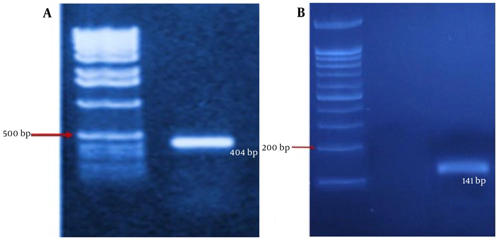
Figure 1.
PCR amplification confirming bacterial identification and purity; A, amplification of the 16S rRNA gene specific for P. gingivalis; B, amplification of the 16S rRNA gene specific for E. coli. M: DNA ladder.
4.2. Cell Culture and Treatment With Bacteria
Cultured spindle-shaped DPS-7 dental pulp stem cells were observed adhering to the bottom of the plate under an inverted microscope (Figure 2).

Figure 2.
Morphology of cultured DPS-7 dental pulp stem cells observed under an inverted microscope. DPS-7 cells exhibited spindle-shaped fibroblast-like morphology during culture.
4.3. Expression Levels of c-Myc and p53 Genes in Cells Treated with P. Gingivalis Bacteria and E. Coli
4.3.1. Quantitative and Qualitative Confirmation of RNA Extracted From DPS-7 Cells After Treatment with P. gingivalis Bacteria and E. coli
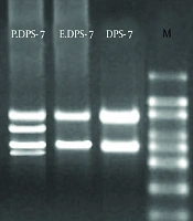
In the P.DPS-7 group, the presence of four ribosomal bands (two ribosomal bands corresponding to DPS-7 cells and two bands associated with P. gingivalis) suggested bacterial association in exposed DPS-7 cell samples following incubation with P. gingivalis (Figure 3). In contrast, the E.DPS-7 control group displayed only two ribosomal bands from DPS-7 cells, indicating that E. coli does not influence oral cancer development. The quantity and purity of the extracted RNA were 452.3, 319.7, and 286.5 ng/µL for DPS-7, E.DPS-7, and P.DPS-7, respectively.
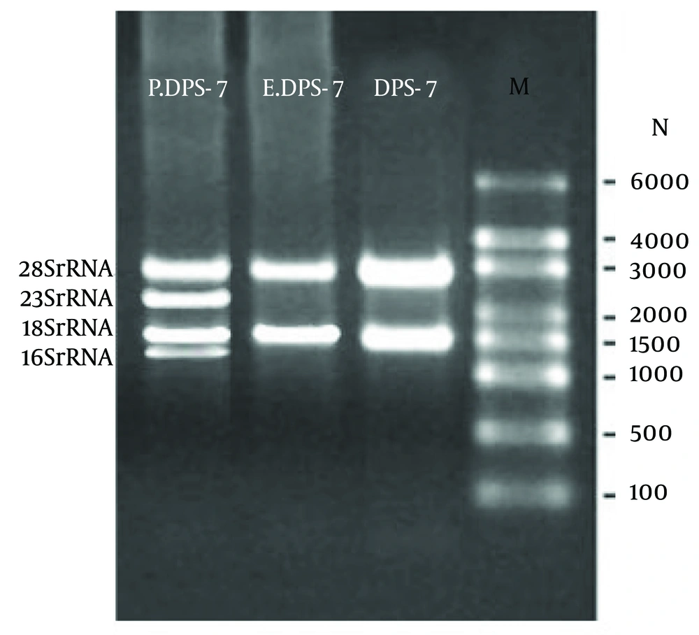
Figure 3.
Agarose-gel electrophoresis of extracted RNA samples from DPS-7 cells following bacterial exposure. The presence of 18S and 28S ribosomal RNA bands confirmed RNA integrity in DPS-7 cell samples. Additional bacterial ribosomal RNA bands were observed in samples exposed to P. gingivalis. M: RNA marker ladder.
4.3.2. cDNA Construction and Optimization of PCR Conditions Using the Designed Primers
Using the described method, c-Myc and p53 yielded 128 base pairs, whereas the reference gene, β-actin, yielded 626 base pairs (Figure 4).
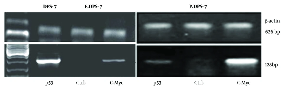
Figure 4.
Agarose-gel electrophoresis of RT-PCR products for c-Myc, p53, and β-actin genes obtained from DPS-7 experimental groups. Amplified products corresponded to the expected fragment sizes for each target gene.
4.3.3. RT-PCR
The Kruskal-Wallis test demonstrated statistically significant differences among the experimental groups for both c-Myc and p53 expression levels. Subsequent pairwise Mann-Whitney U tests showed significantly increased c-Myc expression and significantly decreased p53 expression in the P.DPS-7 group compared with untreated DPS-7 cells and E.DPS-7 cells (P < 0.001). No statistically significant difference was observed between untreated DPS-7 cells and E.DPS-7 cells (P > 0.05) (Figure 5).
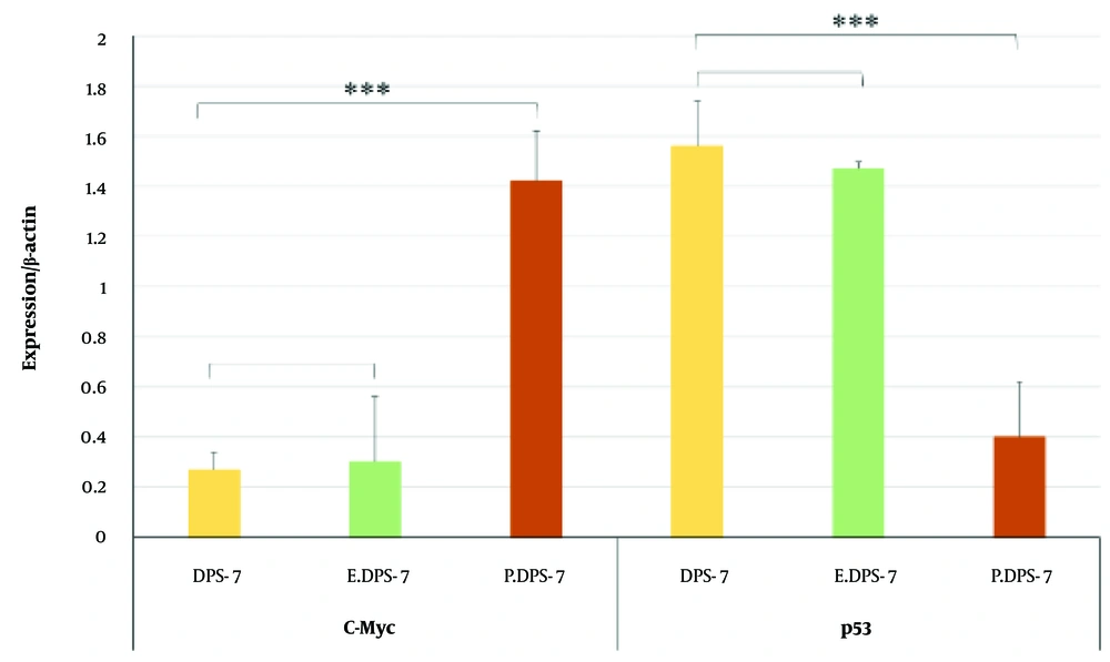
Figure 5.
Relative gene-expression levels of c-Myc and p53 were calculated using the 2-ΔΔCt method after normalization to β-actin and are presented as mean ± SD from three independent experiments (* P < 0.05, ** P < 0.001, *** P < 0.0001, **** P < 0.00001).
5. Discussion
In the present study, the results demonstrated that P. gingivalis increased c-Myc and decreased p53 gene expression in DPS-7 cell lines, supporting the hypothesis that exposure to P. gingivalis may induce molecular alterations associated with carcinogenesis-related pathways. The association between compositional changes in oral bacteria and OSCC, as well as the mechanisms by which oral bacteria influence oral cancer progression, is a critical area for identifying potential strategies for early diagnosis and effective treatment. Therefore, the results of the present study indicate that P. gingivalis may play a role in oral cancer development and could serve as a basis for future clinical development of preventive, early diagnostic, and therapeutic protocols. Several studies have suggested that repeated exposure of cells to P. gingivalis can lead to cancer, especially in the orodigestive system (11-13). Therefore, DPS-7 cells were exposed to P. gingivalis for a continuous 42-hour incubation period to evaluate the molecular response to bacterial exposure and detect the carcinogenic effect of this bacterium. Notably, P. gingivalis uses lipid microdomains of the target cell membrane and cytoskeletal functions to facilitate its internalization into cells (14). Because two ribosomal bands, 18S rRNA and 28S rRNA, are associated with DPS-7 cells, whereas two bands, 16S rRNA and 23S rRNA, are associated with P. gingivalis, the presence of all four ribosomal bands in the P.DPS-7 group suggested bacterial association with the exposed DPS-7 cells. This finding suggests the involvement of P. gingivalis in oral cancer progression and indicates increased oncogene expression and decreased tumor suppressor gene expression in P. gingivalis-infected DPS-7 cells.
To the best of our knowledge, these findings suggest that P. gingivalis may contribute to molecular changes associated with oral carcinogenesis. Although the association of P. gingivalis with OSCC and other orodigestive cancers has been frequently reported, several mechanisms have been suggested in cellular and clinical studies (11, 12). Most studies have suggested that prolonged exposure to P. gingivalis not only contributes to oral cancer through chronic inflammation but also promotes cancer migration and invasiveness and induces resistance to chemotherapy. This has been documented through the upregulation of matrix metalloproteinases (MMPs-1 and MMPs-2d) and inflammatory cytokines such as interleukin-8 in OSC-20 and SAS cells (15, 16). Geng and colleagues exposed human immortalized oral epithelial cells to P. gingivalis and found that persistent exposure to the bacterium caused morphological changes, increased proliferative ability (higher S-phase fraction), enhanced cell migration, and promoted invasive properties, with multiple tumor-related genes identified as involved (17, 18). These results are consistent with the findings of the present study regarding the associative role of P. gingivalis in OSCC; however, none of the previous studies specifically focused on the two tumor suppressor genes and oncogenes examined here.
Other studies have investigated human and murine cells and pre-established cell lines to elucidate the mechanism of P. gingivalis in OSCC. A review of studies indicated its role in three phases, including epithelial-mesenchymal transition of malignant cells, neoplastic proliferation, and tumor invasion through different mechanisms (19). Another review highlighted mechanisms including sustained proliferative signaling through activation of the PI3K/AKT pathway; evasion of apoptosis through activation of the Jak/STAT, PI3K/AKT, and nucleoside diphosphate kinase pathways; evasion of growth suppressors by inactivating phosphatase and tensin homolog and p53; sustained angiogenesis through induction of vascular endothelial growth factor and Ephrin B2; avoidance of immune destruction through apoptosis of T and B cells; and invasion/metastasis by inducing epithelial-mesenchymal transition (20).
Wild-type p53, known as the guardian of the genome, is a tumor suppressor protein that protects genomic integrity by regulating the cell cycle and controlling cell death genes in response to DNA damage. It is widely used as an important tumor marker in clinical practice across different cancers (21). It has also been suggested as a useful marker for diagnosing premalignant lesions and OSCC and for predicting a poor prognosis in such patients (22). Other studies have documented that suppression and downregulation of the guardian of the genome, p53, by any factor can lead to OSCC by blocking apoptosis and through other mechanisms (23, 24). This finding is consistent with the present study, in which we provided evidence that P. gingivalis is the contributing factor. Additional evidence supports the role of p53 in gingival epithelial cell progression. As determined in the review by Kuboniwa and colleagues, P. gingivalis induced changes in the levels and phosphorylation of p53 and PI3K in gingival epithelial cells (25), which is consistent with the results of the present study, although that review did not focus exclusively on OSCC.
Another important factor investigated in the present study was c-Myc, an oncoprotein and transcription factor that drives activating mutations in cancer-causing proteins. c-Myc is not only involved in tumor initiation and growth but also plays a critical role in tumor progression, proliferation, and invasion (26). Studies on OSCC have also revealed that c-Myc overexpression significantly affects cell survival, proliferation, and tumor growth (27). Among the various factors suggested to contribute to c-Myc overexpression or upregulation in OSCC pathogenesis, P. gingivalis had not been previously evaluated; we documented this here for the first time. In another study, Xu et al. reported that c-Myc was upregulated following stimulation of oral and esophageal squamous cell carcinoma with P. gingivalis, which, along with NF-κB (P65) in the nucleus, showed the vital role of P. gingivalis in the progression and development of esophageal squamous cell carcinoma (28).
The present study was novel in examining c-Myc and p53 in human-extracted DPS-7 cells after exposure to P. gingivalis, compared with E. coli and a control group. It demonstrated molecular alterations associated with carcinogenesis-related pathways following exposure to P. gingivalis. However, this study also had some limitations. We did not directly measure apoptosis using flow cytometry, but comparisons of apoptosis levels between contaminated and control groups could provide further insights. Additionally, including other bacteria, such as F. nucleatum, would have allowed a comparative assessment to determine which bacterial species exerted a greater influence on OSCC. In addition, intracellular bacterial viability and internalization were not directly confirmed using invasion assays, fluorescence microscopy, or antibiotic protection assays. Therefore, the observed gene-expression changes may reflect responses to bacterial exposure rather than exclusively intracellular bacterial activity. Finally, this was an in vitro study, and further in vivo studies in animal models, followed by clinical studies in humans, are required to reach definitive conclusions.
5.1. Conclusions
The present study demonstrated that exposure of DPS-7 dental pulp stem cells to P. gingivalis altered the expression of c-Myc and p53 genes, suggesting a possible role for this bacterium in molecular pathways associated with oral carcinogenesis. These findings help clinicians better understand the role of this oral microbiota in OSCC, an aggressive tumor, and provide a foundation for future studies investigating the clinical significance of this association. These findings may also provide a basis for future investigations exploring the relationship between oral microbiota and carcinogenesis-related molecular pathways.