
1. Background
Based on cumulative statistics from the Iranian Ministry of Health during 1986 - 2011, 23,153 people with human immunodeficiency virus (HIV) were registered in Iran; of these, 91.5% were men, 8.5% were women, and nearly 46.5% were in the 25 - 34-year age group. Of these individuals, 3,053 later acquired acquired immunodeficiency syndrome (AIDS), and 4,311 died. Based on the epidemiological model of the Joint United Nations Programme on HIV/AIDS (UNAIDS) and the World Health Organization (WHO), which uses a special formula for the "estimation of real cases based on recorded cases," 80,000 HIV-positive people are estimated to live in Iran. However, unofficial estimates suggest that this number may be as high as 120,000 (1, 2). Individuals with AIDS are highly susceptible to opportunistic infections, including those caused by actinomycetes, because HIV impairs humoral and cellular immune mechanisms.
Actinomycetes are prokaryotes that exist as free-living or saprophytic microorganisms and sometimes form symbiotic relationships with plants. These microorganisms can be isolated from various environmental ecosystems, such as soil, and constitute a significant component of the human body microflora. These bacteria include important genera such as Actinomyces, Nocardia, Streptomyces, and Micromonospora. Among these bacteria, Streptomyces is particularly important, accounting for nearly 60% of all actinomycetes. Genera within the actinomycetes, such as Actinomyces and Nocardia, can cause various infectious diseases, including actinomycosis, nocardiosis, granulomatosis, multiple abscesses, fibrosis, central nervous system infections, and disseminated infections. Furthermore, bacteria such as Actinomyces spp. can directly or indirectly cause oral and dental diseases (3, 4). However, treatment outcomes in patients with HIV are often suboptimal.
For example, the frequency of nocardiosis in HIV-positive patients has increased from 0.3% to 1.85%. Moreover, the adverse effects of current antimicrobials have raised serious public health concerns. Therefore, interest in herbal medicine for the treatment of infections in HIV-positive patients and patients with AIDS has increased in recent years. Thyme and ajwain are two widely studied medicinal herbs with antimicrobial properties. These herbs are widely used in Iranian cuisine and traditional medicine.
Thyme, or Shirazi thyme (Zataria multiflora), belongs to the order Lamiales and the family Lamiaceae and is primarily cultivated in Southwestern Asia. The dried, powdered leaves of the plant are commonly used as a spice for culinary, ornamental, and medicinal purposes (5). The major chemical constituents of thyme include various EOs, flavonoids, phenolic acids, and triterpenes. The EOs of thyme are primarily composed of thymol and carvacrol, which exhibit strong antimicrobial and anti-inflammatory activities. These flavonoids have antioxidant, anti-inflammatory, and antimicrobial properties (6). Other primary components of thyme include triterpenes, tannins, and saponins. These compounds have medicinal benefits (7).
Ajwain (Trachyspermum ammi), also known as ajowan, ajowan caraway, bishop's weed, carom, and thymol seeds, belongs to the order Apiales and the family Apiaceae (8) and is mostly cultivated in Asia (9). The plant leaves and seed-like fruits are used as spices for culinary, medicinal, and preservative purposes. The EO of ajwain fruits primarily contains thymol, γ-terpinene, p-cymene, and terpenoids with antimicrobial, anti-inflammatory, antifungal, and anti-Trichophyton activities.
2. Objectives
Given the importance of oral infections in HIV-infected patients and the lack of antimicrobial susceptibility schemes for oral actinomycetes in Iran, this study aimed to evaluate the susceptibility of actinomycetes isolated from the oral cavity of HIV-infected patients in Tehran, Iran, to 14 commonly used antimicrobial agents and two traditional herbal essential oils (EOs).
3. Methods
3.1. Samples
This study included 285 oral samples. Of these, 200 samples were collected from HIV-infected participants with CD4 lymphocyte counts of 200 cells μL-1 or greater and no recent history of antimicrobial or antifungal use before sampling. In addition, 85 samples were collected from healthy participants who met similar inclusion criteria. Samples were previously collected from the oral cavities of the participants, including lesions, gums, tooth grooves, periodontal areas, and periapical areas, at the Southern Health Center, Tehran University of Medical Sciences, Tehran, Iran (unpublished data).
3.2. Isolation and Phenotypic Identification
Briefly, samples were cultured on blood agar (BA) and brain-heart infusion agar (BHIA) (Merck, Germany) and incubated aerobically and anaerobically using type-A gas packs at 37°C for at least 7 days in a shaker incubator. For Nocardia spp., isolates were subcultured onto paraffin agar and incubated for 14 days. Suspected colonies were then stained using the Gram and Ziehl-Neelsen methods. All isolates were further identified using biochemical assays and confirmed using molecular methods, including polymerase chain reaction (PCR) and nucleotide sequencing targeting 16S rRNA genes.
Amplification was performed using the universal primers 27F (5'-AGA GTT TGA TCC TGG CTC AG-3') and 1492R (5'-TAC GGG TAC CTT GTT ACG ACT T-3'). Edited sequences were submitted to GenBank (https://www.ncbi.nlm.nih.gov/genbank) under accession numbers PQ621735.1, PQ621739.1, PQ6333383.1, PQ634341.1, PQ634379.1, PQ634397.1, and PQ634819.1.
3.3. Antimicrobial Susceptibility Test
Antimicrobial susceptibility testing (AST) of the isolates was performed against 14 commonly used antimicrobial agents, including amikacin, amoxicillin, amoxicillin-clavulanic acid (co-amoxiclav), ciprofloxacin, clindamycin, erythromycin, gentamicin, linezolid, nalidixic acid, nitrofurantoin, penicillin G, rifampin, tetracycline, and trimethoprim-sulfamethoxazole (cotrimoxazole), as well as the EOs of two medicinal herbs, thyme and ajwain.
3.4. Kirby-Bauer Method
In this study, the Kirby-Bauer disk diffusion method on Mueller-Hinton agar (Merck, Germany) was used to assess the antimicrobial susceptibility of the isolates to the selected antimicrobials, based on guidelines from the Clinical and Laboratory Standards Institute (CLSI, USA). Antimicrobial disks were provided by Padtan Teb, Iran. Results were recorded as susceptible (S), intermediately resistant (I), or resistant (R), based on the literature and CLSI-reported zone-diameter breakpoints.
The CLSI document M24, Susceptibility Testing of Mycobacteria, Nocardia, and Other Aerobic Actinomycetes, provides standardized guidelines for susceptibility testing of Nocardia spp., primarily based on the broth microdilution method. However, standardized disk diffusion breakpoints are not available for several genera of actinomycetes in this study, such as Streptomyces, Nocardiopsis, and Saccharopolyspora. In the absence of specific breakpoints, interpretive criteria were adopted from available CLSI guidance, where applicable, and from previously published studies on aerobic actinomycetes. These interpretations were applied cautiously. Based on these susceptibility categorizations, MDR was defined as non-susceptibility to at least one agent in three or more antimicrobial classes.
3.5. Agar Well Diffusion Method
First, reference strains of Staphylococcus aureus PTCC 1917, Klebsiella pneumoniae PTCC 1859, Acinetobacter baumannii PTCC 1919, and Enterococcus faecium PTCC 1821 were cultured on the surface of agar plates with 6 - 8-mm punched wells. A suspension of the isolated actinomycetes in brain-heart infusion (BHI) broth was then transferred into the wells. Plates were incubated at 37°C for 24 hours (2, 10).
3.6. Checkerboard Assay
The checkerboard assay was used to investigate possible synergistic or antagonistic effects of various combinations of antimicrobials with the herbal EOs. These interactions were assessed using the fractional inhibitory concentration (FIC) index, calculated as FICA + FICB = FIC index. In general, FIC values of 0.5 or less indicated synergistic effects, values between 0.5 and 1 indicated additive effects, values between 1 and 4 indicated indifferent effects, and values of 4 or greater indicated antagonistic effects.
In this study, the EO of each herb, Z. multiflora and T. ammi, was assessed in various combinations with five antimicrobials: amikacin, ciprofloxacin, gentamicin, linezolid, and nalidixic acid. Therefore, 10 experimental groups, consisting of 5 antimicrobials × 2 EOs, were assessed in the checkerboard assay. Standard strains of N. asteroides and S. griseus were used as positive controls (11).
3.7. Preparation of the Essential Oils
In this study, standard EOs of Z. multiflora and T. ammi were purchased from Barij Essence, Iran. All EOs were provided with certificates of chemical analysis. Dimethyl sulfoxide (DMSO) was used as the solvent for the EOs. A series of solubility trials was conducted to determine the optimal concentrations for complete dissolution of the EOs in DMSO. Then, 10 μL of each Z. multiflora and T. ammi EO was transferred into sterile tubes, mixed with 900 μL of DMSO, and vortexed until a clear solution formed. These stock solutions were stored until use.
3.8. Cultivation of Bacteria on Culture Media
The isolated actinomycetes were cultured on Mueller-Hinton agar for 24 hours. Colonies were regularly assessed for purity to prevent contamination. The broth macrodilution technique was used to assess the minimum inhibitory concentration (MIC) of the EOs against the isolated actinomycetes.
3.9. Assessment of the Minimum Inhibitory Concentration of the Plant Essential Oils
First, a series of EO dilutions was prepared in 96-well microplates from a stock solution at a concentration of 0.99 μL mL-1. Then, 100 μL of the bacterial suspension adjusted to the 0.5 McFarland standard was added to each well. Plates were incubated at 37°C for 24 hours, and the MICs of the EOs were assessed. The MIC was defined as the lowest concentration of EO in the first well with no visible turbidity.
3.10. Assessment of the Minimum Bactericidal Concentration of the Plant Essential Oils
To assess the minimum bactericidal concentration (MBC) of the EOs, 10-μL samples were collected from the wells immediately before and after the MIC well and subcultured onto BA plates. After 24 hours of incubation at 37°C, plates showing no visible bacterial growth were recorded. The MBC was defined as the lowest EO concentration that resulted in no growth on subcultures.
3.11. Assessment of the Minimum Inhibitory Concentration and Minimum Bactericidal Concentration of the Antimicrobials Against Actinomycetes
In this study, five antimicrobial agents, amikacin, ciprofloxacin, gentamicin, linezolid, and nalidixic acid, were used to assess their MICs and MBCs against the isolated actinomycetes. Briefly, bacterial suspensions were adjusted to the 0.5 McFarland standard and inoculated into 96-well microplates containing serial dilutions of the antimicrobials. The microplates were then incubated at 37°C for 24 hours, and the MIC and MBC of each antibiotic were calculated. To assess the MBC, 10 μL of the bacterial dilution from the well before the MIC well and 10 μL from the well after the MIC well were cultured on BA plates. After 24 hours of incubation at 37°C, plates were examined, and the antibiotic concentration without bacterial growth or colony production was reported as the MBC of the antibiotics for the isolated actinomycetes.
3.12. Statistical Analysis
In this case-control study, data were analyzed using SPSS version 26 (IBM, USA). The Shapiro-Wilk test was used to assess the normality of the distribution. Categorical variables were compared using the chi-square test or Fisher exact test, and continuous variables were analyzed using analysis of variance (ANOVA). A P value of ≤ 0.05 was considered statistically significant.
4. Results
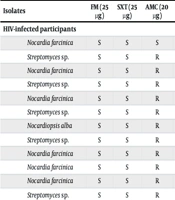
The AST was performed on 19 Actinomycete isolates from HIV-infected participants, including 6 strains of Streptomyces, 6 strains of N. farcinica, 6 strains of Nocardiopsis (including 2 strains of N. alba), and 1 strain of Saccharopolyspora, as well as 3 isolates from healthy participants, including 1 strain each of Streptomyces, N. flavescens, and Nocardiopsis. These isolates were tested against 14 commonly used antimicrobial agents. The antimicrobials included amikacin, amoxicillin, amoxicillin-clavulanic acid, ciprofloxacin, clindamycin, erythromycin, gentamicin, linezolid, nalidixic acid, nitrofurantoin, penicillin G, rifampin, tetracycline, and trimethoprim-sulfamethoxazole (cotrimoxazole) (Table 1).
Table 1.
Antimicrobial Susceptibility Test of the Actinomycetes Isolated from All Participants to 14 Antimicrobials Using the Disk Diffusion Method a
| Isolates | FM (25 µg) | SXT (25 µg) | AMC (20 µg) | LZ (30 µg) | CC (30 µg) | AN (30 µg) | CP (50 µg) | GM (15 µg) | NA (30 µg) | RA (5 µg) | TE (30 µg) | E (15 µg) | AMX (25 µg) | PEN (10 IU) |
|---|---|---|---|---|---|---|---|---|---|---|---|---|---|---|
| HIV-infected participants | ||||||||||||||
| Nocardia farcinica | S | S | S | S | R | S | S | S | R | S | S | R | R | R |
| Streptomyces sp. | S | S | R | S | R | S | S | S | R | R | S | R | R | R |
| Nocardia farcinica | S | S | R | S | R | S | S | S | R | R | R | R | R | R |
| Streptomyces sp. | S | S | R | S | R | S | S | S | R | I | R | R | R | R |
| Nocardia farcinica | S | S | R | S | R | S | S | S | R | I | R | R | R | R |
| Streptomyces sp. | S | S | R | S | R | S | S | S | R | I | R | R | R | R |
| Nocardiopsis alba | S | S | R | S | R | S | S | S | R | I | R | R | R | R |
| Streptomyces sp. | S | S | R | S | R | S | S | S | R | I | R | R | R | R |
| Nocardia farcinica | S | S | R | S | R | S | S | S | R | I | R | R | R | R |
| Nocardia farcinica | S | S | R | S | R | S | S | S | R | R | R | R | R | R |
| Nocardia farcinica | S | S | R | S | R | I | S | S | R | R | R | R | R | R |
| Streptomyces sp. | S | S | R | S | R | I | S | S | R | R | R | R | R | R |
| Nocardiopsis sp. | S | S | R | S | R | S | S | S | R | R | R | R | R | R |
| Nocardiopsis sp. | S | S | R | S | R | S | S | S | R | R | R | R | R | R |
| Nocardiopsis sp. | S | S | R | S | R | S | S | S | R | I | R | R | R | R |
| Streptomyces sp. | S | S | S | S | R | S | S | S | R | S | R | R | R | R |
| Saccharopolyspora sp. | S | S | R | S | R | S | S | S | R | R | R | R | R | R |
| Nocardiopsis sp. | S | S | R | S | R | S | S | S | R | I | R | R | R | R |
| Nocardiopsis alba | S | S | S | S | R | S | S | S | S | I | R | R | R | R |
| Healthy participants | ||||||||||||||
| Streptomyces sp. | S | S | R | S | R | S | S | S | R | R | R | R | R | R |
| Nocardia flavescens | S | S | R | S | R | S | S | S | S | I | R | R | R | R |
| Nocardiopsis sp. | S | S | R | S | R | S | S | S | R | I | R | R | R | R |
a Abbreviations: FM, nitrofurantoin; SXT, trimethoprim-sulfamethoxazole; AMC, amoxicillin-clavulanic acid; LZ, linezolid; CC, clindamycin; AN, amikacin; CP, ciprofloxacin; GM, gentamicin; NA, nalidixic acid; RA, rifampin; TE, tetracycline; E, erythromycin; AMX, amoxicillin; PEN, penicillin G. All antimicrobial units are in micrograms. For nalidixic acid and nitrofurantoin, no basic values are specified in CLSI, and the specified values are based on the experiences of various researchers. Linezolid levels are not listed in CLSI.
As shown in Table 1, all isolates were MDR, exhibiting resistance to at least 5 antimicrobial agents. Specifically, 100% of the isolates were resistant to penicillin G, amoxicillin, erythromycin, tetracycline, and clindamycin. In contrast, consistent susceptibility was observed to nitrofurantoin, trimethoprim-sulfamethoxazole, linezolid, ciprofloxacin, and gentamicin, with all isolates classified as susceptible. Furthermore, amikacin demonstrated high efficacy, as all isolates were susceptible to this agent except for 2 isolates, N. farcinica and Streptomyces sp., which showed intermediate resistance. For nalidixic acid, most isolates were resistant; however, 2 exceptions, Nocardiopsis alba and N. flavescens, showed full susceptibility. Variable responses were observed for rifampin, as the isolates showed susceptibility, resistance, or intermediate resistance to the drug. Similarly, amoxicillin-clavulanic acid produced variable susceptibility patterns, with a few isolates susceptible and most others resistant.
The results of the well diffusion assay, in which microbial supernatants from 19 Actinomycete strains from HIV-positive patients were assessed against 4 pathogens, S. aureus PTCC 1917, K. pneumoniae PTCC 1859, A. baumannii PTCC 1919, and E. faecium PTCC 1821, revealed clear zones of growth inhibition on Mueller-Hinton agar. This finding indicated that microbial supernatants containing secreted metabolites had inhibitory activity against these pathogens. All 19 Actinomycete strains showed inhibitory activity against the pathogens, with the strongest effect consistently observed against S. aureus PTCC 1917. Nocardiopsis alba showed the strongest broad-spectrum activity, producing inhibition zones up to 25 mm against S. aureus PTCC 1917, whereas the weakest activity was generally observed against E. faecium PTCC 1821.
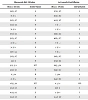
Using the checkerboard assay, investigation of the synergistic or antagonistic characteristics of the 5 selected antimicrobials, gentamicin, ciprofloxacin, amikacin, linezolid, and nalidixic acid, and the EOs, Z. multiflora and T. ammi, against 22 Actinomycete strains, including 19 from HIV-infected and 3 from healthy participants, revealed that most combinations were synergistic, whereas 3 showed additive interactions (FIC = 0.74) (Tables 2 and 3). In the checkerboard assay, significant differences in the mean FIC were reported among the 10 experimental groups using ANOVA (F = 3.62; P = 0.00032). In addition, the Tukey honestly significant difference test was used to more accurately identify significant differences between groups. Synergistic effects were observed for combinations of antimicrobials such as linezolid and amikacin with thyme. Other comparisons showed no statistically significant differences (P > 0.05). In this study, the separate effects of antimicrobials and EOs on FIC were significant (P < 0.05); however, the interactive effects of antimicrobials and EOs on FIC were not statistically significant (P = 0.0813). Furthermore, the Tukey honestly significant difference test was used for pairwise comparison of the mean FIC among the 5 antimicrobials, calculated as the mean of the combinations with the 2 EOs. The mean FIC of linezolid differed significantly from those of the other antimicrobials, including amikacin, ciprofloxacin, gentamicin, and nalidixic acid.
Table 2.
Summary of Fractional Inhibitory Concentration Values for the Combinations of Antimicrobials with Z. Multiflora
| No. | Isolate | Gentamicin | Ciprofloxacin | Linezolid | Amikacin | Nalidixic Acid |
|---|---|---|---|---|---|---|
| 1 | Nocardia farcinica | 0.5 | 0.5 | 0.5 | 0.749 | 0.495 |
| 2 | Streptomyces sp. | 0.496 | 0.496 | 0.497 | 0.5 | 0.498 |
| 3 | Nocardia farcinica | 0.5 | 0.5 | 0.498 | 0.499 | 0.495 |
| 4 | Streptomyces sp. | 0.5 | 0.5 | 0.497 | 0.5 | 0.499 |
| 5 | Nocardia farcinica | 0.479 | 0.479 | 0.497 | 0.499 | 0.490 |
| 6 | Streptomyces sp. | 0.5 | 0.5 | 0.498 | 0.499 | 0.495 |
| 7 | Nocardiopsis alba | 0.499 | 0.499 | 0.5 | 0.749 | 0.495 |
| 8 | Streptomyces sp. | 0.496 | 0.496 | 0.495 | 0.5 | 0.495 |
| 9 | Nocardia farcinica | 0.5 | 0.5 | 0.498 | 0.5 | 0.498 |
| 10 | Nocardia farcinica | 0.5 | 0.5 | 0.497 | 0.499 | 0.498 |
| 11 | Nocardia farcinica | 0.475 | 0.475 | 0.497 | 0.499 | 0.499 |
| 12 | Streptomyces sp. | 0.47 | 0.47 | 0.498 | 0.5 | 0.495 |
| 13 | Nocardiopsis sp. | 0.475 | 0.475 | 0.498 | 0.499 | 0.490 |
| 14 | Nocardiopsis sp. | 0.5 | 0.5 | 0.497 | 0.5 | 0.498 |
| 15 | Nocardiopsis sp. | 0.496 | 0.495 | 0.497 | 0.499 | 0.498 |
| 16 | Streptomyces sp. | 0.499 | 0.5 | 0.498 | 0.499 | 0.490 |
| 17 | Saccharopolyspora sp. | 0.499 | 0.498 | 0.5 | 0.748 | 0.498 |
| 18 | Nocardiopsis sp. | 0.48 | 0.475 | 0.497 | 0.5 | 0.490 |
| 19 | Nocardiopsis alba | 0.499 | 0.5 | 0.49 | 0.499 | 0.498 |
| 20 | Streptomyces sp. | 0.49 | 0.496 | 0.0498 | 0.499 | 0.490 |
| 21 | Nocardia flavescens | 0.499 | 0.5 | 0.499 | 0.499 | 0.495 |
| 22 | Nocardiopsis sp. | 0.5 | 0.5 | 0.497 | 0.499 | 0.490 |
Table 3.
Summary of Fractional Inhibitory Concentration Values for the Combinations of Antimicrobials with T. Ammi
| No. | Isolate | Gentamicin | Ciprofloxacin | Linezolid | Amikacin | Nalidixic Acid |
|---|---|---|---|---|---|---|
| 1 | Nocardia farcinica | 0.499 | 0.499 | 0.0627 | 0.312 | 0.364 |
| 2 | Streptomyces sp. | 0.495 | 0.495 | 0.377 | 0.499 | 0.497 |
| 3 | Nocardia farcinica | 0.499 | 0.499 | 0.497 | 0.498 | 0.364 |
| 4 | Streptomyces sp. | 0.499 | 0.499 | 0.497 | 0.499 | 0.498 |
| 5 | Nocardia farcinica | 0.48 | 0.48 | 0.0377 | 0.498 | 0.5 |
| 6 | Streptomyces sp. | 0.499 | 0.499 | 0.497 | 0.498 | 0.364 |
| 7 | Nocardiopsis alba | 0.499 | 0.499 | 0.499 | 0.312 | 0.364 |
| 8 | Streptomyces sp. | 0.495 | 0.495 | 0.495 | 0.499 | 0.497 |
| 9 | Nocardia farcinica | 0.499 | 0.499 | 0.497 | 0.499 | 0.497 |
| 10 | Nocardia farcinica | 0.499 | 0.499 | 0.377 | 0.498 | 0.497 |
| 11 | Nocardia farcinica | 0.48 | 0.48 | 0.377 | 0.498 | 0.498 |
| 12 | Streptomyces sp. | 0.48 | 0.48 | 0.497 | 0.499 | 0.364 |
| 13 | Nocardiopsis sp. | 0.48 | 0.48 | 0.497 | 0.498 | 0.5 |
| 14 | Nocardiopsis sp. | 0.499 | 0.499 | 0.377 | 0.499 | 0.497 |
| 15 | Nocardiopsis sp. | 0.496 | 0.496 | 0.377 | 0.498 | 0.497 |
| 16 | Streptomyces sp. | 0.499 | 0.499 | 0.497 | 0.498 | 0.364 |
| 17 | Saccharopolyspora sp. | 0.499 | 0.499 | 0.499 | 0.312 | 0.497 |
| 18 | Nocardiopsis sp. | 0.48 | 0.48 | 0.377 | 0.499 | 0.5 |
| 19 | Nocardiopsis alba | 0.499 | 0.499 | 0.489 | 0.498 | 0.497 |
| 20 | Streptomyces sp. | 0.49 | 0.49 | 0.497 | 0.498 | 0.5 |
| 21 | Nocardia flavescens | 0.499 | 0.499 | 0.498 | 0.498 | 0.364 |
| 22 | Nocardiopsis sp. | 0.5 | 0.5 | 0.496 | 0.498 | 0.364 |
5. Discussion
In this study, the antimicrobial susceptibility of 19 Actinomycetes isolates from the oral cavity of HIV-infected participants and 3 additional isolates from healthy participants was assessed against various concentrations of 14 antimicrobials, including amikacin, amoxicillin, amoxicillin-clavulanic acid, ciprofloxacin, clindamycin, erythromycin, gentamicin, linezolid, nalidixic acid, nitrofurantoin, penicillin G, rifampin, tetracycline, and trimethoprim-sulfamethoxazole, using the Kirby-Bauer method. Although nitrofurantoin is primarily used for urinary tract infections and is not routinely prescribed for systemic or oral infections, it was included to provide a broader AMR profile. Therefore, the reported susceptibility should be interpreted cautiously and does not necessarily imply clinical applicability for oral Actinomycetes infections. Furthermore, the MIC and MBC values of these isolates for 5 antimicrobials (amikacin, ciprofloxacin, gentamicin, linezolid, and nalidixic acid) were assessed.
The MIC and MBC values of the EOs of Z. multiflora and T. ammi against these Actinomycete isolates were also investigated. The bacterial isolates were exposed to each antimicrobial with various concentrations of Z. multiflora or T. ammi EOs, and FIC values were then calculated. The finding that all isolates were resistant to penicillin G, amoxicillin, erythromycin, tetracycline, and clindamycin should be interpreted carefully. Actinomycetes such as Nocardia, Streptomyces, and Nocardiopsis are known to naturally possess resistance mechanisms as part of their biological characteristics. Because several members of this group produce antimicrobial compounds, they commonly carry self-protection genes that confer resistance to various antimicrobial classes. Resistance to β-lactam antibiotics in these bacteria has been associated with β-lactamase production, structural characteristics of the cell wall, and alterations in penicillin-binding proteins. In addition, resistance to macrolides, lincosamides, and tetracyclines has been associated with ribosomal modification and efflux systems (12-16). Therefore, the high resistance rates observed in this study may partly reflect the intrinsic characteristics of these organisms rather than exclusively acquired resistance resulting from antimicrobial exposure.
Compared with previous reports on actinomycetes isolated from HIV-infected patients, the resistance profile in this study appears relatively high, particularly for β-lactams and macrolides. Similar resistance patterns have been reported in clinical Nocardia isolates from Europe and Asia, where high resistance to penicillin and erythromycin has been documented. However, the consistent susceptibility to amikacin, linezolid, and trimethoprim-sulfamethoxazole in this study is consistent with global reports, in which these agents remain effective treatment options. These similarities suggest that the observed resistance profile may reflect intrinsic resistance mechanisms and global antimicrobial use patterns rather than a purely local phenomenon (10, 12, 15).
In a 6-month study by Eshraghi et al. (17) on 100 patients with periodontal infections who showed signs of gingivitis and periodontitis, isolates of A. viscosus and A. naeslundii were identified. In a study by Abtahi et al. in Arak, Iran, in 2019, the prevalence of nocardiosis in patients with pulmonary infections was investigated, and an infection rate of 4.32% was reported (18). In a 2018 study by Khatibi et al. (19) on the pathogenicity of filamentous bacteria in denture-associated stomatitis, a condition affecting 24% - 60% of denture wearers, the primary causative agents were identified as Candida albicans and filamentous bacteria, particularly Actinomyces spp. Results showed that actinomycetes were isolated from 5 of 15 control samples (33.3%) and 11 of 15 patient samples (73%). Findings from the study by Abbasian et al. (20) demonstrated that combined treatment with intravenous antimicrobials, such as imipenem, trimethoprim-sulfamethoxazole, and amikacin, along with oral medications, was effective in treating disseminated nocardiosis. Khoroushi et al. (21) reported that the MICs of bell pepper and eggplant skin were 250 mg mL-1 for Streptococcus mutans and 125 mg mL-1 for S. sobrinus and S. sanguinis. For eggplant caps, the MIC was 500 mg mL-1 for S. mutans and S. sobrinus and 125 mg mL-1 for S. sanguinis. Findings by Zakerbostanabad and colleagues indicated that the MIC and MBC of ginger extract against Actinomyces spp. were 0.02 and 0.04 mg mL-1, respectively. Therefore, ginger extract could be used in the preparation of antimicrobial mouthwashes.
In this study, 19 actinomycete isolates from the oral cavity of HIV-infected participants and 3 actinomycete isolates from the oral cavity of control participants were exposed to pathogenic bacteria, including S. aureus PTCC 1917, K. pneumoniae PTCC 1859, A. baumannii PTCC 1919, and E. faecium PTCC 1821, to investigate their antimicrobial characteristics. The results showed that all 22 Actinomycetes isolates exhibited antimicrobial characteristics in the presence of pathogenic bacteria, producing clear zones of inhibition on Mueller-Hinton agar. Although this assay was not the primary aim of the study, it provided complementary insight into the antimicrobial potential of Actinomycetes isolates. In a study by Ebadi et al. (22), 52 Actinomycete isolates were characterized. Isolate 28 showed an Rf value similar to that of gentamicin, whereas isolates 4 and 34 showed Rf values similar to that of streptomycin. The 16S rRNA genes of the isolates were sequenced, revealing that isolate 28 shared 99.93% similarity with S. youssoufiensis and isolate 4 shared 99.93% similarity with S. cyaneofuscatus. Selvin et al. (23) investigated the role of Streptomyces Btl7 in the biosynthesis of antibacterial agents from Dendrilla nigra. These researchers reported an MIC of 44 μg mL-1 and an MBC of 88 μg mL-1.
In a study by Zakerbostanabad and colleagues on the antimicrobial characteristics of actinomycetes isolated from Iranian deserts against S. aureus, K. pneumoniae, A. baumannii, and E. faecium, 22 of 300 isolates showed antimicrobial activity against these bacteria. Isolates from the Lut Desert in Central Iran generally showed better antimicrobial characteristics. Results of the study by Nasri et al. (24) demonstrated that, among 51 isolates of halophilic Actinomyces spp., 3 isolates produced active antimicrobial metabolites against Bacillus cereus, S. aureus, and C. albicans, whereas Pseudomonas aeruginosa and Escherichia coli were resistant to these metabolites. The isolated bacteria were identified as S. flavidofoscus HBUM 17405, N. dansvili OK-22, and Actinomyces Nd28. Soofiani and colleagues characterized Streptomyces isolates from the soil of East Azerbaijan Province, Iran, and found that 44 of 310 Actinomycete isolates showed antibacterial activity against Shigella flexneri ATCC 1290, Listeria monocytogenes ATCC 3390, B. cereus ATCC 1431, E. coli ATCC 1399, and K. pneumoniae ATCC 1234.
Another study by Ezeonwumelu et al. (25) was conducted on actinomycetes experimentally isolated from the oral cavity of HIV-positive patients. They reported that trimethoprim-sulfamethoxazole had the weakest inhibitory activity against S. aureus but was most effective against E. coli ATCC 2592 and P. aeruginosa ATCC 27853. A study by Bandari et al. (26) on cytotoxic compounds of Actinomyces from the Persian Gulf showed that 15 Actinomyces isolates produced protease enzymes, and the anticancer characteristics of the isolates against 2 malignant leukemia cell lines were demonstrated at high concentrations. In a study by Mirsonbol et al. (11) on the antimicrobial activity of S. tendae 944 against B. cereus, P. aeruginosa, Salmonella Typhimurium, Proteus mirabilis, S. aureus, and Micrococcus luteus using the agar well diffusion method, the findings revealed that S. tendae strain 944 had strong antimicrobial activity against all investigated pathogens.
In a 2003 study by Sweeney et al. (27), the antibacterial activity of linezolid was assessed against a range of bacterial strains, including vancomycin-susceptible and vancomycin-resistant E. faecalis, methicillin-susceptible and methicillin-resistant S. aureus, penicillin-susceptible and methicillin-resistant E. faecalis, penicillin-sensitive and methicillin-resistant S. pneumoniae, E. coli, and K. pneumoniae. Among 35 antimicrobials assessed in combination with linezolid, the FIC analysis revealed synergistic effects with 6 antibiotics, including amoxicillin, erythromycin, imipenem, sparfloxacin, teicoplanin, and tetracycline, whereas antagonistic effects were observed with ofloxacin and sparfloxacin. Detailed demographic characteristics are provided in the Supplementary Data (Supplementary Text S1). The lack of strict matching between the groups may represent a potential confounding factor. In addition, the lack of standardized disk diffusion breakpoints for several Actinomycetes genera represents a methodological limitation and may affect the interpretation of susceptibility categories.
5.1. Conclusions
Overall, AST of the isolated actinomycetes in this study revealed frequent AMR patterns among the bacterial isolates. Moreover, the MDR profiles of all Actinomycetes isolates highlighted possible difficulties or failures in the effective treatment of these infections. Notably, any delay or failure in the treatment of infections in patients with HIV/AIDS can result in death. Therefore, AST profiling of microbial infections in HIV-infected individuals can contribute to improved infection management and longer patient survival.
The findings also demonstrated that the isolates were MDR to various commonly used antimicrobials, including amoxicillin, clindamycin, erythromycin, nalidixic acid, penicillin G, and tetracycline. Some of these agents are listed by the WHO as first-line medicines for major bacterial infections. In addition, the reported AMR to previously effective antimicrobials such as co-amoxiclav increases concerns about the rapid AMR development of actinomycetes. In conclusion, the findings highlight the importance of early diagnosis and treatment of infections caused by actinomycetes and suggest that thyme and ajwain EOs may have potential as adjunctive agents. However, further in vivo and clinical studies are needed to verify their therapeutic applicability. Because this was the first study of its kind in Iran, further studies on the prevalence of actinomycetes in HIV-infected patients and the AST of these bacteria are strongly recommended.