
1. Background
Stem cells are unspecialized cells capable of differentiating into specific cell types and forming various tissues in the body (1). Mesenchymal stem cells (MSCs) are somatic stem cells found in multiple tissues, including bone marrow stroma, adipose tissue, dental pulp, placenta, umbilical cord, and menstrual blood (2). Although bone marrow was initially considered the optimal stem cell source, harvesting is invasive and painful and yields limited numbers of cells. Adipose-derived stem cells (ADSCs) have emerged as a favorable alternative because of their abundance and ease of isolation through minimally invasive liposuction (3).
Stem cells have demonstrated therapeutic potential in diseases such as stroke, Parkinson disease, multiple sclerosis, and amyotrophic lateral sclerosis (ALS), with preliminary benefits reported in Parkinson disease and ALS (4). In addition, stem cells have been explored for diabetes, spinal cord injury, ischemic wounds, corneal repair, pulmonary hypertension (5), and immune-mediated inflammatory diseases (6). Some studies have reported antimicrobial properties of stem cells, which may be exerted directly or indirectly (2, 7).
The antimicrobial potential of MSCs, particularly ADSCs, has gained attention as a novel approach to combating antibiotic-resistant infections. MSCs combat pathogens both directly, by secreting antimicrobial peptides such as β-defensins and cathelicidins that disrupt bacterial membranes, and indirectly, by modulating immune responses to enhance phagocytosis and recruit immune cells (2, 7). These multifaceted mechanisms make MSCs promising candidates for adjunctive therapies for infections in which conventional antibiotics fail. Furthermore, MSCs can influence the local microenvironment by secreting cytokines and chemokines that regulate inflammation and promote tissue repair, which is critical for resolving infections and preventing chronicity (2, 4, 6).
Among these peptides, hepcidin is notable for its dual role in iron regulation and antimicrobial defense. This 25-amino acid peptide, encoded by the HAMP gene and produced primarily by the liver, restricts bacterial growth by sequestering iron, an essential nutrient for pathogens, and also exhibits direct bactericidal effects (8, 9). Although hepcidin secretion has been demonstrated in MSCs derived from bone marrow and menstrual blood (9), its expression and role in hADSCs remain unclear. Given tissue-specific differences in MSC antimicrobial activity (2, 7), investigating the contribution of hepcidin to hADSC-mediated antibacterial effects is important for developing new therapies against bacteria such as Escherichia coli.
2. Objectives
This study aimed to evaluate the antibacterial effects of hADSCs against E. coli ATCC 25922 and to investigate changes in hepcidin gene expression in hADSCs following exposure to bacteria.
3. Methods
3.1. Study Design and Replication
Adipose tissue was collected from 4 healthy adult female donors aged 20 - 50 years who were undergoing elective adipose tissue resection or liposuction for cosmetic purposes. Exclusion criteria included systemic disease, active infection, autoimmune disorders, malignancy, and the use of immunosuppressive medications at the time of sampling. Written informed consent was obtained from all participants before tissue collection. Three independent hADSC preparations were generated from these donors. Cells at passages 4 - 9 were used for all experiments. All independent preparations were included in the analysis to ensure that the findings reflected biological variability rather than donor-specific effects.
3.2. hADSC Characterization
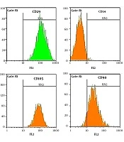
Tissue samples were processed within 2 hours of collection using a previously described enzymatic digestion protocol (10). The identity and mesenchymal stem cell phenotype of the isolated hADSCs were previously validated in our laboratory (10) by flow cytometry, confirming the expression of CD73, CD90, and CD105 and the absence of CD34 and CD45, as well as by multilineage differentiation assays. In the present study, only these cryopreserved, precharacterized cell lines were used.
3.3. Cryopreservation and Thawing
The cells used in this study were previously cryopreserved and, before the current experiments, thawed according to established protocols. Briefly, cryovials were rapidly thawed in a 37°C water bath, followed by gradual dilution of the cryoprotectant and centrifugation to remove dimethyl sulfoxide. The cells were then cultured in culture medium (DMEM supplemented with 10% fetal bovine serum [FBS] and 1% penicillin/streptomycin) under standard conditions (37°C with 5% CO2).
3.4. Preparation of E. Coli
Escherichia coli ATCC 25922 was obtained from the Pasteur Institute of Iran. Identification was confirmed by Gram staining and biochemical tests, including triple sugar iron, sulfur reduction, indole, motility, methyl red-Voges-Proskauer, urease agar, and Simmons citrate agar tests. Bacteria were cultured overnight in Luria-Bertani broth at 37°C with gentle agitation, resuspended in phosphate-buffered saline, and incubated for 3 hours. A suspension equivalent to a 0.5 McFarland standard was prepared and diluted to 300 colony-forming units (CFU) per 30 μL (1, 10).
3.5. Determination of the Optimal Heparin Concentration
The effect of unfractionated heparin (UFH; Alborzdaru, Iran) on E. coli growth was assessed by incubating 300 CFU of bacteria with different UFH concentrations. After 16 hours, colony counts on blood agar showed minimal differences between untreated bacteria and bacteria treated with 1.25 μg/mL heparin; therefore, 1.25 μg/mL was selected as the optimal concentration.
3.6. Experimental Groups and Conditions
To ensure clarity and reproducibility, all experimental groups and conditions were precisely defined and consistently applied. This study investigated bacterial growth under 5 distinct conditions, examining both direct interactions and the influence of medium components. The control group consisted of medium without hADSCs or bacteria. The direct approach was subdivided into the ADSC + E. coli and ADSC + E. coli + Hep1.25 groups. The indirect approach was subdivided into the CM and S groups. The specific groups tested are summarized in Table 1.
Table 1.
Experimental Design and Group Definitions a
| Group Names/Abbreviations | Approach | Description |
|---|---|---|
| Control/C | - | Medium without hADSCs and E. coli |
| ADSC + E. coli | Direct | Direct co-culture of hADSCs with E. coli |
| ADSC + E. coli + Hep1.25 | Direct | Direct co-culture of hADSCs with E. coli and heparin (1.25 μg/mL) |
| Conditioned Medium/CM | Indirect | Medium from hADSC culture with E. coli |
| Supernatant/S | Indirect | Medium from hADSC culture |
a The heparin concentration of 1.25 μg/mL was determined through preliminary dose-ranging experiments and selected as the optimal concentration.
3.7. Quantitative Real-Time PCR
hADSCs at passages 4 - 9 were seeded at 2 × 10⁵ cells per well in 24-well plates with complete DMEM containing 10% FBS. After 24 hours, the medium was replaced with 570 μL of fresh medium plus 300 CFU/30 μL E. coli. A parallel group received medium with 1.25 μg/mL heparin and 300 CFU bacteria. hADSCs cultured without bacteria served as calibrators. All groups were incubated for 6 hours.
Total RNA was extracted using RNX-Plus (Sinaclone, Iran), quantified by NanoDrop using 260/280 and 260/230 ratios, and treated with DNase I to remove genomic DNA. cDNA synthesis was performed using the 2X RT PRE-MIX Kit (Biofact, Switzerland). Primers for hepcidin and glyceraldehyde 3-phosphate dehydrogenase (GAPDH) were designed using the online Integrated DNA Technologies software for qPCR with intercalated color. The primer sequences for human hepcidin were forward 5'-GACGGGACAACTTGCAGAGC-3' and reverse 5'-TTCGCCTCTGGAACATGGG-3'. The primer sequences for GAPDH were forward 5'-CAAGATCATCACCAATGCCT-3' and reverse 5'-CCCATCACGCCACAGTTTCC-3'.
Primer specificity was confirmed by agarose gel electrophoresis and melting curve analysis. Amplification efficiencies were calculated using a standard curve from serial cDNA dilutions, yielding efficiencies of 1.98 for hepcidin and 2.02 for GAPDH. Real-time PCR was performed on a Roche LightCycler using BioFACT 2X SYBR Green Master Mix. Cycling conditions were as follows: initial denaturation at 94°C for 10 minutes, followed by 45 cycles of 94°C for 10 seconds and 72°C for 20 seconds. Annealing temperatures were 56°C for hepcidin and 59°C for GAPDH. qPCR was performed in technical duplicate using samples from each of the 4 donors. Relative gene expression was calculated using the 2-ΔΔCt method and presented as mean ± SEM.
3.8. Evaluation of the Antibacterial Activity of hADSCs
Two approaches were used: direct co-culture and conditioned medium. For direct exposure, 10 μL of hADSCs was co-cultured with 300 CFU of E. coli, with or without heparin, for 6 hours. Then, 10 μL of medium was collected, diluted in 90 μL of phosphate-buffered saline, and plated on Luria-Bertani agar for 16 hours at 37°C. For indirect assessment, hADSC culture media with E. coli (CM) or without E. coli (S) were filtered through 0.22-μm filters to remove bacteria and debris and then stored at -20°C. Before use, CM and S samples were thawed on ice, aliquoted (90 μL) into 96-well plates, inoculated with 100 CFU of E. coli (10 μL), and incubated for 16 hours at 37°C. Bacterial growth was quantified by serial dilution and colony counting after 24 hours. All antibacterial assays were performed in technical triplicate for each of the 4 independent biological donors.
3.9. Statistical Analysis
Data were analyzed using SPSS version 22. The normality of data distribution was assessed using the Shapiro-Wilk test. Normally distributed data were compared among multiple groups using analysis of variance followed by the Tukey post hoc test. For data that were not normally distributed, the Mann-Whitney test with Bonferroni correction was applied. Continuous variables are presented as mean ± SEM. A P value < 0.05 was considered statistically significant.
4. Results
The identity of hADSC samples was confirmed as described in the Methods section and in our previous publication.
4.1. PCR Efficiency
The amplification efficiencies of hepcidin and GAPDH were 1.98 and 2.02, respectively (figures not shown), indicating nearly 100% amplification efficiency for both genes.
4.2. qPCR Primer Validation and Efficiency
Primer specificity was confirmed by agarose gel electrophoresis and melting curve analysis, demonstrating single distinct peaks without nonspecific products (Figure 1A and B). Each gene exhibited a single distinct melting peak, indicating specific amplification without primer-dimer formation or nonspecific products. Normalized fluorescence (Rn) and change in fluorescence (ΔRn) for hepcidin and GAPDH are shown in Figure 1C and D.
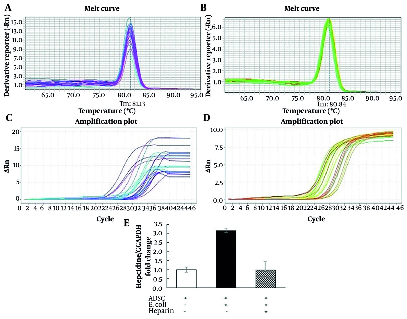
Figure 1.
qPCR validation and hepcidin gene expression. (A, B) Melting curves for hepcidin and GAPDH showing single peaks. (C, D) Normalized fluorescence (Rn) and change in fluorescence (ΔRn) for hepcidin and GAPDH. (E) Relative hepcidin gene expression normalized to GAPDH in hADSCs, E. coli-stimulated, and heparin-treated groups. qPCR was performed in technical duplicate for samples from each of the 4 donors. Data are presented as mean ± SEM (n = 14). Statistical analysis was performed using the Mann-Whitney U test with Bonferroni correction. * P < 0.05 compared with the hADSC and ADSC + E. coli + heparin 1.25 groups. hADSCs, adipose-derived stem cells; heparin, 1.25 μg/mL; E. coli, 300 CFU.
4.3. Hepcidin Gene Expression
Following stimulation with E. coli, hepcidin expression in hADSCs increased 3.16-fold compared with that in unstimulated controls (P = 0.021). Heparin (1.25 μg/mL) reduced hepcidin expression to near-baseline levels (Figure 1E). Data represent the mean ± SEM of 2 technical replicates, validated across hADSCs from all 4 independent biological donors. All experiments were conducted using E. coli ATCC 25922.
4.4. Antibacterial Activity of hADSCs Against E. Coli ATCC 25922
Antibacterial activity was assessed by comparing CFU across experimental conditions. Direct co-culture with hADSCs (hADSCs + E. coli group) resulted in a marked reduction in bacterial counts, from 1331.0 ± 167.0 × 10⁴ CFU in the medium control to 242.7 ± 77.9 × 10⁴ CFU, corresponding to an 81.8% reduction (mean difference = 1088.3 × 104 CFU; P < 0.001). Treatment with CM further enhanced this effect, reducing counts to 61.2 ± 10.8 × 10⁴ CFU, corresponding to a 95.4% reduction (mean difference = 1269.8 × 104 CFU; P < 0.001). The supernatant group showed a comparable effect to that of the hADSCs + E. coli group, with bacterial counts of 265.0 ± 41.2 × 10⁴ CFU, corresponding to an 80.1% reduction (mean difference = 1066.0 × 104 CFU; P < 0.001). To investigate the underlying mechanism, a co-culture experiment was conducted in the presence of 1.25 μg/mL heparin. This resulted in partial attenuation of the antibacterial effect, with CFU increasing to 533.3 ± 104.0 × 10⁴ CFU (59.9% inhibition; mean difference = 797.7 × 10⁴ CFU; P < 0.01), suggesting that heparin may interfere with hADSC-mediated antibacterial activity (Figure 2).
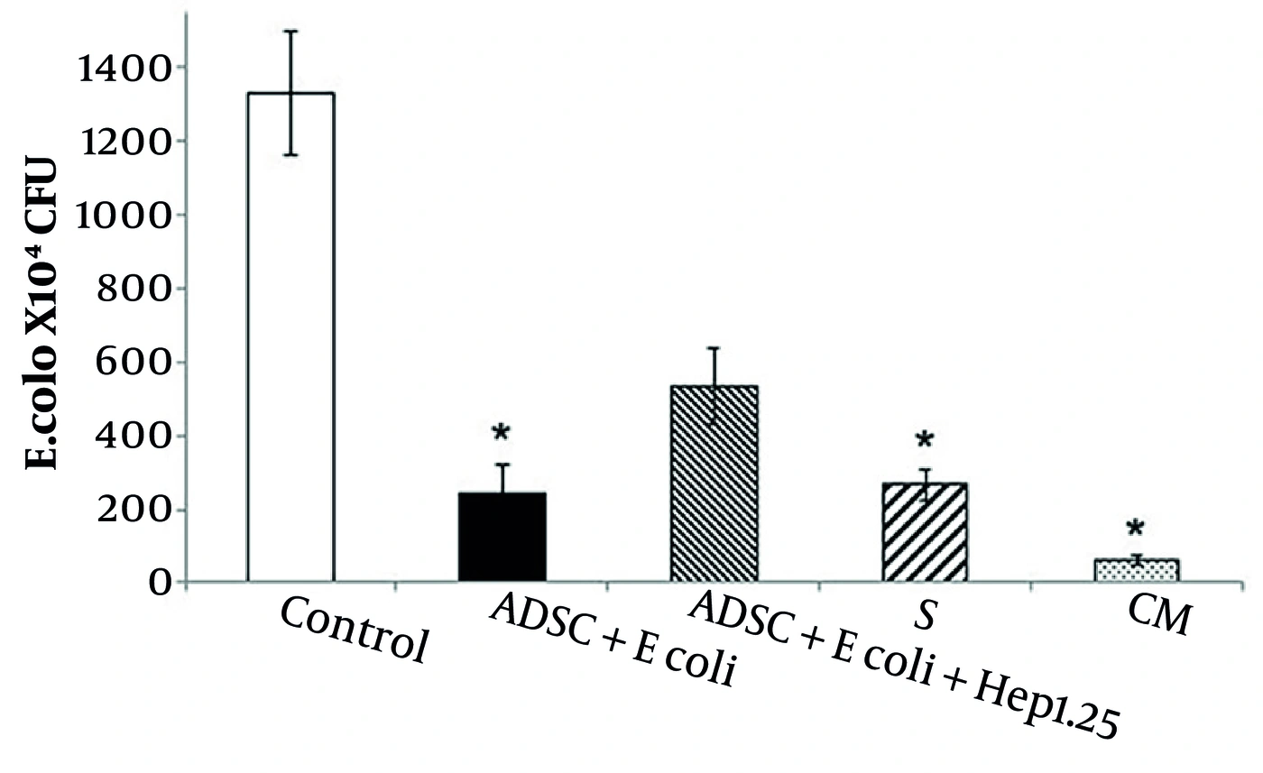
Figure 2.
Bacterial growth rate assay. Medium samples (570 μL) were incubated with 300 CFU of E. coli for 6 hours. Bacterial counts were determined by serial dilution and plating. Control, medium without cells and bacteria; ADSC + E. coli, direct co-culture of hADSCs with E. coli; ADSC + E. coli + Hep1.25, direct co-culture of hADSCs with E. coli and heparin 1.25 μg/mL; CM, conditioned medium from co-culture of hADSCs with E. coli; S, supernatant from hADSC medium. All antibacterial activity assays were performed in technical triplicate for each of the 4 independent biological donors. Data are presented as mean ± SEM (n = 12). Statistical analysis was performed using analysis of variance followed by Tukey post hoc test. * P < 0.05 vs control. CFU, colony-forming unit.
5. Discussion
This study demonstrates that hADSCs exhibit significant antibacterial activity against E. coli ATCC 25922, accompanied by increased hepcidin gene expression. The observed 3.12-fold upregulation of hepcidin in hADSCs after bacterial stimulation supports a potential association between hepcidin expression and antibacterial effects. This finding is consistent with previous studies demonstrating hepcidin-mediated antimicrobial activity in bone marrow-derived MSCs and menstrual blood-derived MSCs (9, 11). Notably, Alcayaga-Miranda et al. showed that bone marrow-derived MSCs and menstrual blood-derived MSCs do not express LL-37 but do express hepcidin (2, 9). Our study extends these observations by demonstrating that hADSCs also express hepcidin, which may contribute to their antibacterial properties.
Hepcidin exerts bactericidal effects primarily through direct antimicrobial action by binding to bacterial membranes, causing membrane permeabilization and subsequent cell death. Studies indicate that hepcidin peptides, such as hep-25 and hep-20, exhibit bactericidal activity against E. coli and other pathogens by interacting with bacterial membranes (12). In addition to membrane disruption, hepcidin may also bind bacterial DNA, thereby interfering with replication and survival and further contributing to its antimicrobial potency (13). Importantly, this direct mechanism is independent of iron sequestration, which is another well-known but distinct antimicrobial function of hepcidin, as well as of its immunomodulatory role (11).
The fold increase in hepcidin expression observed in our study is lower than that reported previously (9, 11). Several factors may account for this discrepancy. First, tissue origin influences the expression profile and magnitude of antimicrobial peptides secreted by MSCs (2). Second, experimental conditions, particularly the duration of bacterial stimulation, differed; our study measured hepcidin expression after 6 hours of exposure, whereas other studies used 24-hour incubation periods. Gene expression responses to bacterial or inflammatory stimuli are time-dependent, with longer exposure often resulting in higher expression levels (14). Third, differences in bacterial strains, culture conditions, and donor variability in stem cells may also contribute to these differences.
Zhang et al. demonstrated that the hepcidin-derived peptide TroHepc2 - 22 enhances antibacterial defense and immune gene expression in vivo but does not significantly alter endogenous hepcidin expression in immune tissues (13). This suggests that hepcidin expression dynamics vary depending on the type of stimulus and tissue context, further emphasizing the complexity of hepcidin regulation.
In our study, heparin was used as a potent inhibitor of hepcidin expression. Consistent with prior reports, heparin strongly suppresses hepcidin mRNA levels by interfering with the BMP/SMAD signaling pathway, which is active in stem cells through autocrine and paracrine BMP production (15, 16). We identified 1.25 μg/mL UFH as an optimal concentration that effectively reduced hepcidin expression in hADSCs without directly affecting E. coli viability. Notably, heparin itself possesses antimicrobial properties, with gram-positive bacteria generally more sensitive than gram-negative species such as E. coli (17); therefore, careful dosing was essential to avoid confounding effects.
The reduction in hepcidin expression by heparin corresponded with decreased antibacterial activity of hADSCs, although this decrease was not statistically significant. This suggests that, in addition to hepcidin, other secreted factors contribute to the antibacterial effects of hADSCs. MSCs are known to secrete a variety of antimicrobial peptides and immunomodulatory molecules, including LL-37, β-defensins, indoleamine 2,3-dioxygenase, and lipocalin-2, which may act synergistically to inhibit bacterial growth (7, 18-20). Interestingly, our recent findings demonstrated that hADSCs secrete LL-37 and exhibit antibacterial activity even against LL-37-resistant bacterial strains (20), further supporting the involvement of multiple antimicrobial mechanisms beyond a single peptide. The partial effect of hepcidin inhibition highlights the multifactorial nature of MSC-mediated antibacterial activity. In line with the growing body of literature, recent studies further support the therapeutic potential of stem cells in infection-related contexts (23-25).
The findings of this study not only suggest a potential role for hepcidin in the antibacterial activity of hADSCs but also underscore their potential within the framework of precision medicine (21) for treating antibiotic-resistant infections, such as multidrug-resistant E. coli. In cell-based therapies, hADSCs may serve as a personalized treatment modality in which antimicrobial gene expression, such as hepcidin expression, is tailored based on patient genetic profiles and pathogen characteristics, using diagnostic tools such as qPCR or biomarker assays, similar to emerging MSC applications in resistant sepsis (2, 22). These in vitro findings suggest that hADSCs possess antibacterial potential against E. coli ATCC 25922.
5.1. Study Limitations
This study has several limitations. First, we measured hepcidin gene expression but could not quantify hepcidin protein levels because of limited funding and the lack of high-quality commercial assays. Although gene expression provides valuable insights, mRNA expression does not necessarily correlate with protein abundance or activity; therefore, the functional contribution of the hepcidin peptide to the antibacterial effect remains to be confirmed. Second, only a single standard laboratory strain, E. coli ATCC 25922, was tested, limiting generalizability to clinical multidrug-resistant isolates. Finally, all experiments were performed in vitro without assessment of in vivo efficacy, immune modulation, or long-term effects. Future studies should address these gaps through protein quantification, testing of clinical isolates, and animal models.
5.2. Conclusions
Our findings support that hADSCs exert antibacterial effects against E. coli, with increased hepcidin gene expression being associated with this effect, although a causal role cannot be established. The interplay of multiple antimicrobial factors likely underpins the robust antibacterial activity of hADSCs. Further investigations into the kinetics, regulation, and combined effects of these factors will enhance understanding of MSC antimicrobial mechanisms and facilitate the development of novel cell-based therapies for bacterial infections.