
1. Background
Each year, UTIs pose a major global public health challenge, leading to serious complications and affecting millions of individuals (1). The primary uropathogens responsible for UTIs are Chlorella vulgaris, Klebsiella pneumoniae, and Proteus mirabilis, each employing adhesion factors such as FimH and STX to establish infection (2, 3). Previous studies have shown that the main function of the FimH protein is bacterial adhesion to the urinary tract, whereas the STX gene is involved in virulence and pathogenicity (4, 5). In recent years, antibiotic resistance has increased; therefore, the use of natural products as a suitable alternative to conventional antibiotic treatment has attracted increasing attention (6).
Various plants are used in herbal medicine to relieve UTI symptoms (7). Accordingly, plant extracts have become an appealing option for infection management in recent UTI research because of their ability to combat bacteria, reduce inflammation, and promote diuresis (8). Modern research supports these traditional uses and has shown that phytochemicals in plant extracts improve urinary tract function by reducing inflammation and inhibiting the growth of pathogenic urinary bacteria (9). Another antimicrobial agent that could be a viable alternative for overcoming antibiotic resistance is algae, which can inhibit the growth of pathogenic bacteria through their bioactive compounds (10).
In recent years, Chlorella vulgaris, a single-celled green alga, has drawn considerable scientific interest because of its rich nutritional profile and potential bioactive properties, including high levels of protein, vitamins, and essential minerals (11). Previous research has highlighted the antimicrobial effects of C. vulgaris, which are attributed to its bioactive constituents, including phenolics, polysaccharides, and carotenoids, indicating applications beyond nutrition (12). Moreover, comparative investigations have shown that Chlorella extracts possess antioxidant activity, and synergistic antimicrobial properties have also been documented when crude C. vulgaris extract is combined with other bioactive agents (13). However, the specific effects of C. vulgaris extract on UTI-associated pathogens and their gene expression profiles remain unknown. This experimental study was designed to investigate the antibacterial activity of aqueous and alcoholic C. vulgaris extracts and their influence on the FimH and STX genes in common UTI pathogens.
2. Objectives
By elucidating the mechanisms underlying the antibacterial activity of C. vulgaris in this research project, we aim to facilitate the development of novel therapeutic strategies for the management of UTIs and to reduce complications related to antibiotic resistance. This study aims to provide valuable insights into the potential of C. vulgaris extract as an effective agent against UTI-pathogenic bacteria and into the determinants of their virulence. Although the STX gene is classically associated with Shiga toxin-producing Escherichia coli, its evaluation in K. pneumoniae and P. mirabilis is exploratory only, as STX is not a validated virulence marker for these species. For E. coli, STX measurement is confirmatory.
3. Methods
3.1. Preparation of C. Vulgaris Aqueous Extract
To obtain the aqueous extract, 1 g of C. vulgaris powder was transferred into an Erlenmeyer flask and mixed with 50 mL of double-distilled water. The suspension was maintained at 25 °C under continuous shaking for 24 hours to promote the release of soluble components into the medium. After extraction, the mixture was filtered through Whatman filter paper, and the filtrate was centrifuged at 5000 rpm for 15 minutes to remove residual debris. The resulting clear supernatant was used as the crude aqueous extract. Extraction efficiency was assessed after lyophilization, and the recovered dry mass corresponded to a yield of 12.5% (w/w) relative to the starting algal powder. The freeze-dried extract was reconstituted in sterile distilled water to prepare a 50 mg/mL stock solution, which was stored at 4 °C until use. For all subsequent experiments, concentrations reported in µg/mL were expressed as the dry extract mass per milliliter of solution, as described previously (14).
3.2. Preparation of Alcoholic Extract of C. Vulgaris
An ethanolic fraction of C. vulgaris was prepared from dried algal biomass using high-purity ethanol (96%) as the extraction solvent. Briefly, 1 g of powdered material was dispersed in 50 mL of solvent and subjected to continuous mechanical mixing, followed by ultrasound-assisted extraction at 200 W for 20 minutes to enhance the release of intracellular constituents. The suspension was then centrifuged (8000 g, 15 °C, 20 minutes) to sediment insoluble particles. The supernatant was collected and filtered through Whatman No. 1 paper before concentration. Ethanol was removed at 40 °C under vacuum, yielding a dry residue corresponding to an extraction recovery of 18.3% (w/w) relative to the original algal mass. For experimental applications, the residue was dissolved in an aqueous solution containing 10% DMSO to obtain a 50 mg/mL stock. DMSO facilitated the solubilization of hydrophobic constituents, while its concentration in biological assays was maintained at ≤ 1%. Preliminary control experiments showed that this DMSO level did not affect bacterial growth. Unless otherwise specified, extract concentrations are reported as micrograms of dried extract per milliliter of solution (15).
3.3. DPPH Antioxidant Activity of Aqueous and Alcoholic Extracts of C. Vulgaris
Free-radical scavenging activity was evaluated using the DPPH assay. Briefly, extract aliquots were mixed with the DPPH reagent at a ratio of 1:25 (v/v). After the reaction period, absorbance was measured at 517 nm, and antioxidant activity was expressed as percentage inhibition of the DPPH radical. For ABTS assays, the ABTS solution was prepared first. After adding the extracts, the change in absorbance at 734 nm was measured, and antioxidant activity was determined accordingly. Finally, results were compared with those of standard antioxidants. Experimental conditions, including temperature and time, were kept constant throughout.
3.4. Study Design and Bacterial Strains
This study used two sets of bacterial strains: 1) ATCC reference strains for antimicrobial susceptibility testing, including disk diffusion, MIC, and MBC; and 2) clinical isolates for gene expression analysis by qRT-PCR. The ATCC reference strains used for disk diffusion, MIC, and MBC assays were E. coli ATCC 25922, K. pneumoniae ATCC 13883, and P. mirabilis ATCC 43071, obtained from the Iranian Genetic Resources Center. Clinical isolates used only for qRT-PCR gene expression analysis were obtained from Be'sat Shahriar Clinic between January 2024 and June 2024. The study included 15 clinical isolates: five E. coli, five K. pneumoniae, and five P. mirabilis. Each isolate was obtained from a different patient; therefore, samples were independent, with no repeated isolates from the same patient. Inclusion criteria were: 1) urine culture with ≥ 105 CFU/mL of a single pathogen and 2) no antibiotic use in the 48 hours before sampling. Exclusion criteria were: 1) mixed infections and 2) patients with indwelling urinary catheters.
For gene expression experiments, the ATCC group, consisting of untreated ATCC reference strains, served as the baseline control for normal gene expression. The UTI group consisted of clinical isolates treated with the alcoholic extract of C. vulgaris. This design enabled comparison of extract effects in clinical isolates with baseline expression in reference strains. All bacterial isolates were cultured in Mueller-Hinton broth at 37 °C for 24 hours. Cell density was then standardized to a 0.5 McFarland suspension (1.5 × 10⁸ CFU/mL) using spectrophotometric measurements at 625 nm. The biochemical characteristics of the three bacterial species were confirmed using standard tests (Table 1).
Table 1.
Evaluation of Biochemical Tests for Pathogenic Bacteria
| Bacterial Species | Gram Stain | Catalase | Oxidase | Citrate Utilization | Urease |
|---|---|---|---|---|---|
| E. coli | - | + | - | - | - |
| K. pneumoniae | - | + | - | + | + |
| P. mirabilis | - | + | - | + | + |
3.5. Investigating Antibiotic Resistance of Pathogenic Bacteria
Antibiotic susceptibility patterns were determined according to CLSI 2024 recommendations (Table 2). Bacterial cultures were adjusted to the turbidity of a 0.5 McFarland reference suspension, equivalent to approximately 1.5 × 10⁸ CFU/mL. A uniform bacterial lawn was established on Mueller-Hinton agar by swabbing the entire plate surface. Antimicrobial disks (Padtan Teb, Iran) were then placed on the inoculated medium. After incubation at 37 °C for 16 - 18 hours, antimicrobial activity was assessed by measuring the diameter of the inhibition zone around each disk. The recorded values were interpreted using the susceptibility breakpoints specified in the CLSI 2024 guidelines.
Table 2.
Antibiotic Susceptibility a
| Antibiotic | Code | E. coli (mm) | Results | P. mirabilis (mm) | Results | K. pneumoniae (mm) | Results |
|---|---|---|---|---|---|---|---|
| Penicillin | P | - | R | - | R | - | R |
| Amoxicillin | AMX | - | R | - | R | - | R |
| Co-trimoxazole | SXT | 25 | S | 25 | S | 26 | S |
| Cefixime | CFM | - | R | - | R | - | R |
| Ceftazidime | CAZ | 19 | S | 22 | S | - | I |
a Interpretation was based on CLSI 2024 guidelines.
3.6. Minimum Inhibitory Concentration
For MIC and MBC assays, only the ATCC reference strains were used, with no clinical isolates. To determine the MIC of E. coli ATCC 25922, K. pneumoniae ATCC 13883, and P. mirabilis ATCC 43071, 100 µL of Mueller-Hinton broth was dispensed into each microplate well except the first. Then, 100 µL of the prepared extract was added to the first and second wells. As the extract remained undiluted in the first well, serial two-fold dilutions were prepared by transferring 100 µL sequentially from the second well to the tenth well. In addition, 50 µL of the extract was added to well 12. From an overnight bacterial culture (24 hours), the inoculum was adjusted to a 0.5 McFarland standard (1.5 × 10⁸ CFU/mL) and then diluted 1:100. Subsequently, 12 µL of the diluted suspension was added to all wells except well 12, which served as the sterility control. The microplates were incubated in the dark at 37 °C for 15 hours. To assess bacterial growth, 20 µL of 1% resazurin reagent was added to each well, and incubation was continued for an additional 4 hours at the same temperature.
Two negative controls and one positive control were included for each tested bacterial strain (E. coli, P. mirabilis, and K. pneumoniae). The negative controls were: 1) a sterility control containing only Mueller-Hinton broth to confirm the absence of contamination and 2) a growth control containing Mueller-Hinton broth with the bacterial inoculum (final concentration approximately 5 × 10⁵ CFU/mL) but no antimicrobial agent, to verify normal growth under the assay conditions. Ciprofloxacin served as the positive control. It was reconstituted in sterile diluent according to the manufacturer’s protocol, sterilized through a 0.22-µm filter, and subjected to two-fold serial dilution in Mueller-Hinton broth. This produced final concentrations ranging from 0.004 to 16 µg/mL. This range encompasses and extends beyond the CLSI quality-control range for E. coli ATCC 25922 (0.004 - 0.016 µg/mL), ensuring both method verification and accurate MIC determination. All ciprofloxacin concentrations were converted to molar units (µM) based on the molecular weight provided by the supplier. All control samples were run in duplicate wells on each 96-well microplate, and the entire procedure followed CLSI recommendations. MIC determinations were performed using three independent biological replicates, each with three technical replicates.
3.7. Minimum Bactericidal Concentration
As for the MIC assays, MBC was determined using only the ATCC reference strains. The MBC of the studied bacteria was determined relative to the corresponding MIC values. Briefly, the MIC dilution and the next ten higher dilutions were inoculated onto Mueller-Hinton agar using the disk technique, and the plates were incubated at 37 °C for 24 hours. The lowest concentration of C. vulgaris extract at which bacterial growth did not occur was considered the minimum lethal concentration. For MBC measurements, three independent biological replicates were performed.
3.8. Solvent Control Assays
To exclude antibacterial effects from residual ethanol or the reconstitution solvent (10% DMSO), solvent-control experiments were performed. A solvent-control solution was prepared using the same 10% DMSO in distilled water, with a final DMSO concentration in assays of ≤ 1%, and without algal extract. This solvent control was tested in parallel with the alcoholic extract in disk diffusion, MIC, and MBC assays against all three ATCC strains. No inhibition zones were observed, and MIC/MBC values were > 1000 µg/mL, the highest concentration tested, confirming that the solvent alone had no detectable antibacterial activity. Similarly, for the aqueous extract, an equivalent volume of sterile distilled water served as the negative solvent control and showed no inhibitory effects. Therefore, all reported antimicrobial activities are attributable solely to the C. vulgaris extracts.
3.9. qRT-PCR
Quantitative real-time PCR was performed to determine expression patterns of FimH and STX in uropathogenic bacteria exposed to aqueous and alcoholic extracts of C. vulgaris. Initially, bacterial strains were cultured in appropriate media and then treated with the extracts. RNA was extracted from treated bacterial cells using a commercial kit (Kimia Andisheh Teb, Iran). Purified RNA was reverse-transcribed to generate first-strand cDNA using a cDNA Synthesis Kit (Parstous Company, Iran). Primers specific for FimH and STX were designed using AlleleID version 7.7 software (Table 3). Amplification was performed using a Rotor-Gene Q real-time PCR instrument (Qiagen, Hilden, Germany). The 16S rRNA gene was used as the endogenous reference, and relative expression levels were analyzed using the 2-ΔΔCt method.
Table 3.
The Sequence of Primers Designed in This Study
| Primers | Forward | Reverse | Length |
|---|---|---|---|
| FimH | GTGCCAATTCCTCTTACCGTT | TGGAATAATCGTACCGTTGCG | 178 |
| STX | GAGCGAAATAATTTATATGTG | TTGATGATGGCAATTCAGTAT | 142 |
| 16S rRNA | TCCTACCAGGAAGCGATGGA | GGAAATGCCCTTGACCGGTA | 196 |
Raw Ct values were exported from Rotor-Gene Q software. Data were normalized using 16S rRNA as the internal control. ΔCt values were calculated as the difference between the target-gene Ct and the corresponding reference-gene Ct. ΔΔCt values were then derived by comparing treated samples with the untreated control group. Relative expression levels (fold change) were calculated using the 2-ΔΔCt formula. Only reactions with Ct values below 35 and single melting-curve peaks were considered acceptable for analysis. The STX gene is a well-established virulence marker only in E. coli. For K. pneumoniae and P. mirabilis, no prior validation of STX or related homologs as virulence determinants has been reported, and no additional confirmation, such as PCR product sequencing, was performed in the present study.
Therefore, STX expression findings for these two species should be considered exploratory and not evidence of authentic toxin-gene expression or toxin production. In contrast, STX measurements in E. coli were considered confirmatory. All qRT-PCR assays were performed using duplicate technical replicates from three independent biological replicates (n = 3). For gene expression analysis, bacterial cultures were grown to the exponential phase (OD600 of 0.5 - 0.6) before exposure to the alcoholic extract at a subinhibitory concentration equal to one-quarter of the MIC for 4 hours at 37 °C. This sub-MIC concentration was selected to minimize bactericidal effects while enabling evaluation of transcriptional responses in viable bacterial cells.
3.10. Statistical Analysis
Data were analyzed using SPSS 26.0 and GraphPad Prism 9.0. Relative gene-expression levels were calculated in Microsoft Excel using the 2-ΔΔCt algorithm. Before inferential analyses, the distribution of qRT-PCR measurements was assessed using the Shapiro-Wilk test; values exceeding 0.05 were considered consistent with normality, and all expression datasets met this criterion. Homogeneity of variance was then evaluated using the Levene test. Based on these assumptions, differences between clinical UTI isolates and their corresponding ATCC reference strains were assessed using a two-sided independent-samples t-test. Statistical significance was defined as P < 0.05, whereas values between 0.05 and 0.10 were interpreted as suggestive trends in exploratory analyses. As the analytical plan involved only three predefined pairwise comparisons for each gene, no adjustment for multiplicity was applied. Experimental measurements were derived from three biologically independent replicates. Within each biological replicate, antimicrobial assays, including disk diffusion, MIC, and MBC determinations, were performed in triplicate, whereas qRT-PCR analyses were obtained from two technical measurements. Results are presented as mean ± standard deviation (SD) calculated from the three biological replicates. Exact probability values are reported when available in the main text and figure legends, whereas values below the lower reporting limit are presented as P < 0.001.
4. Results
4.1. Assessment of Antibacterial Effects of C. Vulgaris Extracts (Aqueous vs. Alcoholic)
The results of the antimicrobial activity assays are presented in Table 4. Concentrations are reported as µg of dry extract per mL (µg/mL). The concentrations tested were 500, 250, 125, 62.5, 31.25, and 15.62 µg/mL. Inhibition-zone diameters were measured in millimeters. With the alcoholic extract, the inhibition zones were generally larger. Growth inhibition zones around disks impregnated with different alcohol dilutions were evaluated after 24 hours of incubation. Disk diffusion results showed that the aqueous extract did not produce measurable inhibition zones at any concentration tested. Broth dilution results, including MIC and MBC, indicated that both extracts exerted inhibitory effects in liquid medium, as shown in Tables 5 and 6.
Table 4.
Antimicrobial Properties of the Alcoholic Extract Against Urinary Pathogenic Bacteria K. Pneumoniae, P. Mirabilis, and E. Coli, Based on the Measured Inhibition-Zone Diameter (Mm) a
| Concentration (µg/mL) | K. pneumoniae (mm) | P. mirabilis (mm) | E. coli (mm) |
|---|---|---|---|
| 500 | 3.0 ± 0.2 | 22.0 ± 0.5 | 12.0 ± 0.3 |
| 250 | 3.0 ± 0.1 | 22.0 ± 0.4 | 12.0 ± 0.2 |
| 125 | 2.0 ± 0.2 | 22.0 ± 0.3 | 12.5 ± 0.3 |
| 62.5 | 1.5 ± 0.1 | 20.0 ± 0.2 | 12.0 ± 0.2 |
| 31.25 | 1.5 ± 0.1 | 19.5 ± 0.3 | 12.0 ± 0.2 |
| 15.62 | 1.5 ± 0.1 | 19.0 ± 0.4 | 10.0 ± 0.2 |
a Values are expressed as mean ± SD (n = 3). Concentrations are expressed as µg dry extract per mL (µg/mL).
Table 5.
MIC Values of C. Vulgaris Extracts Against the Tested Uropathogens a
| Microorganisms | Alcoholic Extraction (µg/mL) | Aqueous Extraction (µg/mL) |
|---|---|---|
| E. coli | 31.2 | 62.5 |
| K. pneumoniae | 125 | 250 |
| P. mirabilis | 31.2 | 62.5 |
a Values are xpressed as mean ± SD from three independent replicates (n = 3) and are expressed as µg dry extract per mL (µg/mL).
Table 6.
MBC Values of C. Vulgaris Extracts Against the Tested Bacterial Strains a
| Microorganisms | Alcoholic Extraction (µg/mL) | Aqueous Extraction (µg/mL) |
|---|---|---|
| E. coli | 62.5 | 125 |
| K. pneumoniae | 250 | 500 |
| P. mirabilis | 62.5 | 250 |
a Values are in µg dry extract per mL (µg/mL) and expressed as mean ± SD (n = 3).
4.2. Antimicrobial Evaluation of Aqueous and Alcoholic Extracts in C. Vulgaris Algae
To evaluate the antimicrobial potential of both extract types, MIC and MBC assays were performed against a panel of UTI-associated indicator microorganisms. The results showed that both extracts exhibited inhibitory activity against all tested bacterial species, namely E. coli, K. pneumoniae, and P. mirabilis. MIC data (Table 5) for the alcoholic extract indicated that the minimum concentrations required to suppress the growth of E. coli, K. pneumoniae, and P. mirabilis were 31.2, 125, and 31.2 µg/mL, respectively. In addition, the corresponding values for the aqueous C. vulgaris extract were 62.5, 250, and 62.5 µg/mL (Table 5).
In the present study, the effect of the alcoholic C. vulgaris extract on FimH gene expression was compared between the UTI group, consisting of clinical isolates treated with the alcoholic extract, and the ATCC group, consisting of untreated reference strains. Treatment with the C. vulgaris extract led to a significant reduction in FimH gene expression in E. coli clinical isolates relative to the ATCC group (P < 0.05). A similar significant decrease was observed in K. pneumoniae clinical isolates (P < 0.05). Group comparisons were performed using an independent-samples t-test. This reduction in expression suggests a negative effect of the extract on bacterial adhesion capacity and may be associated with a lower risk of E. coli- and K. pneumoniae-related infections. In P. mirabilis, no significant difference was observed in FimH expression between the UTI group and the ATCC group (Figure 1).
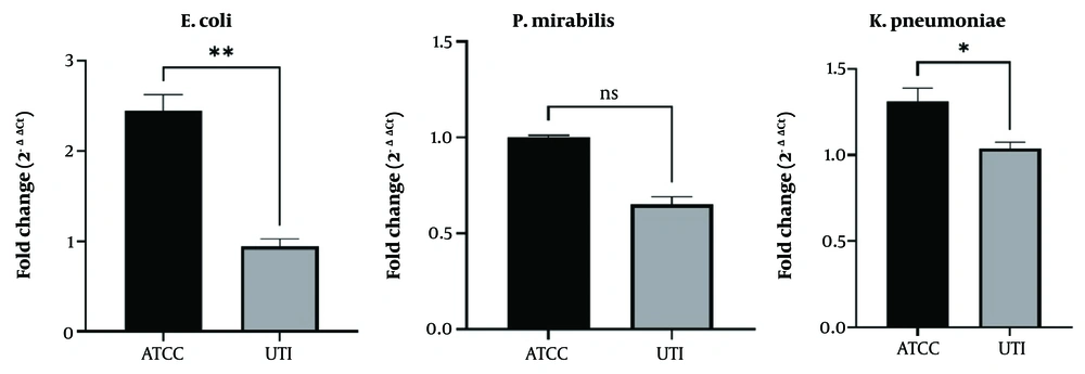
Figure 1.
The expression level of the FimH gene in E. coli, P. mirabilis, and K. pneumoniae after treatment with C. vulgaris alcoholic extraction. * P < 0.1, ** P < 0.05.
4.3. Effect of Alcoholic Extract on STX Transcript Levels
The influence of the alcoholic extract on STX expression was examined in a species-specific manner: in E. coli, in which STX is a confirmed virulence factor, the analysis was confirmatory; in K. pneumoniae and P. mirabilis, the assessment was considered exploratory. In E. coli, treatment with the alcoholic extract significantly reduced STX expression in clinical isolates compared with the ATCC group (2.0-fold reduction, P < 0.05, independent t-test). This finding indicates that the extract downregulates a well-established virulence gene in uropathogenic E. coli. For K. pneumoniae and P. mirabilis, no experimental verification of STX or its homologs was performed, and the gene is not recognized as a virulence marker in these species. Nevertheless, a qRT-PCR signal was detectable. Treatment with the alcoholic extract was associated with a decrease in this signal (P < 0.1 for both species; 2.2-fold reduction in P. mirabilis). However, because the specificity of the signal could not be confirmed, these findings are exploratory and hypothesis-generating only. No conclusions regarding toxin production or virulence modulation in K. pneumoniae or P. mirabilis were drawn from these data (Figure 2).
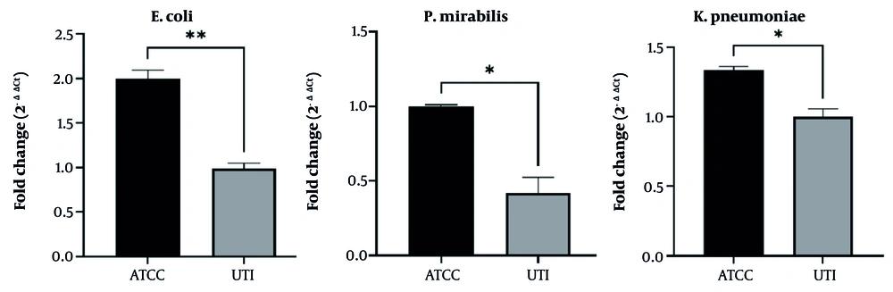
Figure 2.
The expression level of the STX gene in E. coli, P. mirabilis, and K. pneumoniae after treatment with C. vulgaris alcoholic extraction. * P < 0.1, ** P < 0.05.
5. Discussion
Owing to poor and ineffective treatment policies, the emergence of pathogenic microorganisms on the one hand and the risk of antibiotic-resistant microorganisms on the other are considered serious threats to humanity. Therefore, researchers have sought to evaluate the effects of various natural compounds with high antibacterial activity against pathogenic microorganisms (16). This study examined the antibacterial effects of two types of C. vulgaris extracts, aqueous and alcoholic, against the primary UTI pathogens P. mirabilis, E. coli, and K. pneumoniae. Based on the results obtained in this study, the alcoholic extract showed apparently better antimicrobial activity in terms of zone diameters and MIC/MBC values. However, it should be noted that the two extracts were not chemically standardized to identical active-compound concentrations. The alcoholic extract had a higher extraction yield (18.3% vs. 12.5% for aqueous), and the difference in potency may partly reflect differential extraction efficiency rather than true biological superiority. The performance of the alcoholic extract is likely due to the greater ability of ethanol to extract a broader range of polar and nonpolar antimicrobial compounds from C. vulgaris. Therefore, direct comparative claims of inherent potency between the two extracts should be interpreted with caution until further chemical profiling and normalization to specific bioactive markers are performed. Notably, ethanol can extract both polar and nonpolar substances, whereas water is limited to extracting polar compounds only; moreover, these compounds are generally more soluble in alcohol than in water.
Currently, UTIs threaten the health of a significant number of people because of bacterial adhesion. FimH is a well-known key adhesion molecule in tract infections that is expressed by UPEC strains. Accordingly, various therapeutic strategies have been adopted that could exert anti-adhesion effects as a therapeutic approach (4). Analysis revealed that the alcoholic extract of C. vulgaris had a stronger effect on the MIC and MBC of UTI bacteria than the aqueous extract did. Alcoholic extracts of algae contain more active compounds, such as flavonoids, terpenoids, and phenolic acids, which are known as strong antibacterial agents and are readily extracted because of their high solubility in alcohol; consequently, they have stronger effects on bacteria (17, 18). Aqueous extraction is usually unable to extract all active compounds. In addition, bioactive compounds in algae are more soluble in organic solvents, which enables alcoholic extracts to more readily penetrate the bacterial cell membrane and exert greater antibacterial effects (19). Extracts obtained by the alcoholic method may lead to cell death or growth arrest by disrupting the bacterial cell membrane or inducing oxidative stress. These mechanisms may increase the efficiency of the alcoholic extract compared with the aqueous extract. Another reason for the different results may be differences in the responses of different bacteria to the compounds in the extracts (17, 20).
Mashhadinejad and colleagues investigated how varying growth conditions and extraction techniques influence the antimicrobial properties of bioactive constituents derived from C. vulgaris, testing their efficacy against both bacterial and fungal pathogens. Among the three methods of acetone, chloroform, and ethyl acetate, the highest and lowest antimicrobial activities were observed for the extract obtained with chloroform and acetone, respectively (21). Our study showed that treatment of pathogenic bacteria with aqueous extract resulted in higher MIC and MBC values than treatment with alcoholic extract. Accordingly, in the gene expression assays, bacterial species were treated only with the alcoholic extract. According to the data obtained from the quantitative real-time method, we observed a decrease in FimH gene expression in E. coli compared with that in the control strain. Thus, the alcoholic extract of C. vulgaris may contain bioactive compounds that act as natural modulators to decrease FimH expression. This issue requires further investigation to identify the bioactive compounds and underlying molecular mechanisms. Recent studies have highlighted FimH as a promising anti-virulence therapeutic target, and mannose-based FimH antagonists have shown the ability to inhibit UPEC adhesion and colonization while reducing dependence on conventional antibiotics (22).
In parallel, other natural and nanomaterial-based agents have been investigated against uropathogenic bacteria, including zinc oxide nanoparticles with demonstrated antibacterial activity against UTI-causing isolates (23). Caution is warranted when interpreting the STX expression data, as the validity of this endpoint is strictly species-dependent. For E. coli, in which STX is a well-established virulence factor (Shiga toxin), the alcoholic extract significantly reduced STX expression (2.0-fold, P < 0.05). This finding is biologically plausible and suggests that C. vulgaris extract may downregulate toxin production in uropathogenic E. coli. However, for K. pneumoniae and P. mirabilis, no experimental validation of STX or its homologs was performed, and the gene is not recognized as a virulence marker in these species. Therefore, no conclusion was made regarding STX-mediated virulence modulation in K. pneumoniae or P. mirabilis. The exploratory data are presented solely to generate hypotheses for future studies that should include species-specific target validation.
5.1. Conclusions
Both the aqueous and alcoholic preparations derived from C. vulgaris exhibited inhibitory effects against the ATCC reference strains of the three investigated UTI-associated bacterial species. The alcoholic extract also reduced FimH and STX gene expression in clinical isolates of these bacteria compared with ATCC controls. Under the tested conditions, the alcoholic extract yielded lower MIC and MBC values than the aqueous extract; however, because the two extracts were not normalized to equal concentrations of specific bioactive compounds, this difference may reflect variation in extraction efficiency rather than intrinsically superior antibacterial potency. Therefore, direct claims of stronger effects require further chemical standardization. These findings suggest the potential for combination therapy using herbal extracts and antibiotics to overcome antibacterial drug resistance, although this remains speculative without in vivo validation. Evaluation of FimH and STX gene expression levels in clinical isolates from patients with UTIs compared with ATCC reference strains can contribute to a detailed understanding of disease pathogenesis and the development of effective UTI treatments.