
1. Background
The COVID-19 pandemic was a global outbreak of coronavirus and has been defined as a global health crisis, resulting in more than a million deaths as of March 2025 (1, 2). Coronaviruses belong to a family of RNA viruses with a crown-like appearance, as they are enveloped (lipid membranes) and have protein spikes on their surfaces (3). Among the viral proteins, the 3-chymotrypsin-like protease (main protease (Mpro)) and papain-like protease (PLpro) play critical roles in viral entry and replication in the host. These proteases are prime targets for designing antiviral compounds (4-6). These proteins are highly conserved in coronaviruses and have no human homologs. They are also essential for viral replication, making them attractive antiviral targets.
Accordingly, inhibitors targeting these enzymes may remain effective against multiple SARS-CoV-2 variants (7, 8). The spike protein of this virus interacts directly with the human Angiotensin Converting Enzyme 2 (ACE2) receptor to initiate viral transmission (9, 10). ACE2, a regulator of the renin-angiotensin system, is broadly expressed in several organs, which may explain multiorgan injury (11, 12). Modulating ACE2 function or expression represents a potential strategy for antiviral intervention (13-15). The interaction between the SARS-CoV-2 spike protein and human ACE2 facilitates viral entry and reduces ACE2 expression, contributing to severe acute respiratory distress syndrome (ARDS) or severe acute respiratory syndrome (SARS) (16). Modulation of ACE2 expression or function may represent an antiviral strategy (17). In the context of antiviral drug development, targeting ACE2 aims to achieve competitive steric blockade of viral entry, which differs from direct-acting antivirals that inhibit viral enzymes such as Mpro. Specifically, small molecules that bind to the receptor-binding domain (RBD) interface on ACE2 (the same region recognized by the SARS-CoV-2 spike protein) are expected to physically prevent the spike-ACE2 interaction, thereby blocking viral attachment and subsequent membrane fusion (18). This host-directed antiviral strategy offers the theoretical advantage of being less susceptible to viral mutations in the spike protein. However, it is critical to emphasize a key distinction: We do not propose direct inhibition of ACE2 enzymatic activity (angiotensin II hydrolysis). Instead, our goal is to identify compounds that sterically occlude the RBD-binding pocket without disrupting ACE2 physiological functions in the renin-angiotensin system. Given ACE2’s essential roles in blood pressure regulation and tissue protection, any potential ACE2-binding agent must be carefully evaluated for unintended modulation of host functions through complementary enzymatic assays.
Remdesivir was the first FDA-approved antiviral drug for the treatment of COVID-19. Nirmatrelvir and ritonavir are Mpro and cytochrome P450 3A inhibitors, respectively (19). Furthermore, various small molecules have been designed to block interactions between the spike protein and ACE2, as well as papain-like protease (20, 21).
Natural compounds are promising sources for pharmaceutical development.
Based on previous studies, numerous natural metabolites have demonstrated antiviral activities (22, 23). For example, apigenin and luteolin were effective against herpes simplex virus and SARS-CoV, respectively. Additionally, kaempferol and myricetin act as chemical inhibitors against SARS-CoV. The drug discovery process has been time-consuming and costly, involving the initial screening of large libraries of potential drugs. However, recent progress has significantly accelerated lead identification and optimization (24). Isoflavonoids are a major subclass of polyphenolic flavonoids. Numerous studies have reported the antiviral properties of various isoflavonoids (25). For instance, Torvanol A, a sulfated isoflavonoid derived from the fruits of Solanum torvum, has shown antiviral activity against herpes simplex virus type I (26). Additionally, genistein, the predominant isoflavonoid in soybean seeds, has been found to inhibit the replication of HSV-1 (KOS and 29R strains) and HSV-2 (333 strain). Consequently, based on these results, isoflavonoids may represent a potential COVID-19 treatment. Molecular docking and virtual screening offer efficient approaches to identify antiviral candidates from large natural compound libraries, saving time and cost in the early stages of drug discovery (27). In this study, we conducted in silico analyses of 82 isoflavonoids to evaluate their predicted binding potential toward SARS-CoV-2 Mpro and ACE2 using molecular docking and complementary computational analyses to identify candidates for further experimental validation. The physicochemical, pharmacokinetic, and ADMET properties were also evaluated.
2. Objectives
This study aimed to evaluate 82 isoflavonoids for their predicted binding potential toward SARS-CoV-2 Mpro and ACE2 using molecular docking and complementary computational analyses and to identify candidates for further experimental validation. The physicochemical, pharmacokinetic, and ADMET properties were also evaluated.
3. Methods
3.1. Ligand and Protein Preparation
The chemical structures of 82 isoflavonoid derivatives were retrieved from the PubChem database (https://pubchem.ncbi.nlm.nih.gov/). Structures were drawn using ChemDraw 21.0.0 (PerkinElmer, Inc.) and exported to Chem3D version 20.1 for initial geometry optimization using the MM2 force field with a convergence threshold of 0.001 kcal/(mol·Å). The target protein structures were downloaded from the Protein Data Bank (http://www.rcsb.org): SARS-CoV-2 main protease (Mpro, PDB ID: 6LU7) and the human ACE2 receptor in complex with the spike RBD (PDB ID: 8E7M). This structure was selected because it reveals the native RBD-binding interface on ACE2. For docking simulations, the binding pocket was defined as the region encompassing residues known to be critical for spike protein recognition (including Lys31, Glu35, Asp38, Tyr41, Gln42, Asn330, and Lys353, depending on residue numbering). This approach enables virtual screening of compounds capable of competitively blocking the spike–ACE2 interaction via steric hindrance, thereby inhibiting viral entry. Notably, this docking strategy targets the protein–protein interaction interface rather than the catalytic site of ACE2. Proteins were prepared using AutoDockTools v4.2.6 as follows: water molecules and heteroatoms (except the native co-crystallized ligand) were removed; polar hydrogen atoms were added; Kollman united-atom charges were assigned; and non-polar hydrogens were merged. The prepared proteins were saved in PDBQT format (28).
3.2. Molecular Docking Method
Docking was performed using AutoDock Vina (29). Self-docking was conducted to validate the docking method. For 6LU7, the native ligand (N3 peptide inhibitor) was re-docked, yielding a root-mean-square deviation (RMSD) of 0.85 Å relative to the crystallographic pose. A reference docking protocol was performed by docking the well-characterized ACE2 ligand MLN-4760 into the selected binding site of 8E7M. Because MLN-4760 is not co-crystallized in the 8E7M structure, this procedure represents reference docking rather than classical redocking. The predicted binding pose was used solely to assess the suitability of the docking protocol and to provide a computational reference for comparison with the screened isoflavonoid derivatives. The docking protocol was considered valid based on an RMSD below 2 Å during validation. For preparation of proteins 8E7M and 6LU7, internal ligands, water molecules, and ions were deleted, and charges and polar hydrogens were added. The search space (grid box) for 6LU7 was defined with dimensions of 44 × 58 × 50 Å, centered at coordinates x = -9.993, y = 11.49, and z = 68.425, with exhaustiveness = 8. The box dimensions for 8E7M were 40 × 40 × 40 Å with a spacing of 1.0 Å, centered at x = 144.335, y = 141.724, z = 143.334, and exhaustiveness was set to 8. Finally, all flavonoid structures were docked to the target proteins. The Discovery Studio 16.0 Client was used to visualize compound interactions and binding poses. To provide a reference for comparing docking scores, MLN-4760, a well-characterized reference compound that binds to the same ACE2 pocket, was docked into the same binding pockets of Mpro and ACE2. Its docking scores served as a baseline to assess relative binding-affinity trends of the isoflavonoid derivatives within an identical computational setup. However, differences in molecular size, flexibility, protonation states, and the fact that this compound was not optimized for the selected targets limit direct mechanistic comparisons. Therefore, comparisons of docking scores between the isoflavonoids and this reference compound should be interpreted strictly as a computational observation, not as a prediction of relative antiviral efficacy.
3.3. DFT Calculations
DFT calculations were performed using the Gaussian 09W program package at the B3LYP/6 - 311++G(d,p) level of theory. Initial geometry optimizations were carried out in the gas phase. We initially attempted to include implicit solvation effects using the Polarizable Continuum Model (PCM) with water as the solvent to better approximate physiological conditions. However, due to computational resource limitations (including memory and disk space constraints on our available high-performance computing cluster), solvated calculations did not converge successfully for all selected compounds. Therefore, all reported electronic properties—including frontier molecular orbital energies, Mulliken charges, molecular electrostatic potential (MEP) maps, and thermochemical parameters—were obtained from gas-phase calculations. Although this approach is adequate for qualitative comparisons of reactivity trends within a structurally related series of compounds, the quantitative values should be interpreted with caution when extrapolated to aqueous biological environments. The geometries used for DFT optimization were generated independently of the molecular docking poses. The outputs were used to calculate frontier energies, ESP maps, Mulliken charges, and thermochemical values using the Gauss View 5.0 graphical interface. The thermochemical parameters, including total energy (E), enthalpy (H°), Gibbs free energy (G°), entropy (S), and softness (σ), hardness (η), electron affinity (EA), and chemical potential (μ), were computed at the B3LYP/6 - 31+G** level and T = 298 K according to the following equations.
E = Et + Er + Ev + Ee
H° = E + kBT
G° = H° − TS
S = St + Sr + Sv + Se
σ = 1/η
EA = − ELUMO
Η = ½ (ELUMO – EHOMO)
Μ = ½ (EHOMO + ELUMO)
Energy change (ΔE) = −μ2 /2η
Maximal charge acceptance (ΔN max) = −μ/η
Where kB is the Boltzmann constant, and the t, r, v, and e subscripts represent the translational, rotational, vibrational, and electronic portions of S or E, respectively. Thermochemical parameters, including total electronic energy (Etot), enthalpy (H°), Gibbs free energy (G°), and entropy (S), were computed at T = 298 K. All energies are reported in Hartree/particle unless otherwise specified. Entropy values are reported in cal/(mol·K). Orbital energies (EHOMO, ELUMO) and derived descriptors are reported in electron volts (eV).
3.4. ADMET Profile
The physicochemical and pharmacokinetic properties and ADMET (Absorption, distribution, metabolism, and toxicity) were predicted using Swiss ADME and pre-ADME online software (30).
4. Results
4.1. Molecular Docking
The main protease in coronavirus is responsible for viral polyprotein processing and maturation (31, 32). Accordingly, this protein is an important target in antiviral drug design to inhibit viral replication by preventing polyprotein cleavage (33, 34). A library containing 82 flavonoid scaffolds was subjected to molecular docking studies against the SARS-CoV-2 main protease (Mpro) (PDB ID: 6LU7) and the human ACE2 receptor (PDB ID: 8E7M) to evaluate binding affinities and binding energies. The structures and their binding energies are illustrated in Figures 1-3 and Table 1. Compounds were ranked based on binding energy values, with more negative values indicating stronger binding affinity.
Table 1.
Binding Energies of the Examined Structures in the Active Site of the SARS-CoV-2 Main Protease (Mpro) (PDB ID: 6LU7) and Human ACE2 Receptor (PDB ID: 8E7M)
| No | R | R’ | Binding Energy (kcal/mol) | No | R | R’ | Binding Energy (kcal/mol) | ||
|---|---|---|---|---|---|---|---|---|---|
| No | R | R’ | 6LU7 | 8E7M | No | R | R’ | 6LU7 | 8E7M |
| 1 | 4-OH | 3-OH | -7.0 | -6.3 | 42 | 4-OH | 5-OMe | -6.8 | -6.7 |
| 2 | 4-OH | 2-OH; 3-OMe | -7.1 | -6.4 | 43 | 2-OH; 4-OMe | 5-OMe | -6.9 | -6.8 |
| 3 | 4-OMe | 3-OH | -6.7 | -6.3 | 44 | 2,4,5-OMe | -- | -7.1 | -5.9 |
| 4 | 3,4-OMe | 2-OMe; 4-OH | -6.8 | -6.5 | 45 | 4-OH | 5-OH; 10-(2-hydroperoxy-3-methylbut-3-en-1-yl) | -7.6 | -7.0 |
| 5 | 3,4-OMe | 2,3,4-OMe | -6.8 | -6.4 | 46 | 2-OMe | 5,6-OMe | -6.9 | -6.5 |
| 6 | 3,5-OMe; 4-OH | 2-OH; 3-Me; 4-OMe | -7.3 | -6.7 | 47 | 3-OH | -- | -8.1 | -7.4 |
| 7 | 2-OH; 4,5-OMe | 2-OH | -7.2 | -6.2 | 48 | -- | 5-OMe; 6-OH | -7.6 | -6.9 |
| 8 | 2,4,5-OMe | 2-OMe; 3-OH | -6.4 | -6.0 | 49 | -- | -- | -7.4 | -7.3 |
| 9 | 3,5-OMe; 4-OH | 2-OH | -7.1 | -6.6 | 50 | -- | -- | -8.2 | -7.6 |
| 10 | 3-CHO; 4-OMe, 5-OH | 2-OH | -7.2 | -6.4 | 51 | -- | -- | -7.9 | -7.2 |
| 11 | 4-OH | 1-OH; 2-OMe | -7.0 | -6.6 | 52 | -- | -- | -7.9 | -6.9 |
| 12 | 4,6-OMe | 2-OMe, 3-OH | -6.4 | -6.1 | 53 | -- | -- | -7.8 | -7.8 |
| 13 | 2,3-OH; 5-OMe | 2-OH | -6.4 | -6.4 | 54 | -- | -- | -7.6 | -7.4 |
| 14 | 2,4-OH | 2-OMe; 3 - 3-methylbut-2-en-1-yl; 4-OH | -7.9 | -6.9 | 55 | -- | -- | -7.6 | -7.6 |
| 15 | 4-OH | 1 - 3-methylbut-2-en-1-yl; 3 - 2-hydroxy-3-methylbut-3-en-1-yl; / 2,4-OH | -6.9 | -6.6 | 56 | -- | -- | -7.5 | -7.3 |
| 16 | 3 - 3-methylbut-2-en-1-yl; 4-OMe | 2,4-OH | -7.4 | -6.6 | 57 | -- | -- | -7.3 | -7.1 |
| 17 | 3-OH; 4-OMe; 5 - 3-methylbut-2-en-1-yl | 2,4-OH | -7.4 | -7.8 | 58 | -- | -- | -8.6 | -7.1 |
| 18 | 2,4-OH | 1 - 3-methylbut-2-en-1-yl; 3 - 2-hydroxy-3-methylbut-3-en-1-yl; / 2,4-OH | -7.3 | -6.5 | 59 | -- | -- | -7.1 | -7.6 |
| 19 | 4 - 2-methoxyhexacosan-1-ol | 1 - 2-hydroxy-3-methylbut-3-en-1-yl; 3 - 3-methylbut-2-en-1-yl; / 2,4-OH | -4.7 | -3.3 | 60 | -- | -- | -7.5 | -6.9 |
| 20 | 4,6-OH | 2-(3-methylbut-2-en-1-yl)oxy; 4-OH | -7.2 | -6.6 | 61 | -- | -- | -8.2 | -7.7 |
| 21 | 4-OH | 1 - 2-methylbut-3-en-2-yl; 2,4-OH | -7.3 | -6.7 | 62 | -- | -- | -7.1 | -6.7 |
| 22 | 4,6-OH | 1 - 3,3-dimethylbut-1-ene; 2,4-OH | -7.4 | -6.8 | 63 | -- | -- | -7.1 | -7.6 |
| 23 | 2-allyl; 3,4-OH; 5-(3-methylbut-2-en-1-yl) | 2,4-OH | -7.0 | -7.2 | 64 | -- | -- | -6.8 | -6.3 |
| 24 | 4-OH | 1-(E)-(2,6-dimethylhepta-1,5-diene-1-yl); 2,4-OH | -7.1 | -7.0 | 65 | -- | -- | -7.8 | -6.8 |
| 25 | 2,4,5-OH; 3-(E)-(3,7-dimethylocta-2,6-dien-1-yl) | 2-OH | -7.4 | -7.3 | 66 | -- | -- | -8.2 | -7.5 |
| 26 | 4-(E)-((3,7-dimethylocta-2,6-dien-1-yl)oxy) | 2-OH; 4-OMe | -7.3 | -7.2 | 67 | -- | -- | -8.1 | -7.1 |
| 27 | 4-(3,7-dimethylocta-2,6-dien-1-yl)oxy) | 2-OH | -7.5 | -7.3 | 68 | -- | -- | -7.2 | -7.1 |
| 28 | 4-OMe | 2-((2E,6E)-8-hydroxy-3,7-dimethylocta-2,6-dien-1-yl)oxy) | -6.8 | -6.7 | 69 | -- | -- | -7.2 | -7.7 |
| 29 | 3,4-OH | 2-(3,7-dimethylocta-2,6-dien-1-yl)oxy) | -7.3 | -6.8 | 70 | -- | -- | -7.8 | -7.0 |
| 30 | 4-OMe | 2-(6,7-dihydroxy-3,7-dimethyloct-2-en-1-yl)oxy) | -6.8 | -6.3 | 71 | -- | -- | -7.5 | -6.5 |
| 31 | 3 - 2-methylbut-3-en-2-yl; 4-OH; 5-(4,8-dimethylnona-3,7-dien-1-yl) | 2,4-OH | -6.6 | -5.4 | 72 | -- | -- | -7.4 | -6.7 |
| 32 | 1 - 4-methylpent-3-en-1-yl; 2-OMe; 3-OH; 4-(4,8-dimethylnona-3,7-dien-1-yl) | 2,4-OH | -6.7 | -4.9 | 73 | -- | -- | -7.9 | -7.3 |
| 33 | 1-(4-methylpent-3-en-1-yl); 2,3-OH; 4-(4,8-dimethylnona-3,7-dien-1-yl) | 2,4-OH | -6.9 | -7.8 | 74 | -- | -- | -7.5 | -6.5 |
| 34 | 3-(3-methylbut-2-en-1-yl); 4,6-OH | 2,4-OH | -7.8 | -7.5 | 75 | -- | -- | -7.4 | -7.3 |
| 35 | 3-(3-methylbut-2-en-1-yl); 4,-OH | 1-(2-methylprop-1-en-1-yl); 2-OMe; 4-OH | -7.6 | -6.8 | 76 | -- | -- | -7.8 | -7.4 |
| 36 | 3,6-(4-methylpent-3-en-1-yl); 4-OH; 5-OMe | 2,4-OH | -7.2 | -7.7 | 77 | -- | -- | -7.1 | -5.8 |
| 37 | 4-OH; 5-OMe | 2,4-OH | -6.8 | -6.0 | 78 | -- | -- | -7.9 | -6.5 |
| 38 | 4-OH; 5-(3-methylbut-2-en-1-yl) | 1-(3-methylbut-2-en-1-yl); 2-OMe; 4-OH | -7.6 | -6.3 | 79 | -- | -- | -7.8 | -7.5 |
| 39 | 3-OMe; 4-OH | 2,3-OMe | -6.9 | -6.4 | 80 | -- | -- | -7.9 | -6.0 |
| 40 | 3-(3-methylbut-2-en-1-yl); 4-OH | 1-(3-methylbut-2-en-1-yl); 2-OMe; 4-OH | -8.2 | -7.6 | 81 | -- | -- | -8.7 | -7.8 |
| 41 | 4-OH; 5-CHO | 5-OH | -7.3 | -6.6 | 82 | -- | -- | -8.6 | -8.2 |
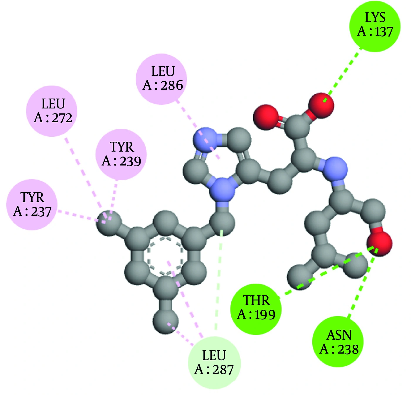
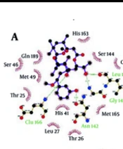
Figure 1.
Two-dimensional interaction diagrams of MLN-4760 as the reference compound with the active site of SARS-CoV-2 main protease (Mpro, PDB ID: 6LU7). Color code: light green = carbon–hydrogen bond; dark green = conventional hydrogen bond; light pink = alkyl and π-alkyl interactions.
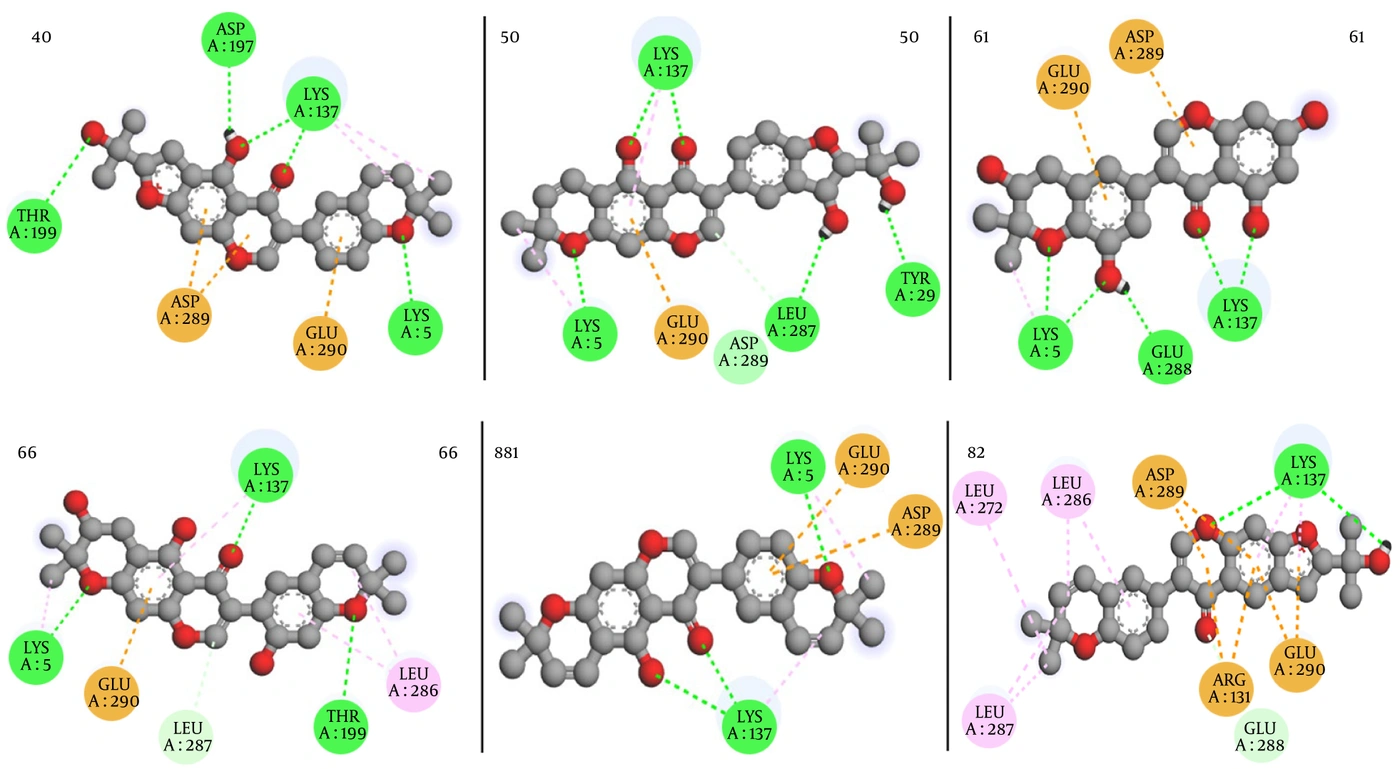
Figure 2.
Two-dimensional interaction diagrams of MLN-4760 as the reference compound with the RBD-binding pocket of human ACE2 (PDB ID: 8E7M). Color code: dark green = conventional hydrogen bond; dark pink = π-π stacking; light pink = alkyl and π-alkyl interactions.
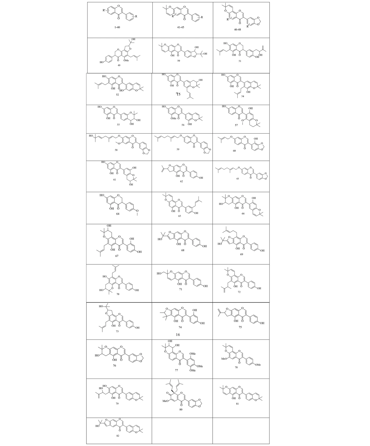
Figure 3.
Structure of examined Isoflavonoids
Structures with favorable binding energies were selected, and their interactions with the targets were examined. Ligands that interacted with key amino acids in the binding site with appropriate orientation were selected. Compounds 81 and 82 had the highest binding energy with both proteins 6LU7 and 8E7M. The interaction profiles of the selected ligands are summarized in Tables 2 and 3. To validate the docking results, comparative docking studies were performed using MLN-4760. However, because MLN-4760 is a validated ACE2 inhibitor rather than an established Mpro inhibitor, comparisons involving Mpro should be interpreted only as computational reference values and not as indicators of relative biological activity. The findings are presented in Figures 1 and 2 and Table 2 and 3.
Table 2.
The Docking Results into Protein (PDB ID = 6LU7) for Studied Compounds
| ID | Binding energy / (kcal/mol) | Amino acid residue | Type of interaction |
|---|---|---|---|
| 40 | -8.2 | Thr 199, Asp 197, Lys 137, Lys 5 / Asp 289, Glu 290 | Hydrogen bond / π-anion |
| 50 | -8.2 | Lys 137, Lys 5, Leu 287, Tyr 239 / Glu 290 | Hydrogen bond / π-anion |
| 61 | -8.2 | Lys 5, Glu 288, Lys 137 / Glu 290, Asp 289 | Hydrogen bond / π-anion |
| 66 | -8.2 | Lys 5, Lys 137, Thr 199 / Glu 290 / Leu 287 | Hydrogen bond / π-anion / Carbon hydrogen bond |
| 81 | -8.7 | Lys 137, Lys 5 / Glu 290, Asp 289 | Hydrogen bond / π-anion |
| 82 | -8.6 | Lys 137 / Leu 272, Leu 286, Leu 287 / Asp 289, Arg131, Glu 290 | Hydrogen bond / π-alkyl / π-anion |
| MLN-4760 | -6.1 | Lys137, Thr199, Asn238 / Tyr237, Tyr239, Leu272, Leu286, Leu287 | Hydrogen bond / Alkyl & π-alkyl |
Table 3.
The Docking Results into Protein (PDB ID = 8E7M) for Studied Compounds
| ID | Binding Energy / (kcal/mol) | Amino acid Residue | Type of Interaction |
|---|---|---|---|
| 40 | -7.6 | Arg 393, Asp 350 / Phe 40, Trp 349 / Ala 348 | Hydrogen bond / π-π stacked / Carbon hydrogen bond |
| 50 | -7.6 | Ser 43, Ala 348, Asp 350 / Met 62 / Trp 349 | Hydrogen bond / Alkyl / Carbon hydrogen bond |
| 61 | -7.7 | Ala 348, Ser 44 / His 378 / Phe 40 / Thr 347, Trp 349 | Hydrogen bond / Alkyl / π-π stacked / carbon-hydrogen bond |
| 66 | -7.5 | Arg 393, Asp 350 / Phe 40, Tyr 385, Trp 349 | Hydrogen bond / Alkyl |
| 81 | -7.8 | Phe 390 / Arg 393, Phe 40, Ala 348, His 378 | π -sigma / Alkyl |
| 82 | -8.2 | Phe 390 / Phe 40, Trp 349 | π-sigma / π-π stacked |
| MLN-4760 | -5.8 | Ala348, Tyr385 / Phe40 / Phe390, Arg393 | Hydrogen bond / π-π stacked / Alkyl & π-alkyl |
Docking results revealed that compounds 40, 50, 61, 66, 81, and 82 had the best binding energies of -8.2, -8.2, -8.2, -8.2, -8.7, and -8.6 kcal/mol, respectively, against the SARS-CoV-2 main protease (Mpro) (6LU7). Under the identical docking protocol, the binding energies of compounds 40, 50, 61, 66, 81, and 82 for Mpro ranged from -8.2 to -8.7 kcal/mol, whereas the reference compound MLN-4760 yielded scores of -5.8 and -6.1 kcal/mol, respectively. Within the context of this in silico screening, these differences indicate more favorable predicted binding affinities for the isoflavonoid derivatives. However, it is essential to interpret this comparison with caution, as the reference drugs were not designed to inhibit Mpro or ACE2, and docking score magnitude alone does not directly translate to biological inhibitory potential. These differences suggest stronger predicted binding under the employed docking protocol, consistent with previous reports highlighting flavonoids as effective protease inhibitors (35-37). As shown in Figure 4 and Table 2, Lys137, Lys5, Glu290, Leu287, Thr199, and Asp289 are important residues that participate in hydrogen bonding and π-interactions between the ligands and the active site of the receptor. As shown in Figure 1, key amino acids, including Lys137, Thr199, and Leu287, participate in hydrogen bonds and π-interactions between the antiviral drugs and the enzyme active site.
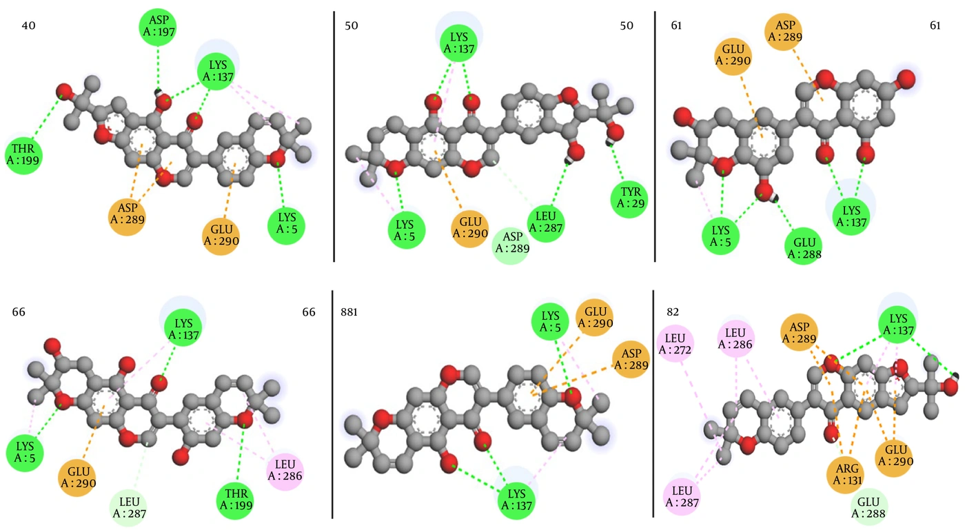
Figure 4.
Two-dimensional interaction diagrams of top-scoring isoflavonoid derivatives with the active site of SARS-CoV-2 Mpro (PDB ID: 6LU7). From left to right: compounds 40, 50, 61, 66, 81, and 82. Color code: green = van der Waals interactions; light green = carbon–hydrogen bond; dark green = conventional hydrogen bond; light pink = alkyl and π-alkyl; orange = π-cation and π-anion.
These interactions are crucial for enzymatic inhibition, as reported in earlier structural studies of coronavirus proteases (38, 39). In contrast, the reference compound exhibited weaker interaction networks, which may explain its comparatively lower binding affinities.
Based on the ACE2 docking results, compounds 40, 50, 61, 66, 81, and 82 had the best binding energies of -7.6, -7.6, -7.7, -7.5, -7.8, and -8.2 kcal/mol, respectively, against the human ACE2 receptor (8E7M). These findings suggest that the binding energies of these compounds were more favorable than the docking energies recorded at -5.8 and -6.1 for MLN-4760, respectively. The key residues involved in hydrogen bonding and π-interactions include Asp350, Phe40, Trp349, Ala348, Ser43, and Ser44. Docking of the selected compounds to the binding site of the human ACE2 receptor indicated that all significant amino acids participated in hydrogen bonds and π-interactions. These results agree with previous studies emphasizing the importance of ACE2 binding modulation in antiviral strategies (40, 41).
It is important to interpret the docking results against ACE2 within a clear mechanistic framework. In this study, we aimed to identify isoflavonoid derivatives that competitively bind to the RBD-interacting interface on ACE2, thereby sterically blocking the spike-ACE2 interaction and preventing viral entry. We do not propose direct inhibition of ACE2’s enzymatic (carboxypeptidase) activity. While compounds 81 and 82 exhibited the highest binding affinities toward the ACE2 RBD-binding pocket (Table 1 and Figure 5), these in silico findings do not directly confirm therapeutic inhibition of viral entry. The proposed mechanism is competitive steric hindrance: the isoflavonoid derivatives are predicted to occupy key residues involved in spike protein recognition (including Lys31, Glu35, Asp38, Tyr41, Gln42, Asn330, and Lys353), thereby reducing or preventing spike-ACE2 interaction. However, ACE2 is a physiologically essential host receptor that regulates blood pressure (via conversion of angiotensin II to angiotensin 1 - 7) and protects against tissue injury. Consequently, compounds that bind to the RBD-binding pocket could potentially interfere with its native enzymatic activity or downstream signaling if the binding site overlaps with the catalytic domain. Notably, the RBD-binding interface on ACE2 is distinct from the catalytic site, which is located in the extracellular peptidase domain. This spatial separation offers a theoretical opportunity to block viral entry without disrupting enzymatic function, but this hypothesis requires experimental verification. Therefore, the present results should be considered hypothesis-generating. Future experimental validation must include not only viral entry inhibition assays (e.g., pseudovirus neutralization tests) but also functional assessments of ACE2 enzymatic activity (e.g., angiotensin II hydrolysis assays) to confirm that the compounds do not adversely affect host physiology.
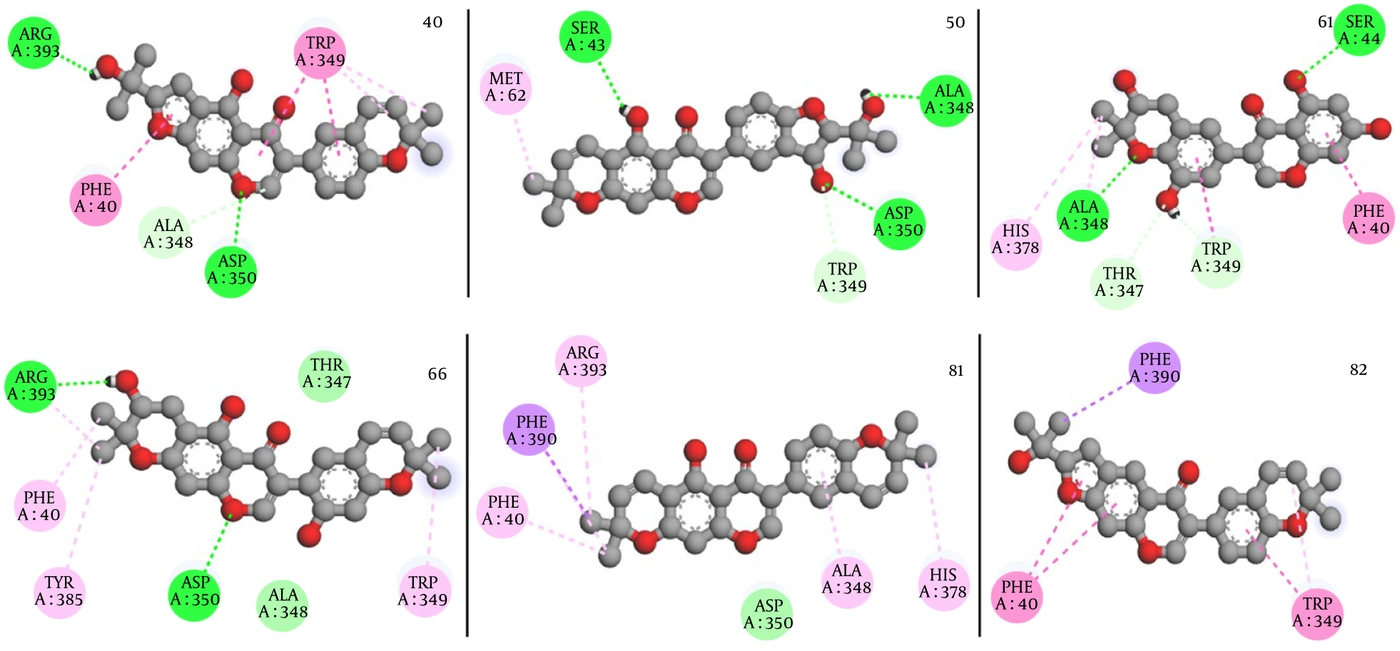
Figure 5.
Two-dimensional interaction diagrams of top-scoring isoflavonoid derivatives with the RBD-binding pocket of human ACE2 (PDB ID: 8E7M). From left to right: compounds 40, 50, 61, 66, 81, and 82. Color code: green = van der Waals interactions; light green = carbon–hydrogen bond; dark green = conventional hydrogen bond; dark pink = π-π stacking; light pink = alkyl and π-alkyl; purple = π-sigma.
A critical mechanistic distinction should be emphasized: our goal is to identify compounds that sterically block the spike–ACE2 interaction without necessarily inhibiting ACE2 enzymatic activity. As documented in the literature, the ACE2 RBD-binding interface is spatially distinct from the catalytic site, which contains the Zn²⁺ ion coordinated by residues His374, His378, Glu402, and His405 (42, 40). The RBD interface is located on the outer surface of the peptidase domain, whereas the catalytic site is more deeply buried. In addition, well-characterized ACE2 enzymatic inhibitors typically contain zinc-chelating pharmacophores (carboxylates or hydroxamates), a feature absent in our isoflavonoid derivatives. However, we acknowledge that allosteric effects cannot be excluded, and experimental enzymatic assays are required to confirm that ACE2 function is not disrupted.
4.1.1. Limitations of Docking-Score Comparisons Across Different Compound Classes
While our docking results suggest that the selected isoflavonoids possess favorable binding energies toward Mpro and ACE2 compared with the reference drugs, several important limitations must be acknowledged. First, docking scores are influenced by molecular size, the number of rotatable bonds, and hydrogen-bonding capacity—properties that differ substantially between the isoflavonoids (MW ~370 - 440 Da, multiple aromatic rings and hydroxyl groups) and the smaller reference drugs (MW ~170 - 430 Da, fewer hydrogen-bond donors/acceptors). Second, binding affinity predicted by AutoDock Vina does not account for solvation effects, entropic penalties, or conformational changes in the protein upon ligand binding. Therefore, the observed differences in docking scores should be interpreted as predicted binding preferences under equilibrium conditions, not as direct measures of antiviral efficacy. Experimental validation using enzymatic inhibition assays (for Mpro) and spike-ACE2 binding competition assays (for ACE2) is required to determine actual inhibitory activity.
4.2. DFT Calculations
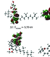
To predict the reactive electrophilic or nucleophilic positions of a molecule, analysis of frontier molecular orbitals (HOMO and LUMO) can be used. Examination of the frontier-orbital energy values and the energy gap provides essential information to predict the biological activity of the studied compounds. A smaller energy gap indicates higher polarizability, which in turn suggests greater biological activity. The HOMO and LUMO energies, as well as the energy gap for compound 82, the most effective compound, and compound 32, the compound with the lowest activity, are illustrated in Figure 6. The frontier orbital gap for the compounds indicated an order of reactivity of 82 > 32. The molecular orbital analysis showed that the LUMO of 32 and 82 was localized on the flavonoid region.
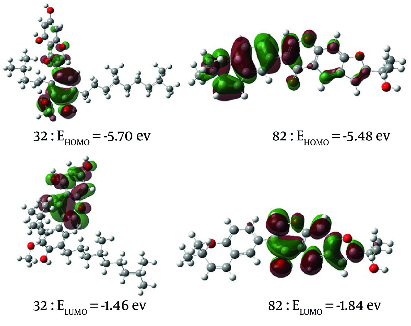
Figure 6.
Frontier molecular orbitals (HOMO and LUMO) of compounds 32 and 82. Top panels: HOMO surfaces (isovalue = 0.02); bottom panels: LUMO surfaces (isovalue = 0.02). Corresponding energies (in eV) are shown. Compound 82 shows a smaller HOMO–LUMO gap (3.64 eV) compared to compound 32 (4.24 eV), indicating higher reactivity.
It should be emphasized, however, that the DFT descriptors reported herein were calculated in the gas phase due to the convergence difficulties encountered with implicit solvation models. Although gas-phase calculations are widely employed for qualitative assessments of electronic structure and relative reactivity among congeners—and are considered acceptable for establishing trends rather than absolute values—the absence of solvation effects represents a limitation. Biological environments are aqueous, and solvation can significantly influence molecular orbital energies, charge distributions, and hydrogen-bonding propensities. Therefore, while our DFT results support the docking findings in terms of relative ordering of reactivity (e.g., compound 82 > compound 32), the absolute values of descriptors such as hardness, chemical potential, and electron affinity should be regarded as semi-quantitative estimates that require further validation using solvated DFT or molecular dynamics-based QM/MM approaches.
The molecular electrostatic potential (MEP) is an appropriate tool for analyzing reactive properties and characterizing intermolecular interactions. Nucleophilic and electrophilic regions of a molecule can be identified using MEP, which can help predict where hydrogen bonds may form. The Mulliken charges for the studied ligands are presented in Figure 7. It can be observed that the total charge of the molecule is equal to zero, and the negative charge is delocalized on oxygen atoms. The electrostatic surface potential (ESP) shows the distribution of positive and negative charges throughout the molecule, based on which sites of electrophilic or nucleophilic attack can be predicted. The ESP maps of the studied structures are shown in Figure 7, where the electron-rich, electron-deficient, slightly electron-rich, and neutral sites are colored red, blue, yellow, and green, respectively.
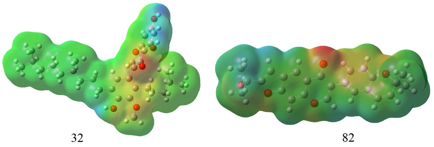
Figure 7.
Molecular electrostatic potential (ESP) maps of compounds 32 and 82 calculated at the B3LYP/6 - 311++G(d,p) level. Red regions: electron-rich (negative potential, nucleophilic); blue regions: electron-deficient (positive potential, electrophilic); yellow: slightly electron-rich; green: neutral. The negative potential is primarily localized on oxygen atoms, supporting their involvement in hydrogen bond formation with amino acid residues.
The MEP analyses confirmed the distribution of electrophilic and nucleophilic regions within the molecules, facilitating the prediction of interaction sites. The negative electrostatic potential localized on oxygen atoms supports their involvement in hydrogen bond formation with amino acid residues, consistent with established structure–activity relationships in flavonoid-based inhibitors (43, 44).
The thermochemical parameters, such as total energy (E), enthalpy (H°), Gibbs free energy (G°), entropy (S), and some reactivity descriptors, including softness (σ), hardness (η), electron affinity (EA), and chemical potential (μ), are listed in Table 4. The tendency of a compound to participate in a charge transfer reaction can be predicted by the ratio of ΔE/ΔNmax. A zero value for this ratio indicates that a molecule is electron-saturated and would not participate in this kind of process. This ratio for all studied compounds is not zero, indicating that they can participate in a charge transfer process. The stability of a compound can be indicated by the Gibbs free energy. If this value is positive, it means that the compound is unstable, whereas a negative value of G indicates that the compound is thermodynamically stable. Positive values for entropy indicate disorder in the system, whereas negative values for S reveal that the system is more ordered. Based on the results, the total energy for compound 32 is more negative than that for compound 82, indicating that 32 is more stable. The order of electron affinity (EA) is 82 > 32, indicating the higher activity of compound 82 in nucleophilic reactions. All calculated hardness values are positive, as expected for stable molecules, with compound 32 (η = 2.12 eV) being harder (less polarizable) than compound 82 (η = 1.82 eV). The lower hardness and higher softness of compound 82 indicate greater polarizability and potentially higher chemical reactivity, consistent with its superior docking scores. The non-zero ΔE/ΔNmax values indicate that all compounds are able to participate in charge transfer interactions, which are essential for ligand–protein binding (45).
Table 4.
The Calculated Thermochemistry Parameters for Studied Structures a
| ID | 32 | 82 |
|---|---|---|
| Etot b | -1735.335 | -1341.227 |
| H b | -1735.292 | -1341.201 |
| G b | -1735.417 | -1341.282 |
| S c | 263.319 | 169.680 |
| Egap | 4.24 | 3.64 |
| IP | 5.70 | 5.48 |
| EA d | 1.46 | 1.84 |
| η d | 2.12 | 1.820 |
| σ | 0.47 | 0.55 |
| μ | -3.58 | -3.66 |
| ΔE | -3.02 | -3.68 |
| ΔNmax | 1.69 | 2.01 |
| ΔE/ΔNmax | -1.79 | -1.83 |
a In eV-1.
b In Hartree/particle.
c In cal/(mol.K).
d In eV.
4.3. Pharmacokinetics and Toxicity Profile
PreADMET software and the SwissADME online server are important tools for anticipating the pharmacokinetics and toxicity of compounds. These tools can help scientists develop orally well-absorbed drugs. In the drug discovery process, the Rule of 5 anticipates that poor absorption or penetration is more likely when the number of H-bond donors is more than 5, acceptors are more than 10, the molecular weight is larger than 500, and the computed LogP is higher than 5. However, this rule specifically emphasizes that the Rule of 5 applies to compounds that are not substrates for active transporters. The physicochemical properties of the top-ranked compounds by docking score (40, 50, 61, 66, 81, and 82) are given in Tables 5-7. As indicated in Table 5, all studied compounds conform to the Lipinski rule of five.
Table 5.
Physicochemical Properties of Compounds
| Entry | MW | n-R | HBA | HBD | TPSA (A2) | MLogP | Lipinski Violation |
|---|---|---|---|---|---|---|---|
| 40 | 420.50 | 6 | 5 | 2 | 79.90 | 2.84 | 0 |
| 50 | 436.45 | 2 | 7 | 3 | 109.36 | 1.10 | 0 |
| 61 | 370.35 | 1 | 7 | 4 | 120.36 | 0.36 | 0 |
| 66 | 436.45 | 1 | 7 | 3 | 109.36 | 1.37 | 0 |
| 81 | 402.44 | 1 | 5 | 1 | 68.90 | 2.64 | 0 |
| 82 | 402.44 | 2 | 5 | 1 | 72.81 | 2.30 | 0 |
| Rule of Lipinski | ≤500 | ≤10 | ≤10 | ≤5 | ≤140 | ≤5 | ≤1 |
The Human Intestinal Absorption (HIA) percentage is defined as the extent to which a drug is absorbed from the intestine after oral administration. As shown in Table 6, all compounds exhibited higher values in the range of 83.7 - 96.5% and were predicted to be well absorbed. Caco-2 cell permeability (CCP) is used to investigate in vitro human intestinal permeability of a compound. All selected compounds showed moderate cell culture permeability. All compounds exhibited weak to moderate MDCK values ranging from 0.044 - 0.076 nm/s, representing drug uptake efficiency. In addition, P-glycoprotein (P-gp) is one of the primary barriers to drug transfer, and it excludes toxins and xenobiotics from cells. As indicated in Table 6, all compounds can inhibit P-gp except 50 and 61 analogues.
Table 6.
In Silico ADME Prediction of Compounds
| Entry | HIA % | Caco2 (nm/sec) | MDCK/ (nm/s) | P-gp inhibition | in vitro skin permeability (logKp, cm h-1) | PPB% | BBB |
|---|---|---|---|---|---|---|---|
| 40 | 95.037940 | 26.3786 | 0.0449867 | Inhibitor | -2.19612 | 97.158152 | 4.5185 |
| 50 | 92.231838 | 2.81439 | 0.0448564 | Non | -2.52048 | 82.388764 | 0.0991487 |
| 61 | 83.707843 | 17.0327 | 0.0767351 | Non | -4.18523 | 84.383915 | 0.100561 |
| 66 | 92.071572 | 2.81494 | 0.0443958 | Inhibitor | -2.78844 | 88.666303 | 0.183573 |
| 81 | 96.306327 | 31.1371 | 0.0455693 | Inhibitor | -2.09294 | 91.030133 | 0.17835 |
| 82 | 96.538546 | 44.6644 | 0.0492143 | Inhibitor | -1.9145 | 93.510626 | 0.0533213 |
All selected compounds had negative skin permeability values. Plasma protein binding (PPB) can affect the pharmacodynamic and pharmacokinetic behavior of a drug. As shown in Table 6, the PPB% of compounds 50, 61, and 66 is low (< 90), and for compounds 40, 81, and 82, the PPB% was > 90. Finally, the blood-brain barrier (BBB) values of all compounds are low (Cbrain/Cblood < 1), except for compound 40. Therefore, they may not have neurotoxic side effects.
The toxicity profile of the selected compounds is shown in Table 7. As shown in Table 7, the Ames test predicted compound 82 as mutagenic, while all other selected compounds were nonmutagenic. None of the compounds showed predicted carcinogenicity. All six compounds exhibited medium risk for hERG inhibition, indicating a potential cardiotoxicity concern that should be evaluated experimentally. No neurotoxicity (BBB penetration) was predicted for compounds 50, 61, 66, 81, and 82, while compound 40 showed potential BBB permeability (46-49).
Table 7.
Toxicity Prediction of Compounds
| Entry | Ames Test | Carcino Mouse | hERG Inhibition |
|---|---|---|---|
| 40 | nonmutagenic | negative | medium risk |
| 50 | nonmutagenic | negative | medium risk |
| 61 | nonmutagenic | negative | medium risk |
| 66 | nonmutagenic | negative | medium risk |
| 81 | nonmutagenic | negative | medium risk |
| 82 | mutagen | negative | medium risk |
The toxicity predictions raise important safety concerns that must be addressed before any experimental studies. Compound 82's predicted Ames positivity suggests a potential mutagenic mechanism, which may be related to the presence of specific aromatic hydroxylation patterns or conjugated systems capable of undergoing metabolic activation to reactive intermediates. To mitigate this liability, we propose that future medicinal chemistry efforts focus on bioisosteric replacement of hydroxyl groups with less redox-active moieties (e.g., methoxy, fluorine, or trifluoromethyl groups) or reduction of the number of aromatic hydroxyl groups to minimize oxidative bioactivation. Alternatively, the introduction of electron-withdrawing groups at positions susceptible to metabolic oxidation may reduce the formation of reactive quinone-like intermediates. Regarding the universal medium hERG risk, we suggest that reducing overall lipophilicity, breaking molecular planarity through the introduction of sp³-hybridized carbons, or masking basic nitrogen centers (e.g., as amides or esters) could reduce hERG affinity without compromising target binding. We emphasize that these structural modifications are purely speculative at this stage and require iterative medicinal chemistry optimization coupled with experimental validation (hERG patch-clamp assays and Ames testing) to confirm whether safety can be improved while maintaining antiviral activity.
Overall, the integration of docking, electronic structure analysis, and pharmacokinetic evaluation demonstrates that flavonoid derivatives, particularly compound 82, are candidates for further antiviral drug development. These findings are consistent with previous studies highlighting the therapeutic potential of flavonoids against viral targets (50-53). It is important to note that these ADMET predictions are based on computational models and do not replace experimental pharmacokinetic and toxicological studies. The absence of predicted neurotoxicity and favorable intestinal absorption values should be considered encouraging indicators for prioritization, not confirmed safety profiles.
5. Discussion
In this study, a library of isoflavonoids was evaluated as potential inhibitors of SARS-CoV-2 Mpro and ACE2 using in silico approaches. Isoflavonoid derivatives, particularly Ulexin C (compounds 81 and 82), were identified as computationally prioritized candidates with favorable predicted binding to SARS-CoV-2 Mpro and the ACE2 spike-binding interface. The docking results indicated hydrogen bonding and π-π stacking interactions with Lys 137, Lys 5, Glu 290, Leu 287, Thr 199, and Asp 289 in Mpro. In addition, Asp 350, Phe 40, Trp 349, Ala 348, Ser 43, and Ser 44 were identified as key residues in the ACE2 active site that may contribute to stable ligand binding. The predicted binding energies of the top isoflavonoid derivatives ranged from -7.6 to -8.7 kcal/mol for Mpro and -7.5 to -8.2 kcal/mol for ACE2, which were more negative than those calculated for MLN-4760 (range, -5.8 to -6.1 kcal/mol) under the same docking protocol. DFT calculations indicated favorable electronic properties and thermodynamic stability for Ulexin C (compounds 81 and 82). However, it is critical to emphasize that these computational findings do not directly predict antiviral efficacy. Docking scores reflect predicted binding affinities rather than functional inhibition. Therefore, biochemical and cellular validation—including enzymatic inhibition assays for Mpro and spike–ACE2 competition assays—is essential to confirm the actual inhibitory activity of the identified compounds. Importantly, compound 82 carries a predicted mutagenicity risk, and all compounds show medium hERG inhibition potential, representing major safety liabilities. Therefore, while compounds 50, 61, 66, and 81 may be considered starting points for further optimization (with compound 82 requiring careful safety evaluation and structural modification to eliminate mutagenicity), none of these compounds should be considered direct drug leads without extensive experimental safety profiling and rational medicinal chemistry optimization. The favorable ADMET profiles, particularly the absence of predicted neurotoxicity, support further in vitro evaluation; however, safety pharmacology must be prioritized alongside efficacy studies.
5.1. Limitations and Required Experimental Validation
The present study is entirely computational. No enzymatic, biophysical, cell-based, or antiviral assays were performed. Therefore, the docking scores, DFT descriptors, and ADMET predictions presented here are hypothesis-generating and do not constitute evidence of antiviral activity or safety. The following experimental assays are required to validate or refute these in silico predictions. In addition, the DFT calculations were performed in the gas phase, as solvated calculations (PCM with water) could not be completed due to computational resource limitations. Although gas-phase DFT is widely used for qualitative comparisons of reactivity trends within a series of structurally related compounds, it does not fully capture the electronic redistribution and stabilization that occur in aqueous physiological environments. Solvation can influence frontier orbital energies, charge distributions, and hydrogen-bonding propensities, which may affect predictions of ligand–protein interactions. Therefore, the absolute values of electronic descriptors (e.g., hardness, chemical potential) should be regarded as semi-quantitative estimates. Future studies using solvated DFT or QM/MM approaches are recommended to refine these values under physiological conditions.
A critical limitation of this study is that the predicted toxicity profiles—particularly the mutagenicity of compound 82 and the medium hERG risk across all compounds—are derived from computational models and have not been experimentally confirmed. These predictions may overestimate or underestimate actual toxicity, and experimental validation is essential. Furthermore, even if confirmed, the mutagenicity of compound 82 represents a severe liability that would require extensive structural modification. We therefore recommend that future studies prioritize compounds 50, 61, 66, and 81 for initial experimental evaluation, and that compound 82 be considered only after rational structural modifications have been designed and tested to eliminate its predicted mutagenic potential.
To validate the computational predictions, we recommend the following experimental studies:
- Mpro enzymatic inhibition assay: Using a fluorogenic substrate (e.g., a FRET-based assay) to measure the half-maximal inhibitory concentration (IC50) of compounds 81 and 82 against recombinant SARS-CoV-2 Mpro.
- Spike–ACE2 binding competition assay: Using surface plasmon resonance (SPR) or ELISA-based methods to assess whether Ulexin C derivatives competitively block the interaction between the spike protein RBD and ACE2.
- Cytotoxicity assessment: Standard MTT or CCK-8 assays in human cell lines (e.g., Vero E6, HEK293T, or Calu-3) to determine the half-cytotoxic concentration (CC50) and selectivity index (SI = CC50/IC50).
- Antiviral activity in cell culture: Plaque reduction assay or qRT-PCR-based viral load quantification using live SARS-CoV-2 (BSL-3 required) or pseudovirus particles expressing the spike protein.