
1. Background
The intensive care unit (ICU) is a specialized setting that provides continuous monitoring of critically ill patients by multidisciplinary teams (1). Many patients require mechanical ventilation until adequate spontaneous breathing returns, with an estimated 20 million patients receiving this support annually worldwide (2, 3). These patients often require invasive monitoring and multisystem support because of complex conditions (4). Age-related changes, comorbidities, and multiorgan dysfunction increase the risk of adverse outcomes and require coordinated multidisciplinary management (5).
A major challenge in the management of critically ill patients is controlling pain, agitation, anxiety, sleep disturbances, and intolerance to mechanical ventilation, making sedative and analgesic agents unavoidable in many cases (6). Inadequate control of pain and agitation may increase sympathetic activity, lead to accidental removal of endotracheal tubes and catheters, and, in the long term, cause chronic pain, post-traumatic stress disorder (PTSD), and reduced health-related quality of life (7). Commonly used sedatives include benzodiazepines, opioids, barbiturates, and nonbenzodiazepines such as propofol and dexmedetomidine, with opioids receiving particular attention because of their combined analgesic and sedative properties (8, 9).
Despite their essential role in ICU care, these medications have well-recognized adverse effects, particularly at high doses or with prolonged use, and may affect multiple organ systems in vulnerable patients (10). Deep and prolonged sedation is associated with longer ICU stays, longer durations of mechanical ventilation, delirium, ICU-acquired weakness, higher mortality, and increased hospital workload (11). In addition, because of their immunomodulatory effects, these agents may increase the risk of infection, microaspiration, gastrointestinal motility disorders, and healthcare costs (12). For example, Gil Castillejos et al. (13) found that excessive prolonged sedation, compared with light sedation, resulted in longer mechanical ventilation, more pressure ulcers and ventilator-associated pneumonia, and a greater need for tracheostomy. Similarly, AhmadAli et al. (14) reported that excessively deep and inappropriate sedation through continuous intravenous infusion could cause delayed awakening, altered respiratory mechanics, and impaired airway protection.
Conversely, evidence suggests that proactive strategies to minimize sedation-related complications can improve clinical outcomes. One such strategy is DSI. Yousefi et al. (7) demonstrated that implementing this approach could reduce the duration of mechanical ventilation and the incidence of ventilator-associated pneumonia. A systematic review by Vagionas et al. (11) also concluded that protocolized sedation is a safe strategy for facilitating earlier weaning and improving clinical outcomes. Similarly, Anwar Abdel ElAziz et al. (12) reported that patients who underwent DSI had lower delirium rates, shorter ICU stays, and a reduced duration of ventilator dependence. Nurses can play a critical role in this context because of their 24-hour bedside presence, continuous monitoring of sedation levels, and early detection of complications. Their active involvement in implementing protocols such as DSI is essential for reducing ventilator-related complications and improving patient outcomes (7).
Nevertheless, deep and prolonged sedation continues to occur in clinical practice because of factors such as excessive administration of sedative agents without clear medical indications and inadequate implementation of sedation protocols. These factors can complicate recovery from coma and delay patient awakening (8). Therefore, in addition to applying objective protocols for sedative administration, strategies to mitigate complications associated with delayed awakening appear essential.
ICU patients also commonly experience sensory deprivation and overload because of factors such as brain dysfunction, an unfamiliar environment, imbalanced sensory input, heavy sedation, noise, and painful procedures (15). This condition is associated with delirium, anxiety, agitation, reduced consciousness, hemodynamic instability, pain, and hallucinations and may lead to prolonged mechanical ventilation, a longer hospital stay, and increased mortality (16). Conversely, previous studies have demonstrated the beneficial effects of balanced sensory stimulation. For example, Adineh et al. (17) reported that a balanced sensory stimulation program delivered by family members improved levels of consciousness, reduced pain, and enhanced comfort among ICU patients.
2. Objectives
Although DSI and sensory stimulation independently improve outcomes in mechanically ventilated patients, their combined use within a daily awakening protocol has not been evaluated. Given the limited evidence, particularly in Iran, this study assessed the effect of a combined daily awakening protocol on clinical outcomes in mechanically ventilated ICU patients.
3. Methods
3.1. Design and Setting
This two-group randomized clinical trial investigated the effect of implementing a daily awakening protocol on clinical outcomes in mechanically ventilated patients admitted to the general ICUs of Golestan Hospital, a teaching hospital affiliated with Ahvaz Jundishapur University of Medical Sciences in Ahvaz, Iran. The trial was conducted from January to August 2025. Because of the nature of the intervention and the clinical status of the patients, most of whom were mechanically ventilated and receiving sedative medications, patient-level blinding was not feasible or practical. However, outcome assessors and data analysts were blinded to group allocation to minimize potential bias.
The study ICUs comprised 44 active beds and employed 64 nurses, with a nurse-to-bed ratio of approximately 1:4 across most shifts. An intensivist was fully present during the morning shift in both units and conducted daily rounds for all patients. Most patients admitted to these units required level 5 care, indicating the need for mechanical ventilation and advanced life-support interventions.
This study was conducted and reported in accordance with the Consolidated Standards of Reporting Trials (CONSORT) guidelines for clinical trial reporting. Patients or the public were not involved in the design, conduct, reporting, or dissemination plans of this research. No major changes were made to the study protocol after its commencement.
3.2. Population
The sample size was determined using MedCalc statistical software with a power of 90% and a type I error of 5%, resulting in a total sample size of 74 patients (37 in the intervention group and 37 in the control group). These parameters were taken from the study by Anwar Abdel ElAziz et al. (12) for the outcome of duration of mechanical ventilation. Finally, considering the potential for dropout, 90 patients were selected (45 per group) after adding 20% to the calculated sample size of 74 (74 × 1.2 = 88.8, rounded to 90).
z1-α/2 = 1.96, z1-β = 1.645, SD1 = 2.56, SD2 = 1.91, μ1 = 6.91, μ 2 = 5.43
Eligible participants were initially selected using convenience sampling based on the inclusion criteria and were then allocated to the intervention and control groups using permuted block randomization. All patients who met the inclusion criteria during the study period were enrolled, and none declined; therefore, no patients were excluded before randomization. The randomization sequence was generated by a biostatistician who was not involved in patient enrollment, using computerized random number generation with permuted block randomization. A total of nine blocks of 10 were used to reach the target sample size of 90, accounting for potential dropout. All possible sequences of 10 assignments consisting of group A (intervention) and group B (control) were generated, and one sequence was randomly assigned to each block by the biostatistician.
To ensure allocation concealment, the sequence was placed in sequentially numbered, opaque, sealed envelopes by an independent research assistant who had no role in patient screening or enrollment. Envelopes were opened only after a patient met all inclusion criteria and the legal guardian provided written informed consent. Assignment proceeded sequentially within each block. In block 1, patients 1 to 10 were assigned to groups A, B, A, B, A, B, A, B, A, and B, respectively; in block 2, they were assigned to groups A, B, B, A, A, B, B, A, A, and B, respectively; and so forth. In the ninth block, the first patient received group B, the second group A, the third group B, continuing to the tenth patient, who received group A.
The inclusion criteria were age 20 to 60 years, mechanical ventilation at study initiation, an initial Glasgow Coma Scale (GCS) score of 6 to 12, an initial Richmond Agitation-Sedation Scale (RASS) score of ≥ -3 (scores of -3, -2, -1, 0, or higher; patients with RASS scores of -4 or -5 were not eligible), an Acute Physiology and Chronic Health Evaluation II (APACHE II) score of 30 to 40, receipt of continuous sedation infusion, and written informed consent provided by the patient’s legal guardian.
The exclusion criteria were discharge or death before 7 days after intervention initiation. Patients were also excluded if sedation infusion was completely discontinued before day 7, irrespective of the cause, including successful weaning, because the protocol required 7 consecutive days of pre- and postintervention GCS and RASS measurements. Patients who were weaned after completing the 7-day protocol remained in the analysis. Other exclusion criteria included receipt of end-of-life care, administration of neuromuscular blocking agents during the study period, any condition deemed by the attending physician to contraindicate sedative interruption, prior ICU admission within the previous 30 days to avoid duplicate sampling, withdrawal of consent by the legal guardian, anticipated transfer to another ICU or hospital during the study period, and concurrent participation in other interventional studies.
3.3. Intervention
In this study, patients in the intervention group received a daily awakening protocol during their ICU stay for 2 hours per day, between 4:00 and 6:00 PM. This timing was chosen to align with the natural circadian trough in arousal and to minimize interference with morning rounds and nighttime sleep. During these 2 hours, after coordination with the ICU attending physician and ward nurses, continuous sedative infusion was discontinued. Simultaneously, the principal investigator or a trained research assistant administered the sensory stimulation program. Throughout this period, the patient’s clinical and hemodynamic status was continuously monitored. In the event of any complication, the ICU physician was promptly notified to initiate necessary therapeutic and supportive measures, including resumption of sedative infusion, if needed. Both the principal investigator and the research assistant acquired adequate proficiency in delivering sensory stimulation before the study began and performed the intervention in full coordination according to a unified protocol.
The sensory stimulation program used in this study was adapted from Adineh et al. (18), originally developed for ICU patients. The procedure was as follows. First, arousal stimulation was provided by speaking the patient’s name, current time, place, and date near the patient’s ear, repeated three times within 1 hour. Second, auditory stimulation was provided by playing preferred music or voices of family members or acquaintances, either speaking directly to the patient or conversing with each other, for 10 minutes. Third, visual stimulation was provided by holding family photographs, videos, a mirror, colored paper, or a 40-W red, blue, or green light bulb in front of the patient’s eyes for 10 minutes. If the eyes were closed, they were gently opened by hand. Fourth, olfactory stimulation was provided by holding familiar scents, such as perfume, spices, pickled items, medicinal herbs, orange or lemon peel, garlic, or onion, in front of the patient’s nose for 10 seconds. Fifth, tactile stimulation was provided through hand pressure, massage, and rubbing of the extremities, first on one side of the body and then on the other, once per hour. Sixth, motor stimulation was provided by moving the joints of the hands, feet, wrists, hips, and shoulders through their normal range of motion by alternating flexion/extension and raising/lowering of the limbs, performed 15 times per limb per hour.
Patients in the control group had their sedative medication infusion discontinued at the same specified hours under the same conditions as the intervention group. However, no planned sensory stimulation program was implemented for these patients; they received routine ICU care only.
At enrollment, all patients were receiving continuous sedation infusion as part of routine ICU care. The sedative regimen consisted of fentanyl (0.5 - 2 μg/kg/h) and/or midazolam (1 - 5 mg/h), administered either in combination or as single agents and adjusted by the attending physician based on clinical judgment. No specific RASS target was mandated because the study aimed to evaluate the effect of the daily awakening protocol on sedation-agitation status.
3.4. Data Collection Methods and Tools
Data were collected using a structured two-part form. The first part captured demographic and baseline information, including age, sex, clinical diagnosis, type of sedative and analgesic medications used, and initial GCS, APACHE II, and RASS scores. These data were obtained through interviews with family members and nurses, medical record review, and direct physical examination of patients. The second part recorded clinical outcomes, including ICU length of stay, duration of mechanical ventilation, ICU mortality, occurrence of delirium, and the patient’s level of consciousness and sedation-agitation status before and after the intervention.
Data were collected by trained research assistants who were blinded to group allocation. All outcome assessors were experienced ICU nurses with at least 2 years of experience and received standardized training. Throughout the study, they participated in regular coordination meetings to ensure protocol adherence and accurate data collection. To maintain blinding despite the fixed daily intervention time of 4:00 to 6:00 PM, outcome assessors were different from the intervention providers and did not enter ICU rooms during the intervention window. Because both groups received DSI at the same time, stopping sedation did not reveal allocation; the additional sensory stimulation was delivered behind closed curtains or while the assessor was absent. Assessors were also instructed not to discuss sedation schedules with staff or review patient charts for group assignment. Before the study, all outcome assessors completed training on RASS and CAM-ICU. Inter-rater reliability was assessed using 10 video-recorded patient scenarios, demonstrating excellent agreement (intraclass correlation coefficient for RASS = 0.92; Cohen kappa for CAM-ICU = 0.89).
The primary outcomes were sedation-agitation scores and level of consciousness, analyzed as mean differences between the two groups. Secondary outcomes included ICU length of stay, defined as days from ICU admission to ICU discharge; duration of mechanical ventilation, defined as days from initiation of respiratory support to successful weaning; ICU mortality; and occurrence of delirium.
Disease severity at ICU admission was assessed using the APACHE II scale, introduced by Knaus in 1985. This instrument includes 12 physiological variables that evaluate the status of major body systems. According to the standard scoring table, the corresponding mortality rates for scores of 0 to 15, 16 to 19, 20 to 30, and > 30 are approximately 10%, 15%, 35%, and 75%, respectively (19). In the study by Rahmatnejad et al. (20), the area under the curve for this tool was reported as 0.775, indicating high predictive power for mortality risk in ICU patients.
Patients’ level of consciousness was assessed using the GCS immediately before and after the intervention during the first 7 days. The reliability of this instrument in Iran was confirmed by Adinehvand et al. (21), with a test-retest correlation coefficient of 0.86.
Patients’ sedation-agitation level was assessed immediately before and after the intervention during the first 7 days using the RASS. This tool has 10 levels: four levels for agitation (+1 to +4), one level for alert and calm (score 0), and five levels for different depths of sedation (-1 to -5). The RASS was developed by Sessler and colleagues in 2002, and its validity and reliability have been confirmed. In Iran, the scale was translated by Ghabimi et al. (22), who assessed its content validity and reported a reliability intraclass correlation coefficient of 0.95.
Delirium occurrence was assessed twice daily, during the early morning and late evening shifts, throughout the ICU stay using the CAM-ICU. This instrument provides a rapid clinical method for diagnosing delirium in ICU patients, including those who are intubated or unable to speak. The CAM-ICU evaluates four features: acute onset or fluctuating course, inattention, altered level of consciousness, and disorganized thinking. The presence of the first two features plus either of the remaining two indicates delirium (23). In 2019, Arbabi et al. (24) reported a sensitivity of 75%, specificity of 96%, positive predictive value of 92%, negative predictive value of 85%, and kappa coefficient of 0.74 for the CAM-ICU.
In this study, harms were defined as any adverse event associated with implementation of the evidence-based care protocol, such as disconnection of medical devices or hemodynamic instability. These events were systematically monitored through daily clinical assessments by the ICU team and documented in research records. No predefined threshold for harm severity was established; all events were reported descriptively.
3.5. Statistical Analysis
Data were analyzed using descriptive and inferential statistics with SPSS version 22. Continuous variables are reported as mean ± standard deviation, and categorical variables are reported as frequency (percentage). The Shapiro-Wilk test was used to assess normality. Accordingly, the Mann-Whitney U test was used to compare continuous variables between groups, the chi-square test was used for categorical variables, and repeated-measures analysis of variance was used to examine changes in variables over time. The significance level was set at 0.05. No adjustment was made for multiple comparisons. There were no missing data for primary or secondary outcomes among the 83 patients who completed the study. A per-protocol analysis was conducted because 7 patients were excluded after randomization due to death before day 7 (n = 3) or complete discontinuation of sedation infusion before day 7 (n = 4).
4. Results
Of the 90 enrolled patients, 45 were allocated to the intervention group and 45 to the control group. Seven patients were excluded from the final analysis. In the intervention group, 1 patient died, and 2 patients had complete discontinuation of sedation infusion before day 7. In the control group, 2 patients died, and 2 patients had complete discontinuation of sedation infusion before day 7. Thus, data from 83 patients, including 42 in the intervention group and 41 in the control group, were analyzed. These 83 patients comprised the same population presented in Table 1 and included in all subsequent analyses (Figure 1).
Table 1.
Baseline Demographic and Clinical Characteristics of Participants (N = 83) a
| Variables and Groups | No. | Values | Mean Rank | U/χ2 | P Value |
|---|---|---|---|---|---|
| Age | 837.50 | 0.830 | |||
| Intervention | 42 | 38.19 ± 14.98 | 42.56 | ||
| Control | 41 | 38.63 ± 14.57 | 41.43 | ||
| APACHE II | 744.50 | 0.286 | |||
| Intervention | 42 | 36.00 ± 3.17 | 44.77 | ||
| Control | 41 | 35.34 ± 3.35 | 39.16 | ||
| Initial GCS | 839.00 | 0.837 | |||
| Intervention | 42 | 7.66 ± 1.46 | 41.48 | ||
| Control | 41 | 7.68 ± 1.36 | 42.54 | ||
| Initial RASS | 750.50 | 0.300 | |||
| Intervention | 42 | -0.02 ± 1.58 | 44.63 | ||
| Control | 41 | -0.34 ± 1.45 | 39.30 | ||
| Gender | 0.607 | 0.436 | |||
| Male | |||||
| Intervention | 30 | 30 (71.4) | - | ||
| Control | 26 | 26 (63.4) | - | ||
| Female | |||||
| Intervention | 12 | 12 (28.6) | - | ||
| Control | 15 | 15 (36.6) | - | ||
| Initial Diagnosis | 0.969 | 0.325 | |||
| Traumatic | |||||
| Intervention | 27 | 27 (64.3) | - | ||
| Control | 22 | 22 (53.7) | - | ||
| Medical | - | ||||
| Intervention | 15 | 15 (35.7) | - | ||
| Control | 19 | 19 (46.3) | - | ||
| Type of sedative drug | 0.004 | 0.998 | |||
| Fentanyl + midazolam | |||||
| Intervention | 31 | 31 (73.8) | - | ||
| Control | 30 | 30 (73.2) | - | ||
| Fentanyl alone | |||||
| Intervention | 8 | 8 (19.0) | - | ||
| Control | 8 | 8 (19.5) | - | ||
| Midazolam alone | |||||
| Intervention | 3 | 3 (7.1) | - | ||
| Control | 3 | 3 (7.3) | - |
a Values are expressed as mean ± SD or No. (%) unless otherwise indicated.
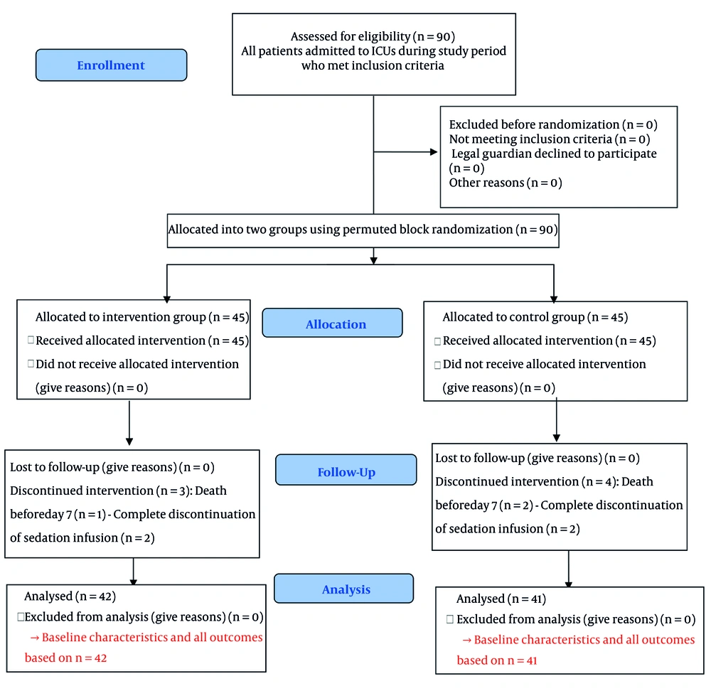
Figure 1.
CONSORT flow diagram of patients participating in the study
4.1. Baseline Characteristics
The Mann-Whitney U test showed no significant baseline differences between the intervention and control groups in age (P = 0.830), APACHE II score (P = 0.286), initial GCS (P = 0.837), or initial RASS (P = 0.300). The chi-square test revealed no significant differences in sex (P = 0.436) or initial diagnosis (P = 0.325). There was also no significant difference between the groups in sedative drug type (P = 0.998). The most common regimen was combined fentanyl and midazolam infusion (37.3% in the intervention group and 36.1% in the control group), followed by fentanyl alone and midazolam alone (Table 1).
4.2. Primary and Secondary Outcomes
Table 2 presents the repeated-measures analysis of variance results for GCS and RASS scores over 7 days. For GCS scores, the main effect of time was significant (P < 0.001, partial η2 = 0.476), indicating that GCS scores improved over time regardless of group assignment. The time × group interaction was also significant (P < 0.001, partial η2 = 0.129). Within-group changes were significant in both the intervention group (P < 0.001) and the control group (P < 0.001), with a larger effect size in the intervention group (partial η2 = 0.527 vs. 0.465; Figure 2). For RASS scores, the main effect of time was not significant (P = 0.399), whereas the time × group interaction was significant (P < 0.001, partial η2 = 0.072). Within-group changes were significant in the intervention group (P < 0.001) and the control group (P = 0.015), with a larger effect size in the intervention group (partial η2 = 0.098 vs. 0.057). Specifically, patients in the intervention group became more awake and calmer, with scores shifting toward 0 or -1 after each intervention. In contrast, patients in the control group became more awake but somewhat more agitated, with scores shifting toward positive values, although agitation rarely exceeded +1, indicating restlessness without aggression (Figure 3).
Table 2.
Repeated-Measures ANOVA Results for GCS and RASS Score Changes Over Seven Days a
| Variables | Groups | Sum of Squares | df | Mean Square | F | P Value | Partial η2 |
|---|---|---|---|---|---|---|---|
| GCS score | |||||||
| Overall model | |||||||
| Time (within subjects) | Both | 86.394 | 6 | 14.399 | 79.865 | < 0.001 | 0.476 |
| Time × group (interaction) | Both | 14.127 | 6 | 2.354 | 13.059 | < 0.001 | 0.129 |
| Error (within) | Both | 95.194 | 528 | 0.180 | - | - | - |
| Group (between subjects) | Both | 9.906 | 1 | 9.906 | 18.450 | < 0.001 | 0.173 |
| Within-group changes | |||||||
| Time | Intervention | 80.743 | 6 | 13.457 | 49.070 | < 0.001 | 0.527 |
| Error | Intervention | 72.400 | 264 | 0.274 | - | - | - |
| Time | Control | 19.778 | 6 | 3.296 | 38.178 | < 0.001 | 0.465 |
| Error | Control | 22.794 | 264 | 0.086 | - | - | - |
| RASS score | |||||||
| Overall model | |||||||
| Time (within subjects) | Both | 3.756 | 6 | 0.626 | 1.038 | 0.399 | 0.012 |
| Overall model | |||||||
| Time × group (interaction) | Both | 24.841 | 6 | 4.140 | 6.869 | < 0.001 | 0.072 |
| Error (within) | Both | 318.260 | 528 | 0.603 | - | - | - |
| Group (between subjects) | Both | 332.959 | 1 | 332.959 | 106.864 | < 0.001 | 0.548 |
| Within-group changes | |||||||
| Time | Intervention | 21.098 | 6 | 3.516 | 4.756 | < 0.001 | 0.098 |
| Error | Intervention | 195.187 | 264 | 0.739 | - | - | - |
| Time | Control | 7.498 | 6 | 1.250 | 2.681 | 0.015 | 0.057 |
| Error | Control | 123.073 | 264 | 0.466 | - | - | - |
a Abbreviation: GCS, Glasgow Coma Scale; RASS, Richmond Agitation-Sedation Scale. Mauchly test of sphericity was significant for most comparisons (P < 0.05); Degrees of freedom were corrected using the Greenhouse-Geisser method. Partial η2 is reported as a measure of effect size.
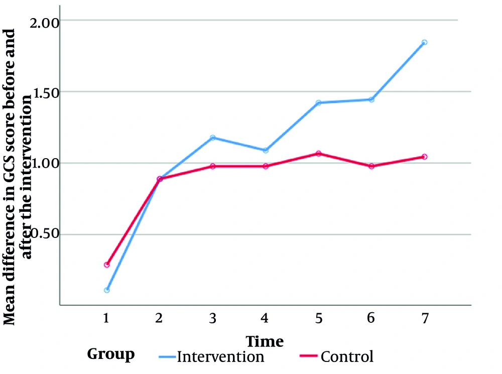
Figure 2.
Trend of the mean difference in GCS score before and after the intervention over seven days in the study groups
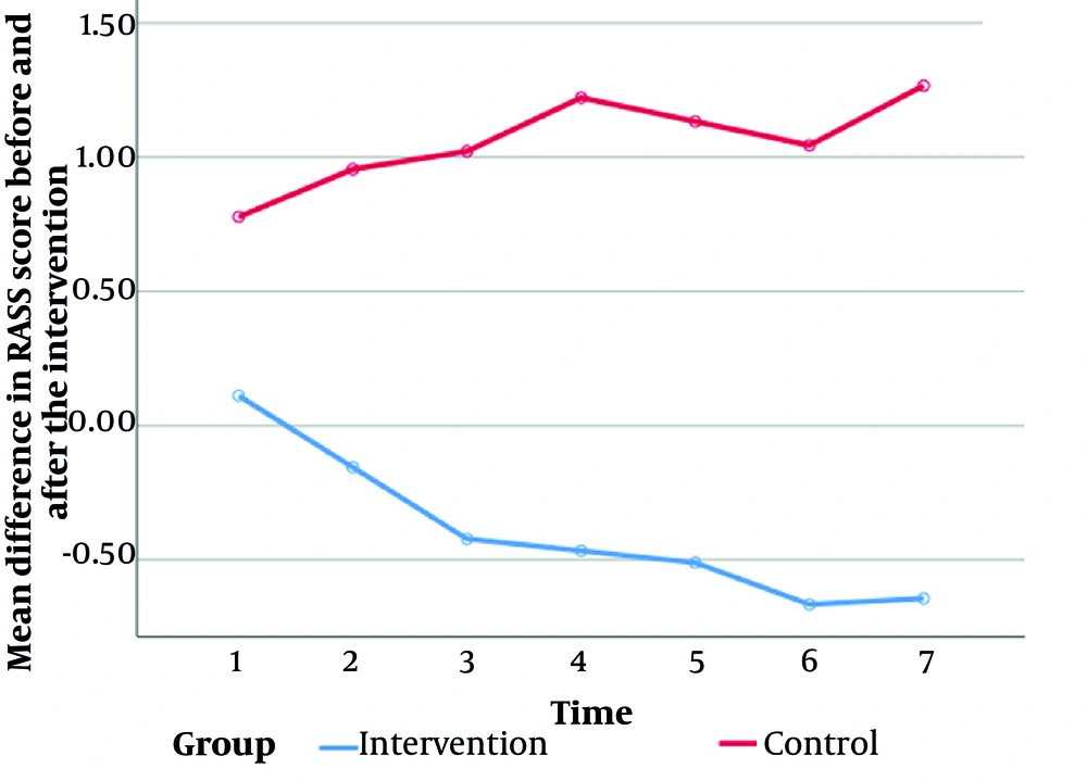
Figure 3.
Trend of the mean difference in RASS score before and after the intervention over seven days in the study groups
Based on Table 3, the Mann-Whitney U test showed that the mean ICU length of stay was significantly lower in the intervention group (18.66 ± 7.20 days) than in the control group (23.24 ± 5.99 days; P < 0.001). Furthermore, the mean duration of mechanical ventilation was significantly shorter in the intervention group (13.73 ± 6.94 days) than in the control group (18.21 ± 4.01 days; P < 0.001).
Table 3.
Comparison of ICU Length of Stay and Mechanical Ventilation Duration Between the Intervention and Control Groups (N = 83)
| Outcomes and Groups | No. | Mean ± SD | Mean Rank | U | P Value |
|---|---|---|---|---|---|
| Length of ICU stay | 478 | < 0.001 | |||
| Intervention | 42 | 18.66 ± 7.20 | 32.88 | ||
| Control | 41 | 23.24 ± 5.99 | 51.34 | ||
| Duration of mechanical ventilation | 424.500 | < 0.001 | |||
| Intervention | 42 | 13.73 ± 6.94 | 31.61 | ||
| Control | 41 | 18.21 ± 4.01 | 52.65 |
As shown in Table 4, the intervention group had a significantly lower delirium rate (26.2%) than the control group (51.2%), with a statistically significant difference (χ2 = 5.48, P = 0.019). However, the difference in ICU mortality between the intervention group (11.9%) and the control group (14.6%) was not statistically significant (χ2 = 0.134, P = 0.714).
Table 4.
Comparison of the Incidence of Delirium and ICU Mortality Between the Intervention and Control Groups (N = 83) a
| Outcomes and Groups | Yes | No | χ2 | df | P Value |
|---|---|---|---|---|---|
| Delirium | 5.48 | 1 | 0.019 | ||
| Intervention | 11 (26.2) | 31 (73.8) | |||
| Control | 21 (51.2) | 20 (48.8) | |||
| ICU mortality | 0.134 | 1 | 0.714 | ||
| Intervention | 5 (11.9) | 37 (88.1) | |||
| Control | 6 (14.6) | 35 (85.4) |
a Values are expressed as No. (%).
5. Discussion
This study evaluated the effect of a daily awakening protocol on clinical outcomes in ICU patients. The following sections discuss the findings for each outcome measure.
5.1. Duration of Mechanical Ventilation and ICU Length of Stay
The significant reduction in both the duration of mechanical ventilation and ICU length of stay in the intervention group is consistent with previous studies of DSI or sensory stimulation alone. Anwar Abdel ElAziz et al. (12) reported that DSI significantly reduced both outcomes in critically ill patients. A systematic review by Vagionas et al. (11) concluded that protocolized sedation, particularly when combined with daily awakening and spontaneous breathing trials, safely facilitated weaning and reduced ventilation days. In addition, a meta-analysis by Chen et al. (25) of 45 trials involving 5493 patients confirmed that DSI significantly reduced both outcomes.
However, the magnitude of reduction in the present study, approximately 5 days for both outcomes, was greater than that reported in studies using DSI alone, suggesting an additive or synergistic effect when sensory stimulation is added. Adineh et al. (18) found that a family-delivered sensory stimulation program in brain-injured patients significantly reduced both outcomes, which is consistent with our findings. Notably, de Wit et al. (26) reported conflicting results: a nurse-implemented sedation algorithm led to shorter ventilation (7.6 vs. 9.3 days) and shorter ICU stays (8 vs. 15 days) than DSI alone. This discrepancy suggests that DSI alone, without complementary measures such as sensory stimulation, may be ineffective or even harmful in some settings. Our protocol, which combined DSI with sensory stimulation, may address this limitation.
5.2. Incidence of Delirium
The significant reduction in delirium incidence in the intervention group (26.2% vs. 51.2%) represents an important clinical improvement. This finding supports Adineh et al. (18), who reported that a family-delivered sensory stimulation program reduced the odds of delirium by up to 94% in brain-injured patients. Furthermore, Anwar Abdel ElAziz et al. (12) demonstrated that DSI significantly reduced delirium incidence. Mechanistically, ICU delirium is multifactorial and involves prolonged sedation, sensory deprivation, and circadian rhythm disruption. Our daily awakening protocol targets all three factors: DSI reduces cumulative sedative exposure, while structured sensory stimulation provides orienting inputs, including auditory orientation by calling the patient's name and stating the time and place, visual stimulation through family photographs, and olfactory stimulation through familiar scents, which may help restore circadian entrainment.
However, not all studies have shown similar reductions. For example, Crew et al. (27) found that adding music therapy to music listening did not significantly reduce delirium incidence in mechanically ventilated patients. This discrepancy may be explained by several factors. First, their intervention was limited to auditory stimulation alone, whereas our protocol incorporated five sensory modalities. Second, their control group also received some form of auditory intervention, potentially attenuating the effect size. Third, their small sample size of 17 patients in the protocol group may have lacked sufficient power. The study by Head et al. (28) supports a multisensory approach, demonstrating that a combined positive stimulation protocol, consisting of enriched sensory stories narrated by family members paired with patient-specific music, reduced agitation and increased alertness.
5.3. ICU Mortality
No significant difference in ICU mortality was observed between the intervention and control groups. This finding aligns with most previous studies. Adineh et al. (18) also reported no significant mortality difference after sensory stimulation. Similarly, a meta-analysis by Chen et al. (25) showed that although DSI improves several clinical outcomes, its effect on mortality was not statistically significant in most included trials.
Mortality in critically ill patients is influenced by multiple factors, including disease severity, comorbidities, and baseline pathology. In our study, both groups had similar APACHE II scores, indicating comparable illness severity. The absence of a mortality benefit may suggest that while the daily awakening protocol improves intermediate outcomes, such as ventilation duration, ICU length of stay, and delirium, it does not alter the underlying disease processes that ultimately determine survival. Alternatively, the study may have been underpowered to detect a small but clinically important mortality difference because the observed absolute risk reduction of 4.5% would require a much larger sample size to achieve statistical significance.
5.4. Level of Consciousness and Sedation-Agitation Status
Although consciousness scores increased in both groups over the 7 days, the intervention group, which received both sensory stimulation and DSI, showed a more pronounced upward trend. To understand this synergistic effect, the individual role of each component should be considered. Previous studies, such as AhmadAli et al. (14), have shown that DSI alone can create a window of alertness by reducing cumulative drug exposure. However, this alertness may be accompanied by agitation, inattention, and stress responses. Evidence for this comes from de Wit et al. (26), in which DSI alone led to tachypnea and severe agitation in some patients. Similarly, in the present study, the control group, which received DSI only, exhibited mild agitation, reflected by positive RASS scores, despite significant GCS improvements.
Conversely, many studies, including Adineh et al. (17) and a systematic review by Uzun (16), have reported that sensory stimulation alone improves consciousness in ICU patients through activation of the ascending reticular activating system. However, the present study showed that combining sensory stimulation with reduced sedative burden through DSI not only produced greater increases in consciousness but also maintained patient calmness.
This finding is consistent with a synergistic effect. Daily sedation interruption first removes the pharmacological inhibitory brake from neural networks by reducing neural inhibition (29). At this critical moment of central nervous system disinhibition, multisensory stimulation, particularly auditory, visual, and olfactory stimulation, acts as a powerful targeted stimulus. These inputs not only activate the ascending reticular activating system but also activate cholinergic neurons in the basal forebrain, including the nucleus basalis of Meynert, through specific pathways, such as the olfactory pathway to the amygdala and the visual pathway to the superior colliculus. This activation increases alertness, attention, concentration, and cognitive processing (30). In other words, DSI sets the stage by eliminating pharmacological noise, while sensory stimulation brings the main actor onto the stage by providing rich and meaningful inputs. The result is active and calm wakefulness, superior both quantitatively, through higher GCS scores, and qualitatively, through RASS scores near zero. This mechanistic analysis explains the observed superiority of the intervention group and indicates that combining DSI with sensory stimulation may create a new qualitative phenomenon that cannot be achieved by either component alone.
The RASS findings are also noteworthy. Although the overall within-group trend over 7 days was not significant, the significant time × group interaction and within-group analyses showed that patients in the intervention group became calmer, with RASS scores shifting toward 0 and -1, whereas patients in the control group experienced mild agitation, with scores shifting toward positive values. This pattern is clinically important and suggests that DSI alone may induce mild agitation, while the addition of sensory stimulation mitigates this undesirable effect. Agitation during sedative interruption can lead to accidental extubation, catheter dislodgement, and increased nursing workload.
Mechanistically, DSI alone in the control group, through sudden removal of drug-induced inhibition, may generate unopposed sympathetic outflow manifested as mild agitation, reflected by positive RASS scores. This condition can increase myocardial oxygen consumption and cardiovascular risk in critically ill patients (26). In contrast, multisensory stimulation in the intervention group, initially through tactile stimulation such as massage and auditory stimulation such as music and familiar voices, may promote endorphin release and reduce cortisol levels. Subsequently, visual and olfactory stimulation engage the limbic system, restoring orientation and a sense of safety. The net result is calm wakefulness, reflected by RASS scores near zero, which is not only nonhazardous but also ideal for weaning and physiotherapy (25). Culshaw et al. (31) support this finding by demonstrating that music reduces sedative requirements during spontaneous awakening trials. However, our protocol incorporated multisensory stimulation, including auditory, visual, olfactory, tactile, and motor modalities, and therefore appears more comprehensive, which may explain the superior sedative effect observed.
5.5. Analysis of the Synergistic Effect
The observed synergistic effect can be explained by several interconnected mechanisms. First, DSI reduces cumulative exposure to benzodiazepines and opioids, which prolong ventilation, increase delirium risk, suppress the central nervous system, and cause ICU-acquired weakness (7, 11, 32). Second, structured sensory stimulation provides exogenous inputs to the ascending reticular activating system, promoting cortical arousal and preservation of circadian rhythms. Auditory stimulation, including calling the patient's name and providing time and place orientation, targets the orientation network impaired in delirium. Visual stimulation through family photographs and colored lights and olfactory stimulation through familiar scents engage limbic and parahippocampal circuits, reducing anxiety and agitation. Tactile and motor stimulation, including massage and passive range of motion, also reduce pain and prevent contractures (16, 18, 25). Third, the intervention timing of 4:00 to 6:00 PM aligns with the natural circadian arousal trough, helping to resynchronize the patient's internal clock. This circadian entrainment may explain the greater GCS improvement and calmer RASS scores in the intervention group. In addition, by reducing delirium and agitation, the protocol may indirectly decrease the need for rescue sedation and physical restraints, creating a positive feedback loop that further improves outcomes.
5.6. Limitations
Several limitations should be acknowledged. First, blinding of patients and intervention providers was not feasible because of the nature of the intervention, which may have introduced performance bias. However, this was minimized by blinding outcome assessors. Second, the study was conducted in two ICUs at a single center in Iran, limiting generalizability to other settings with different patient populations, sedation practices, or nursing protocols. Third, the sample size (n = 90), calculated based on ventilation duration, may have been insufficient to detect smaller but clinically meaningful differences in secondary outcomes, such as mortality, or to perform subgroup analyses by diagnosis or sedative type. Fourth, the cumulative sedative dose, such as midazolam or fentanyl equivalents, was not quantified, which could have provided additional mechanistic insight. Fifth, follow-up was limited to the ICU stay; therefore, outcomes after hospital discharge cannot be commented on. Sixth, the intervention was delivered only in the afternoon, between 4:00 and 6:00 PM. Morning implementation might have produced different effects on weaning and delirium, and the optimal timing of daily awakening protocols remains unknown. Future studies should compare morning and evening protocols. Seventh, while the independent effects of DSI alone and sensory stimulation alone have been reported previously, our two-arm design cannot isolate the independent effect of sensory stimulation. A three-arm trial including sensory stimulation alone is needed to confirm synergy. Of note, DSI was not routine care in our ICUs at the time of study conduct, so the DSI-alone arm served as an active comparator. Eighth, multiple outcomes were tested without adjustment for multiplicity. However, the main findings remained highly significant (P < 0.001), and we prioritized reducing type II error in this exploratory study. Ninth, control group patients received routine ICU nursing care, such as turning, suctioning, and verbal reassurance, which may include some low-level, unstructured sensory input. This could have diluted the true effect size, meaning that our findings may underestimate the actual benefit of the structured sensory stimulation protocol.
5.7. Conclusions
This randomized clinical trial demonstrated that a daily awakening protocol combining DSI with structured multisensory stimulation, compared with DSI alone, significantly reduced the duration of mechanical ventilation, ICU length of stay, and delirium incidence while improving consciousness and sedation-agitation profiles in mechanically ventilated ICU patients. These findings suggest that adding structured sensory stimulation to DSI may provide additional clinical benefits beyond DSI alone, although the independent effect of sensory stimulation without DSI was not compared. Therefore, health policymakers and ICU managers should consider training nurses in structured sensory stimulation techniques and integrating this protocol into routine sedation management to improve patient outcomes and optimize resource utilization. Future research should include three-arm trials comparing DSI alone, sensory stimulation alone, and the combined protocol to determine whether the observed effects are additive or synergistic. Future studies should also compare multisensory protocols with single-modality approaches, such as auditory stimulation alone; optimize intervention timing, such as morning versus evening protocols based on individual circadian rhythms; and investigate long-term outcomes such as quality of life and PTSD.