
1. Background
Cancer remains a major global health challenge, and chemotherapy continues to be an essential therapeutic strategy for many malignancies. Among currently available anticancer drugs, cisplatin is widely prescribed because of its proven efficacy against a broad spectrum of solid tumors. However, despite its clinical benefits, cisplatin administration is associated with several adverse effects, including cardiotoxicity, which may compromise treatment outcomes and long-term cardiovascular health (1). Previous studies have shown that cisplatin-induced cardiac injury is mediated by the excessive production of reactive oxygen species (ROS), mitochondrial dysfunction, inflammation, and cardiomyocyte apoptosis, ultimately resulting in structural and functional alterations of cardiac tissue (1, 2). Therefore, identifying effective strategies to attenuate cisplatin-induced cardiotoxicity has become an important area of investigation.
Regular physical activity is widely regarded as an effective non-pharmacological strategy for preserving cardiovascular function and reducing oxidative stress and inflammation. Exercise-induced adaptations are mediated, in part, by the release of bioactive molecules known as exerkines, particularly myokines, which facilitate communication between skeletal muscle and distant organs, including the heart (3). These molecules regulate several physiological processes, including energy metabolism, mitochondrial function, oxidative stress, and inflammatory responses, thereby contributing to cardioprotection (3, 4).
Fibroblast growth factor 21 (FGF21), an exercise-induced myokine, plays a central role in regulating metabolic balance and adaptive cellular responses to stress. FGF21 exerts cardioprotective effects by regulating glucose and lipid metabolism, enhancing mitochondrial function, and suppressing oxidative stress and inflammation (5, 6). Exercise training has been shown to increase circulating and tissue levels of FGF21, suggesting that this molecule may contribute to the beneficial cardiovascular adaptations induced by physical activity (6, 7). In contrast, myostatin, a cytokine belonging to the transforming growth factor-β (TGF-β) superfamily, suppresses skeletal muscle growth and adversely affects metabolic regulation. Elevated myostatin expression has been associated with inflammation, mitochondrial dysfunction, muscle wasting, and adverse cardiovascular remodeling (4, 8). Previous studies have reported that exercise training can reduce myostatin expression, thereby promoting anabolic signaling and improving metabolic health (6, 7).
Moderate-intensity continuous training (MCT) is one of the most commonly prescribed exercise modalities because of its safety, feasibility, and effectiveness in improving cardiovascular and metabolic function (9, 10). Compared with high-intensity exercise protocols, MCT provides substantial physiological benefits while inducing lower physiological stress, making it particularly suitable for individuals exposed to pathological conditions such as chemotherapy-induced toxicity. Evidence suggests that MCT can enhance antioxidant defense mechanisms, improve mitochondrial adaptations, and attenuate inflammatory responses, all of which may contribute to protection against cardiac injury (5, 10).
In addition to exercise interventions, naturally occurring bioactive compounds have received increasing attention as complementary therapeutic approaches. Quinic acid is a naturally occurring cyclitol with potent antioxidant and anti-inflammatory properties (11-13). Experimental studies have shown that quinic acid can reduce oxidative stress, improve endothelial function, and suppress inflammatory signaling pathways, thereby protecting tissues from oxidative damage (11-13). Given that both exercise training and quinic acid target common mechanisms involved in cardioprotection, their combined application may provide greater protection against cisplatin-induced cardiac injury than either intervention alone.
2. Objectives
To date, limited information is available regarding the combined effects of moderate-intensity continuous training and quinic acid supplementation on cardiac myokine expression under cisplatin-induced cardiotoxicity. Therefore, the present study aimed to investigate the effects of MCT combined with quinic acid administration on the expression of FGF21 and myostatin in the cardiac tissue of rats exposed to cisplatin-induced cardiotoxicity. We hypothesized that the combined intervention would increase FGF21 expression while reducing myostatin expression, thereby promoting a cardioprotective molecular environment.
3. Methods
3.1. Animals
This experimental study was conducted using 15 male Wistar rats aged 8 - 10 weeks, with an initial body weight of 240 ± 23 g. All animals were obtained from the Animal Research Center and housed in a controlled animal facility under standard environmental conditions (22 ± 2 °C, 50 - 60% relative humidity, and a 12-h light/12-h dark cycle). Throughout the experiment, rats had ad libitum access to standard laboratory chow and drinking water.
After a 7-day acclimation period, the animals were randomly assigned to five experimental groups, with 3 rats in each group: 1, Healthy control (CON); 2, cisplatin control (CIS); 3, cisplatin + moderate-intensity continuous training (CIS+MCT); 4, cisplatin + quinic acid (CIS+QA); 5, cisplatin + moderate-intensity continuous training + quinic acid (CIS+MCT+QA) (14, 15).
Animal handling and all experimental procedures were conducted in accordance with the National Institutes of Health Guide for the Care and Use of Laboratory Animals. The study protocol was reviewed and approved by the Institutional Animal Ethics Committee.
After acclimation, animals were allocated to groups using a simple randomization procedure. Western blot analyses were performed using independent biological samples obtained from each animal. Protein expression was normalized to glyceraldehyde-3-phosphate dehydrogenase (GAPDH). Whenever possible, samples from all experimental groups were run on the same gel under identical experimental conditions to minimize inter-gel variability. Densitometric analysis was performed using uniform exposure settings. Although complete investigator blinding during Western blot quantification was not implemented, all analyses were conducted using standardized procedures.
3.2. Experimental Design
This study used a randomized post-test controlled experimental design to investigate the effects of moderate-intensity continuous training (MCT), quinic acid supplementation, and their combined intervention on cardiac expression of FGF21 and myostatin in a rat model of cisplatin-induced cardiotoxicity.
After baseline adaptation, animals assigned to the exercise groups completed an eight-week MCT program. Quinic acid supplementation was administered daily throughout the intervention period. After four weeks of intervention, cardiotoxicity was induced by cisplatin administration in all experimental groups except the healthy control group. Exercise training and quinic acid administration continued for an additional four weeks after cisplatin injection.
3.3. Exercise Familiarization and VO2max Determination
Before the intervention, all rats completed a two-week treadmill adaptation protocol (5 sessions/week, 10 - 15 min/session), during which running speed was gradually increased from 5 to 15 m·min-1. Maximal running capacity was then assessed using an incremental treadmill test adapted from Høydal et al. and Leandro et al. (9, 10). After a 10-min warm-up, animals began running at 15 m·min-1. Treadmill speed was increased by 2 m·min-1 every 2 min until the animals reached exhaustion. Exhaustion was defined as the inability to maintain running despite mechanical encouragement, with the animal remaining at the back of the treadmill for longer than 10 s. The maximal speed achieved was used to estimate exercise intensity and prescribe training workloads.
3.4. Moderate-Intensity Continuous Training Protocol
Rats assigned to the exercise groups completed an eight-week moderate-intensity continuous treadmill training program performed five days per week. Each exercise session comprised three consecutive phases: a 5-min warm-up (30 - 40% VO2max), a 30-min continuous running period at 60 - 65% VO2max, and a 5-min recovery period at 30 - 40% VO2max. The treadmill was maintained at a constant 0° incline throughout all training sessions. Running speed was progressively increased from 10 m·min-1 in week 1 to 25 m·min-1 in week 8 to ensure progressive overload and physiological adaptation (10).
3.5. Quinic Acid Administration
Quinic acid was administered to the designated treatment groups by oral gavage at a daily dose of 100 mg·kg-1 body weight throughout the experimental period.
The 100 mg/kg/day dose was selected based on previous experimental studies demonstrating significant antioxidant, anti-inflammatory, and tissue-protective effects without evidence of toxicity. The use of this dose also facilitates comparison with previous investigations evaluating the biological activity of quinic acid in experimental models of oxidative stress and tissue injury (16, 17).
3.6. Induction of Cisplatin-Induced Cardiotoxicity
To establish the cardiotoxicity model, rats in all treatment groups, except the healthy controls, received an intraperitoneal injection of cisplatin (5 mg·kg-1 body weight) after four weeks of intervention.
3.7. Tissue Collection
At the end of the intervention, animals were euthanized 48 h after the final exercise session and quinic acid administration. Anesthesia was induced with ketamine (50 mg·kg-1) and xylazine (5 mg·kg-1). After thoracotomy, the heart was carefully excised, rinsed with ice-cold normal saline, snap-frozen in liquid nitrogen, and stored at −80 °C until subsequent biochemical analyses.
3.8. Western Blot Analysis
Protein extracts were prepared from cardiac tissue by homogenization in RIPA lysis buffer supplemented with protease inhibitors. Total protein concentration was determined using the Bradford method. For immunoblotting, 30 - 50 μg of protein from each sample was separated on 12% SDS-polyacrylamide gels and electrotransferred to 0.45-μm PVDF membranes.
The membranes were incubated overnight with primary antibodies against myostatin (Proteintech, Cat. No. 19142 - 1-AP, 1:1000), FGF21 (Abcam, Cat. No. ab171941, 1:1000), and GAPDH (Cell Signaling Technology, Cat. No. 5174, 1:1000). After washing, an HRP-conjugated mouse anti-rabbit IgG secondary antibody (Santa Cruz Biotechnology, Cat. No. sc-2357, 1:1000) was applied. Immunoreactive bands were detected using an enhanced chemiluminescence (ECL) detection system and quantified by densitometric analysis using the Fusion FX imaging system (Vilber). Relative protein expression levels were normalized to GAPDH. Each cardiac sample was analyzed in three technical replicates, and the mean of the technical replicates was used as the representative value for each animal in the statistical analysis.
3.9. Statistical Analysis
All statistical analyses were performed using IBM SPSS Statistics version 26.0. Assumptions of normality and homogeneity of variances were assessed using the Shapiro-Wilk and Levene tests, respectively. Given the small sample size (n = 3 per group), these tests have limited reliability; therefore, parametric analyses may be unstable, and the findings were interpreted cautiously as preliminary and hypothesis-generating.
Comparisons among the five experimental groups were conducted using one-way analysis of variance (ANOVA). When a significant overall group effect was observed, Tukey's multiple-comparison test was used for post hoc pairwise comparisons. Results are presented as mean ± SD, and statistical significance was set at P < 0.05.
4. Results
Changes in body weight and heart weight among the experimental groups are summarized in Table 1. Cisplatin-treated rats exhibited lower body weight and heart weight than those in the control group. MCT or quinic acid alone resulted in numerical increases in these parameters, whereas the combined intervention produced the greatest numerical increase among cisplatin-treated groups.
Table 1.
Mean ± SD of Body Weight and Heart Weight in the Experimental Groups a
| Group | Body weight (g) | Heart weight (g) |
|---|---|---|
| Control | 283.70 ± 8.73 | 1.17 ± 0.05 |
| Cis | 225.30 ± 7.63 | 0.87 ± 0.05 |
| Cis+MCT | 236.00 ± 8.18 | 1.00 ± 0.06 |
| Cis+QA | 233.00 ± 11.36 | 1.03 ± 0.09 |
| Cis+MCT+QA | 252.30 ± 8.73 | 1.08 ± 0.09 |
a Values are expressed as mean ± SD (n = 3 per group). Body weight and heart weight represent the final measurements obtained at euthanasia.
4.1. Cardiac Myostatin and FGF21 Protein Expression
One-way ANOVA revealed significant differences among the experimental groups in both cardiac myostatin (F(4, 10) = 99.90, P < 0.0001) and FGF21 protein expression (F(4, 10) = 71.46, P < 0.0001). Tukey’s post hoc analysis identified significant pairwise differences among the experimental groups, as indicated by different superscript letters in Table 2. Descriptive data for both variables are presented in Table 2.
| Group | Myostatin (Fold Change) | FGF21 (Fold Change) |
|---|---|---|
| Control | 1.00 ± 0.13 A | 1.00 ± 0.19 A |
| Cisplatin | 4.62 ± 0.27 B | 2.12 ± 0.36 B |
| Cisplatin + MCT | 3.26 ± 0.22 C | 3.02 ± 0.24 C |
| Cisplatin + Quinic Acid | 3.07 ± 0.16 C | 3.04 ± 0.17 C |
| Cisplatin + MCT + Quinic Acid | 1.84 ± 0.34 D | 4.42 ± 0.27 D |
a Values are expressed as mean ± SD (n = 3 animals per group). Protein expression values are presented as relative fold changes normalized to GAPDH. Statistical comparisons were performed using one-way ANOVA followed by Tukey’s post hoc test.
b Different capital superscript letters indicate statistically significant differences between groups (P < 0.05), whereas groups sharing the same superscript capital letter indicate no statistically significant difference.
4.2. Effect of MCT and Quinic Acid on Cardiac Myostatin Expression
As illustrated in Figure 1, cisplatin administration markedly increased cardiac myostatin protein expression compared with that in the control group (4.62 ± 0.27 vs. 1.00 ± 0.13, P < 0.0001). Moderate-intensity continuous training significantly reduced myostatin expression compared with that in the cisplatin group (3.26 ± 0.22 vs. 4.62 ± 0.27, P = 0.0003). Similarly, quinic acid supplementation significantly decreased myostatin levels relative to those in the cisplatin group (3.07 ± 0.16 vs. 4.62 ± 0.27, P = 0.0001). The combined intervention of MCT and quinic acid produced the greatest reduction in myostatin expression (1.84 ± 0.34), which was significantly lower than that in the cisplatin group (P < 0.0001), the MCT group (P = 0.0002), and the quinic acid group (P = 0.0007).
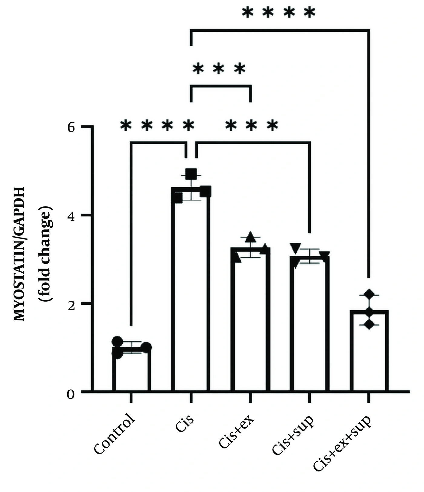
Figure 1.
Relative cardiac myostatin protein expression determined by Western blot analysis in the experimental groups. Rats received moderate-intensity continuous training (MCT), quinic acid supplementation, or their combination following cisplatin administration. Representative Western blot images together with GAPDH-normalized densitometric analysis are shown. Quantitative data are presented as individual values with mean ± SD (n = 3 animals per group). Each data point represents one independent biological replicate (one animal per replicate). Western blot analysis was performed using three technical replicates for each biological sample, and the mean of the technical replicates was used as the representative value for each animal. Statistical comparisons were performed using one-way ANOVA followed by Tukey’s post hoc test. P < 0.05 was considered statistically significant.
4.3. Effect of MCT and Quinic Acid on Cardiac FGF21 Expression
As shown in Figure 2, cisplatin administration significantly increased cardiac FGF21 protein expression compared with that in the control group (2.12 ± 0.36 vs. 1.00 ± 0.19, P = 0.0024). Both MCT and quinic acid supplementation further enhanced FGF21 expression relative to that in the cisplatin group. FGF21 levels reached 3.02 ± 0.24 in the MCT group (P = 0.0115 vs. cisplatin) and 3.04 ± 0.17 in the quinic acid group (P = 0.0105 vs. cisplatin). No significant difference was observed between the MCT and quinic acid groups (P > 0.9999). However, the combined intervention elicited the highest FGF21 expression (4.42 ± 0.27), which was significantly greater than that in the cisplatin group (P < 0.0001), the MCT group (P = 0.0005), and the quinic acid group (P = 0.0005).
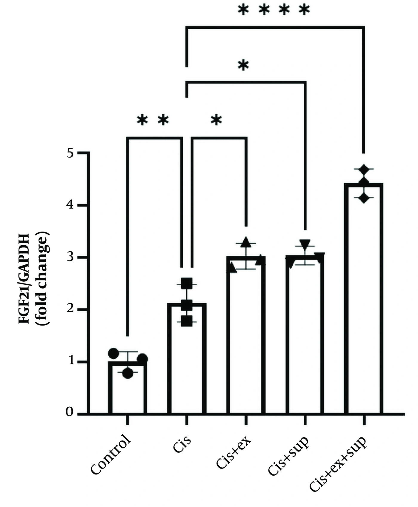
Figure 2.
Relative cardiac FGF21 protein expression determined by Western blot analysis in the experimental groups. Rats received moderate-intensity continuous training (MCT), quinic acid supplementation, or their combination following cisplatin administration. Representative Western blot images together with GAPDH-normalized densitometric analysis are shown. Quantitative data are presented as individual values with mean ± SD (n = 3 animals per group). Each data point represents one independent biological replicate (one animal per replicate). Western blot analysis was performed using three technical replicates for each biological sample, and the mean of the technical replicates was used as the representative value for each animal. Statistical comparisons were performed using one-way ANOVA followed by Tukey’s post hoc test. P < 0.05 was considered statistically significant.
5. Discussion
The present study investigated the effects of eight weeks of moderate-intensity continuous training (MCT), quinic acid supplementation, and their combination on cardiac myostatin and FGF21 protein expression in rats with cisplatin-induced cardiotoxicity. The main findings showed that cisplatin markedly increased myostatin and FGF21 expression in cardiac tissue, whereas MCT and quinic acid attenuated myostatin expression and further enhanced FGF21 levels. The combined intervention produced the most pronounced effects.
Previous studies have suggested that cisplatin-induced cardiotoxicity involves oxidative stress, mitochondrial dysfunction, and inflammatory signaling, which may disrupt cellular homeostasis and promote tissue injury (18, 19). The elevated myostatin expression observed in the cisplatin group may reflect the activation of catabolic pathways in response to cellular stress. Myostatin is recognized as a negative regulator of tissue growth and repair, and its overexpression has been associated with pathological remodeling and impaired cellular adaptation (20).
The reduction in myostatin following MCT is consistent with previous reports of exercise-induced suppression of catabolic signaling pathways. Riahy demonstrated that aerobic training reduced myostatin levels in individuals with metabolic disorders (6), while Bettariga et al. reported similar findings in their meta-analysis of exercise interventions (7). These effects may be associated with anabolic pathways such as IGF-1/PI3K/Akt and TGF-β/SMAD signaling, as suggested by previous studies; however, these pathways were not directly examined in the present study and therefore represent potential mechanisms rather than demonstrated pathways. In contrast, some studies have reported limited effects of exercise on myostatin expression (21), which may be attributable to differences in training protocols, intervention duration, or disease status.
Quinic acid supplementation also reduced cardiac myostatin expression. Although direct evidence linking quinic acid to myostatin regulation is limited, its antioxidant and anti-inflammatory properties have been reported in previous studies (11, 13). Quinic acid may therefore influence pathways related to oxidative stress and inflammation; however, these mechanisms were not directly evaluated in the present study and should be considered hypotheses rather than demonstrated mechanisms.
Recent evidence also supports the use of combined exercise and nutritional supplementation to modulate molecular responses in experimental disease models. Porsesh et al. reported that aquatic exercise combined with coenzyme Q10 improved oxidative stress status and modulated PTX3 and NF200 protein expression more effectively than either intervention alone in an animal model of multiple sclerosis (26). Although conducted in a different pathological context, these findings further support investigating combined exercise and supplementation strategies in the present study.
Another important finding was the elevation of FGF21 expression following cisplatin administration. FGF21 is widely regarded as a stress-responsive metabolic regulator that is activated under conditions of oxidative and mitochondrial stress to support cellular adaptation (22, 23). Therefore, the increase observed in the cisplatin group may represent a compensatory protective response to cardiac injury.
Both MCT and quinic acid further enhanced FGF21 expression. Previous studies have suggested that exercise may enhance FGF21 expression through AMPK-PGC-1α signaling; however, these pathways were not directly examined in the present study and therefore represent potential explanatory mechanisms rather than experimentally demonstrated pathways (6, 24). Similarly, exercise-responsive factors such as FGF21 have been implicated in cardiometabolic protection and cellular stress resistance (3). Although some studies have reported unchanged FGF21 responses after exercise (25), differences in exercise characteristics and pathological conditions may explain these discrepancies.
The elevation of cardiac FGF21 observed following cisplatin administration should not be interpreted solely as a beneficial response. FGF21 is increasingly recognized as a stress-responsive metabolic hormone that is induced under conditions of mitochondrial dysfunction, oxidative stress, and tissue injury as part of an endogenous adaptive response. Therefore, the increased expression observed in the cisplatin group may reflect a compensatory response to cardiac stress rather than direct evidence of improved cardiac health. The further increase in FGF21 following moderate-intensity continuous training and quinic acid supplementation may indicate enhanced adaptive signaling; however, in the absence of a functional cardiac assessment, this finding should not be interpreted as definitive evidence of cardioprotection (22).
The combined intervention produced the greatest observed changes in cardiac FGF21 and myostatin expression among the experimental groups. Although previous studies have suggested that exercise and quinic acid may influence metabolic, oxidative, and inflammatory processes, these pathways were not directly evaluated in the present study. Therefore, their potential contribution to the observed molecular responses remains hypothetical.
These findings indicate that MCT and quinic acid modulated cardiac FGF21 and myostatin expression. The biological pathways responsible for these changes remain uncertain because they were not directly investigated in the present study.
The present experimental design evaluated preventive adaptation before cisplatin exposure. Future investigations should compare preventive, concurrent, and post-treatment exercise interventions. Caution is also warranted when translating the findings of the present study into clinical settings. The use of young, healthy rats in a controlled experimental model does not fully reflect the complexity of conditions experienced by patients undergoing chemotherapy. Therefore, future studies using clinically relevant models and human populations are necessary to confirm the translational significance of these findings.
5.1. Limitations and Future Directions
This study has several limitations. First, the small sample size (n = 3 per group), determined according to institutional ethical regulations based on the 3Rs principles and practical constraints, together with the absence of an a priori power analysis, limits the statistical robustness and generalizability of the findings. Therefore, the results should be considered preliminary and hypothesis-generating.
Second, healthy exercise-only and healthy quinic acid-only groups were not included; therefore, the independent physiological effects of each intervention under normal conditions could not be distinguished from their effects following cisplatin exposure.
Finally, only cardiac FGF21 and myostatin protein expression were evaluated. Cardiac functional assessments, histopathological analyses, conventional cardiac injury biomarkers, and oxidative stress (MDA, SOD, CAT, GPx, GSH, ROS), inflammatory (NF-κB, TNF-α, IL-6), apoptotic (Bax/Bcl-2, Caspase-3), fibrotic (TGF-β and collagen-related proteins), and mitochondrial biomarkers were not assessed. Consequently, the biological significance and underlying mechanisms of the observed molecular changes remain uncertain and should be confirmed in larger studies incorporating comprehensive functional and mechanistic evaluations.
5.2. Conclusions
Cisplatin administration altered cardiac myostatin and FGF21 protein expression in rats. Eight weeks of moderate-intensity continuous training and quinic acid supplementation modulated these molecular responses, with the combined intervention producing the greatest observed changes. These findings are preliminary and require confirmation in larger studies incorporating cardiac functional, histopathological, and mechanistic assessments.