
1. Background
The skin is the body’s largest organ and accounts for approximately 14 - 16% of total body weight (1). It is a complex biological structure comprising two major layers, the epidermis and dermis, supported by a broad range of cellular and structural elements, including epithelial and mesenchymal cells, cutaneous glands, and extensive vascular and neural networks. Derived embryologically from the ectoderm, the epidermis forms the external surface of the skin and serves as the principal boundary between the organism and its surrounding environment (2, 3).
Chronic exposure to solar ultraviolet (UV) radiation can progressively alter the structure and physiology of the skin, ultimately resulting in photoaging. A major component of UV-mediated injury involves remodeling and degradation of the dermal extracellular matrix, which are associated with reductions in elasticity, mechanical strength, and structural integrity that characterize photoaged skin. In addition to contributing to premature cutaneous aging, solar UV radiation is an established environmental carcinogenic factor in humans. Growing evidence concerning the biological responses elicited by different UV wavelengths, particularly UVA and UVB, has encouraged the development of novel photoprotective agents and improved sunscreen delivery systems (4). Alongside conventional approaches to UV protection, increasing attention has been directed toward topical preparations containing plant-derived antioxidants, which may help counteract oxidative and inflammatory processes associated with UV exposure (5).
Rice has a long history of medicinal use and has been incorporated into traditional health care practices, particularly across Asia, to manage various conditions, including diabetes, respiratory and gastrointestinal disorders, and skin-related problems (6-8). Rice bran, a major by-product of rice milling, is a nutritionally valuable source of essential minerals, such as magnesium, calcium, zinc, and copper, as well as proteins containing all essential amino acids (9, 10). Beyond its nutritional value, rice bran contains diverse bioactive and antioxidant constituents, including B-group vitamins, γ-aminobutyric acid, γ-oryzanol, tocopherols, tocotrienols, carotenoids, phytic acid, phenolic compounds, anthocyanins, and, in particular, ferulic acid. This rich phytochemical composition has attracted increasing interest in rice bran oil (RBO) as a promising natural ingredient for topical formulations aimed at protecting the skin against UV-induced damage (11).
The development of nanotechnology-based delivery platforms has provided new opportunities to improve the stability, bioavailability, and biological performance of pharmaceutical and cosmetic ingredients (12-14). Nanoparticulate carriers are particularly attractive because they can protect incorporated compounds from degradation, modulate their release, and facilitate their delivery to specific biological sites (15, 16). Solid lipid nanoparticles (SLNs) are one such delivery platform and are produced by replacing the liquid oil phase of conventional oil-in-water emulsions with a solid lipid or a mixture of solid lipids. The resulting particles contain a lipid matrix that remains solid at temperatures close to room temperature and under physiological conditions, making SLNs suitable carriers for a broad spectrum of bioactive substances.
The incorporation of UV-absorbing organic compounds into lipid-based nanocarriers has previously been associated with enhanced UV absorption and photoprotective activity (17). Nanostructured encapsulation may improve the distribution and retention of UV-protective compounds and, consequently, may allow effective photoprotection with a lower concentration of the active UV-filtering component (18). Although nanotechnology-based approaches have been investigated to improve the UV-protective performance of RBO, the specific use of SLNs as a delivery system to enhance its sunscreen potential remains insufficiently explored (17, 18).
Given the intrinsic photoprotective and antioxidant properties of RBO and the potential of lipid-based nanocarriers to improve the performance of bioactive compounds, incorporating RBO into SLNs represents a promising strategy for developing advanced sunscreen formulations. Such a delivery system could enhance the UV-protective activity of RBO while improving its formulation characteristics and overall performance.
2. Objectives
This study aimed to develop rice bran oil-loaded solid lipid nanoparticles and evaluate their physicochemical characteristics, release behavior, and photoprotective potential.
3. Methods
3.1. Fabrication of Rice Bran Oil-Loaded Solid Lipid Nanoparticles
Rice bran oil-loaded solid lipid nanoparticles (RBO-SLNs) were prepared using an emulsification–probe sonication method previously optimized by the research group (1, 19). Briefly, the lipid phase was prepared by dissolving 100 mg of lecithin, 0.8 mL of Tween 80, 100 mg of cholesterol, and 1 mL of RBO in 10 mL of dichloromethane. The resulting organic phase was then mixed with 10 mL of an aqueous 4% (w/v) polyvinyl alcohol solution. The mixture was sonicated for 3 min using a probe sonicator (Hielscher UP400S, Germany) operated at 400 W, 24 kHz, and 80% amplitude, resulting in a stable white oil-in-water emulsion. Dichloromethane was then removed using a rotary evaporator at 30 °C for 20 min (2).
3.2. Particle Size, Polydispersity Index, and Zeta Potential
The mean particle size, polydispersity index (PDI), and zeta potential of the RBO-SLNs were determined using a Zetasizer (Malvern Instruments, UK). For analysis, 150 mg of the nanoparticle formulation was dispersed in 1 mL of deionized water. Measurements were performed at 25 °C using Zetasizer software version 6.01, with a count rate of 206.3 kcps and a measurement position of 4.65 mm. The PDI was used to characterize the particle-size distribution and assess size uniformity within the nanoparticle dispersion. All measurements were performed in triplicate (n = 3).
3.3. Transmission Electron Microscopy
The morphology and particle dimensions of the RBO-SLNs were examined using transmission electron microscopy (TEM; Zeiss EM10C, 100 kV, Germany). A drop of the nanoparticle dispersion was deposited onto Formvar-coated mesh grids. The specimens were dehydrated sequentially by immersion in ethanol solutions of increasing concentration (50%, 70%, 90%, and 100%), with each step maintained for 10 min. After the ethanol had been completely removed, the grids were dried at 37 °C and examined by TEM at an accelerating voltage of 80 kV.
3.4. Encapsulation Efficiency
The amount of nonencapsulated RBO was determined by centrifugation. Briefly, 500 mg of the RBO-SLN formulation was dispersed in 10 mL of distilled water containing 5% (v/v) Tween 80 and centrifuged at 13,000 rpm for 40 min at 4 °C. The concentration of free RBO in the resulting supernatant (W2) was quantified by ultraviolet (UV)–visible spectrophotometry (Cary 60, Agilent Technologies, Santa Clara, CA, USA) at 313 nm. Encapsulation efficiency (EE) was calculated using the following equation (n = 3):
3.1. EE (%) = (W1 − W2) / W1 × 100
where W1 represents the total amount of RBO initially incorporated into the formulation, and W2 corresponds to the amount of nonencapsulated RBO detected in the supernatant.
Encapsulation efficiency was also verified using an indirect dialysis-based procedure. For this purpose, 500 mg of the nanoparticle formulation was transferred into preactivated dialysis bags and immersed in 2 L of the external medium. Dialysis was continued for 2 h, and a 10 mL aliquot of the external phase was collected. The withdrawn sample was analyzed by UV–visible spectrophotometry at 313 nm, and EE was calculated using the equation above. All measurements were performed in triplicate (n = 3).
3.5. in vitro Release and Release Kinetics
The release of RBO from the RBO-SLNs was investigated using a dialysis method. Briefly, 1.2 g of the formulation was transferred into preactivated dialysis bags with a molecular weight cutoff of 14 kDa (Sigma, Steinheim, Germany). The bags were immersed in 100 mL of receptor medium consisting of distilled water containing 5% (v/v) Tween 80 and maintained at 32 °C under continuous agitation at 100 rpm. At predetermined time points (0, 1, 3, 6, 24, 48, and 72 h), 3 mL samples were withdrawn from the receptor medium and immediately replaced with an equal volume of fresh, prewarmed medium. The absorbance of the collected samples was measured at 313 nm, and all measurements were performed in triplicate. To characterize the release mechanism, the experimental data were fitted to zero-order, first-order, Higuchi, Hixson–Crowell, and Korsmeyer–Peppas kinetic models.
3.6. Fibroblast Cell Compatibility
The cytocompatibility of the RBO-SLNs was evaluated using the MTT assay in HU02 human foreskin fibroblasts obtained from the Iranian Biological Resource Center (IRIBC; C10309). Cells were seeded in 96-well plates at a density of 5 × 10³ cells/cm2 in 0.1 mL of Dulbecco’s modified Eagle’s medium and maintained in a humidified atmosphere containing 5% CO2. After 24 h of incubation, the cells were exposed to RBO-SLNs at concentrations of 5 and 10 mg/mL, while RBO at 10 mg/mL was included as the control treatment. All treatments were prepared in media containing 5% Tween 80.
After 24 h of exposure, MTT solution (5 mg/mL in PBS; Sigma) was added, and the cells were incubated for an additional 3 h at 37 °C. The supernatant was then removed, and the resulting formazan crystals were dissolved in 0.15 mL of dimethyl sulfoxide. Absorbance was measured at 570 nm using a BioTek Epoch™ microplate reader. Experiments were conducted in triplicate (n = 3). Cell viability was normalized to the viability of cells maintained in culture medium containing the formulation dispersant (distilled water with 5% v/v Tween 80).
3.7. Fourier-Transform Infrared Spectroscopy
Attenuated total reflectance Fourier-transform infrared (ATR-FTIR) spectroscopy was performed to characterize the chemical composition of RBO and RBO-SLNs and to investigate potential interactions between RBO and the formulation excipients. Measurements were obtained using an ATR-FTIR spectrometer (PerkinElmer, USA) at a resolution of 4 cm-1 over the spectral range of 400 - 4000 cm-1. The resulting spectra were examined for characteristic absorption bands and evaluated for changes in peak position, intensity, or appearance after RBO was incorporated into the SLN matrix.
3.8. UV Absorption Properties of Rice Bran Oil and Its Solid Lipid Nanoparticles
3.8.1. Dispersion Method
The UV absorbance of RBO and RBO-SLNs was assessed at concentrations of 1%, 3%, and 5% (w/v) using a water/2% Tween 80 dispersion medium. Absorbance was determined spectrophotometrically at 327 nm (UVA), 313 nm (UVB), and 222 nm (UVC). Three independent measurements were performed for each formulation (n = 3). RBO at the corresponding concentrations was used as the control for the respective RBO-SLN formulations.
3.8.2. Transpore® Tape Method
For the Transpore® tape assay, 2 mg/cm2 of each formulation was uniformly applied to Transpore® tape mounted over a quartz cell. The samples were distributed evenly by gently rubbing the surface in circular motions with a gloved finger. After the applied films had dried, absorbance was measured at 327, 313, and 222 nm. Each measurement was performed in triplicate (n = 3).
3.9. Determination of Sun Protection Factor
The sun protection factor (SPF) of RBO, blank SLNs, and RBO-SLNs at concentrations of 1%, 3%, and 5% was determined according to the procedure reported by Sarpietro et al. (3). UV absorbance was recorded from 290 to 320 nm at 5-nm intervals using a BioTek Epoch™ microplate reader. The spectrophotometric SPF was calculated using the Mansur model, as described by Dutra et al. (4):
𝑆𝑃𝐹𝑠𝑝𝑒𝑐𝑡𝑟𝑜𝑝ℎ𝑜𝑡𝑜𝑚𝑒𝑡𝑟𝑖𝑐=
𝐶𝐹×320∑290𝐸𝐸(𝜆)×𝐼(𝜆)×𝐴𝑏𝑠(𝜆)
where CF is the correction factor, 𝐸𝐸(𝜆) represents the erythemal effect spectrum, 𝐼(𝜆) represents the solar intensity spectrum, and 𝐴𝑏𝑠(𝜆) denotes the measured sample absorbance. Because EE(\lambda) and 𝐼(𝜆) values are predefined constants in the Mansur model, SPF was calculated from the wavelength-dependent absorbance of each sample. Three independent determinations were performed for each concentration, and the mean SPF value was calculated.
3.10. Statistical Analysis
Statistical analyses were performed using SPSS software, version 22.0. Differences between two groups were evaluated using an independent-samples t-test, whereas comparisons involving more than two groups were performed using one-way analysis of variance followed by Tukey’s post hoc test. A P value < 0.05 was considered statistically significant. Results with uncertainty values are expressed as means with standard deviations (SDs).
4. Results
4.1. Particle Size, Zeta Potential, and Morphological Characteristics
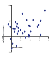
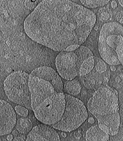
Dynamic light scattering (DLS) analysis showed that the RBO-SLN dispersion had a mean hydrodynamic diameter of 23.53 ± 4.9 nm and a PDI of 0.85. This relatively high PDI indicates a broad particle-size distribution, as also shown in Figure 1A. The particles exhibited a highly negative zeta potential of −48.1 ± 0.63 mV, indicating a strong surface charge that may contribute to electrostatic repulsion among nanoparticles and limit aggregation. Morphological examination by TEM further demonstrated that the nanoparticles were predominantly spherical and relatively uniform, with smooth, well-defined surfaces (Figure 1B).
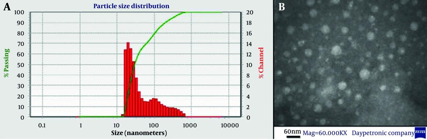
Figure 1.
Characterization of rice bran oil-loaded solid lipid nanoparticles (RBO-SLNs): (A) particle-size distribution determined by dynamic light scattering and (B) transmission electron microscopy image showing nanoparticle morphology.
4.2. Encapsulation Efficiency
The amount of RBO incorporated into the lipid matrix was initially quantified using the centrifugation method. An EE of 84.96 ± 1.51% was obtained, indicating efficient incorporation of RBO into the SLN structure. To verify this finding, EE was independently assessed using the dialysis method, which yielded a comparable value of 86.01 ± 2.11%. No statistically significant difference was detected between the two analytical approaches (P ≥ 0.05), supporting the consistency of the EE results.
4.3. in vitro Release Profile and Kinetic Analysis
The release profile of RBO from the RBO-SLNs is presented in Figure 2. Approximately 39.24% of the incorporated oil was released during the 72-h study period, demonstrating a gradual release pattern throughout the experimental interval. Kinetic modeling showed that the Korsmeyer–Peppas equation provided the best correlation with the experimental release data, yielding the highest coefficient of determination (R2 = 0.943). Based on the calculated release exponent, the release behavior was classified as a Super Case II transport mechanism, suggesting that polymer/lipid matrix relaxation and other matrix-associated processes contributed substantially to RBO release rather than simple Fickian diffusion. The comparative fitting parameters obtained for the different kinetic models are provided in Table S1 in the Supplementary File.
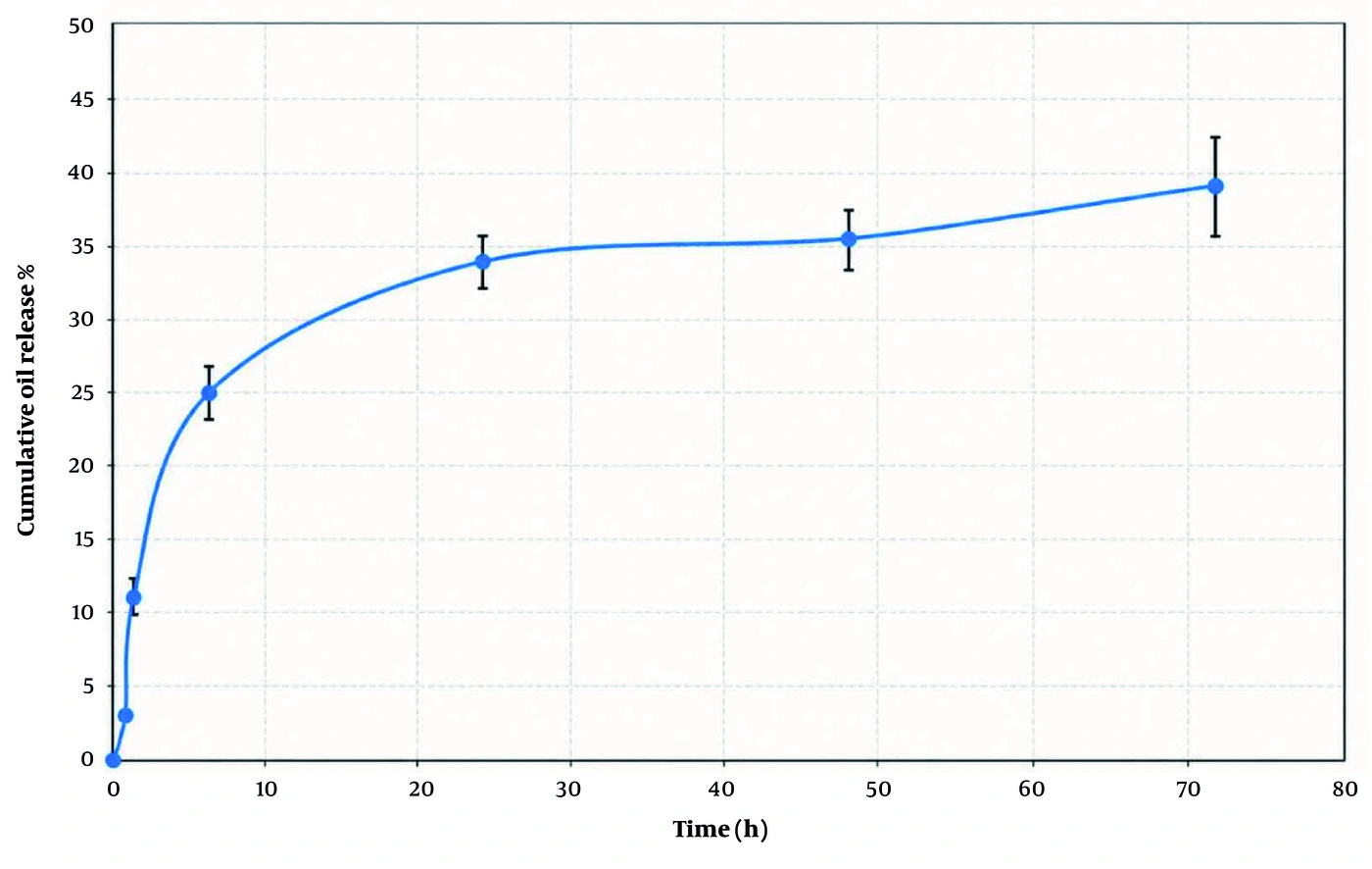
Figure 2.
Cumulative release profile of rice bran oil (RBO) from RBO-loaded solid lipid nanoparticles (RBO-SLNs) over 72 h.
4.4. Fibroblast Cell Viability
The cytocompatibility of RBO-SLNs was investigated in HU02 human foreskin fibroblasts using the MTT assay, with free RBO serving as a comparator (Figure 3). At 5 mg/mL, RBO-SLNs produced a cell viability of 113.8 ± 6.48%. Increasing the nanoparticle concentration to 10 mg/mL reduced cell viability to 71.7 ± 3.98%. In comparison, treatment with RBO at 10 mg/mL resulted in 103.04 ± 9.12% viability.
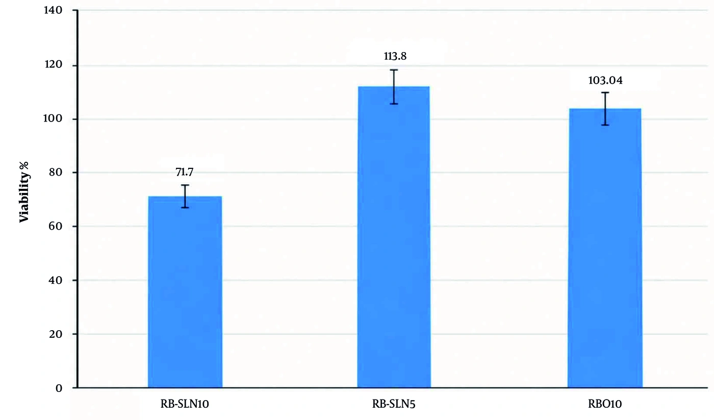
Figure 3.
Viability of HU02 fibroblasts after exposure to rice bran oil-loaded solid lipid nanoparticles (RBO-SLNs) at 5 and 10 mg/mL and rice bran oil (RBO) at 10 mg/mL.
Although cell viability remained above 70% at the highest RBO-SLN concentration tested, suggesting an absence of marked cytotoxicity under the experimental conditions (5), the observed concentration-dependent response should be considered when determining the appropriate formulation concentration. In addition to RBO, excipients such as lecithin, cholesterol, and Tween 80 may influence cellular responses, although their individual contributions were not evaluated in this study. Therefore, the biocompatibility of the formulation should be interpreted in the context of the specific experimental conditions, including the tested concentrations, cell type, and exposure period. Further studies using appropriate excipient controls and additional skin-relevant models are required to clarify the contribution of individual formulation components and confirm the dermal safety of RBO-SLNs.
4.5. Attenuated Total Reflectance Fourier-Transform Infrared Analysis
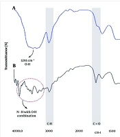
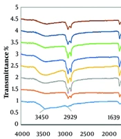
The ATR-FTIR spectrum of RBO exhibited several characteristic absorption bands associated with its lipid constituents (Figure 4, red). The prominent bands detected at 2920 - 2850 cm-1 were assigned to C–H stretching vibrations of the –CH2 and –CH3 groups. The absorption band at approximately 1743 cm-1 corresponded to the C=O stretching vibration of triglyceride ester groups. Additional bands in the 1450 - 1370 cm-1 region were attributed to C–H bending vibrations, whereas absorption features between 1200 and 1000 cm-1 were associated mainly with C–O–C stretching vibrations of ester bonds (6, 20).
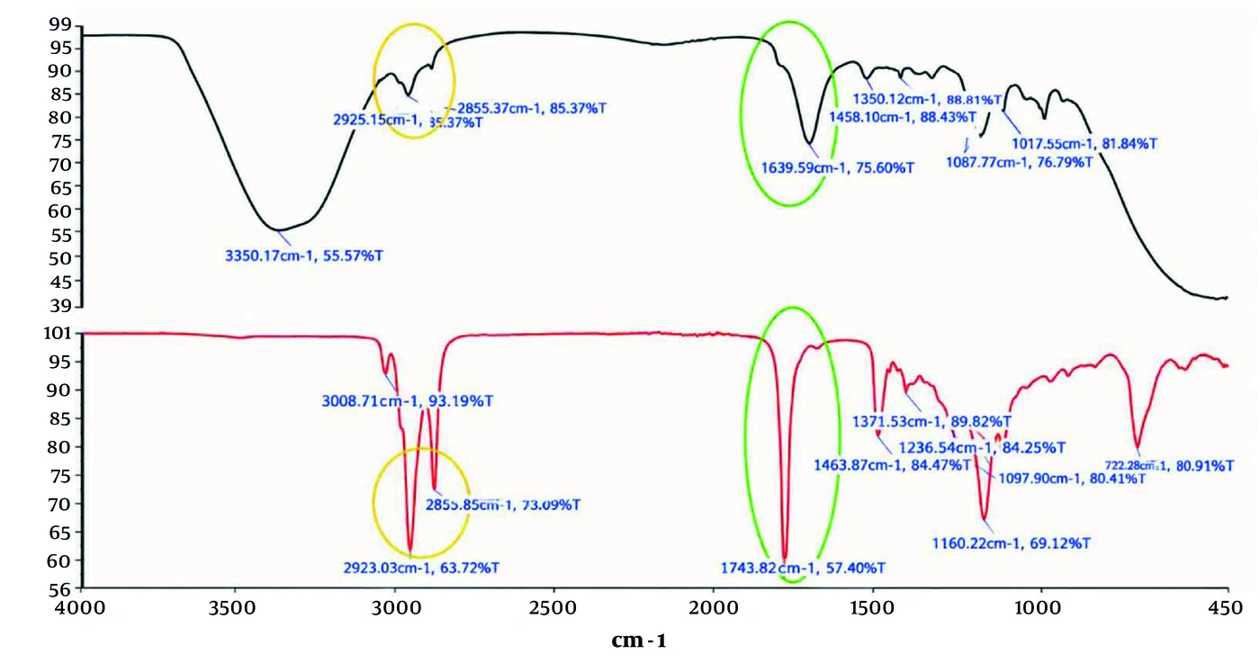
Figure 4.
Attenuated total reflectance Fourier-transform infrared spectra of rice bran oil (RBO; red) and RBO-loaded solid lipid nanoparticles (RBO-SLNs; black), showing the characteristic absorption bands associated with RBO before and after incorporation into the solid lipid nanoparticle system.
After incorporation of RBO into the SLN formulation, the principal characteristic bands of the oil remained detectable in the ATR-FTIR spectrum, indicating the presence of RBO in the formulation. However, ATR-FTIR analysis alone cannot confirm successful encapsulation or establish the location of the oil within the nanoparticles (Figure 4). Nevertheless, the intensity of the band at 1743 cm-1 was reduced in RBO-SLNs relative to free oil. This change may reflect the lower relative proportion of RBO after incorporation into the lipid matrix and the contribution of the other formulation components to the overall spectrum. An increase in spectral intensity around 1639 cm-1 was also observed, which may be associated with contributions from the formulation excipients and changes in the chemical environment accompanying nanoparticle formation.
The absorption band near 3008 cm-1 observed for RBO was not clearly resolved in the RBO-SLN spectrum, potentially because of spectral overlap with the broader absorption feature centered around 3350 cm-1. Overall, preservation of the major RBO-associated absorption bands after nanoencapsulation indicated that the principal chemical characteristics of the oil were retained within the SLN formulation.
4.6. UV Absorption Properties of Rice Bran Oil and Its Solid Lipid Nanoparticles
The UV absorbance of RBO and RBO-SLNs at 1%, 3%, and 5% (w/v) was evaluated at representative wavelengths corresponding to UVA (327 nm), UVB (313 nm), and UVC (222 nm) radiation using both dispersion and Transpore® tape methods. The complete results are summarized in Table 1. Overall, nanoencapsulation resulted in higher absorbance than that observed at the corresponding concentrations of free RBO, although the magnitude and statistical significance of this effect varied by wavelength and analytical method.
Table 1.
UV Absorbance (Mean ± Standard Deviation) of Rice Bran Oil (RBO) and RBO-Loaded Solid Lipid Nanoparticles (RBO-SLNs) Measured Using Dispersion and Transpore® Tape Methods
| Methods and Sample | UVA | UVB | UVC | ||||||
|---|---|---|---|---|---|---|---|---|---|
| 1% | 3% | 5% | 1% | 3% | 5% | 1% | 3% | 5% | |
| Dispersion | |||||||||
| RBO | 0.56 ± 0.016 | 0.705 ± 0.009 | 0.67 ± 4.1E-04 | 0.52 ± 0.036 | 1.12 ± 0.038 | 1.75 ± 0.12 | 0.775 ± 0.016 | 1.48 ± 0.014 | 2.001 ± 0.124 |
| RBO-SLNs | 0.72 ± 0.008 | 0.76 ± 0.011 | 0.84 ± 0.008 | 3.6 ± 0.084 | 4.21 ± 0.05 | 4.86 ± 0.11 | 3.497 ± 0.11 | 4.005 ± 0.18 | 4.41 ± 0.21 |
| Transpore® tape | |||||||||
| RBO | 0.038 ± 0.002 | 0.065 ± 0.004 | 0.044 ± 9.1E-04 | 0.204 ± 1.2E-04 | 0.24 ± 2.5E-04 | 0.23 ± 1.6E-04 | 0.208 ± 8.2E-05 | 0.232 ± 2.6E-04 | 0.216 ± 8.2E-05 |
| RBO-SLNs | 0.062 ± 0.003 | 0.073 ± 0.001 | 0.093 ± 4.5E-04 | 0.25 ± 2.05E-04 | 0.33 ± 2.9E-04 | 0.35 ± 8.16E-05 | 0.236 ± 8.2E-05 | 0.283 ± 4.2E-04 | 0.343 ± 2.5E-04 |
4.6.1. UVA (327 Nm)
At 327 nm, both free RBO and RBO-SLNs exhibited relatively low UVA absorbance. No statistically significant difference in absorbance between the two formulations was observed using the dispersion method. In the Transpore® tape assay, all recorded optical density (OD) values were below 0.1, limiting the ability of this method to reliably distinguish between the formulations in the UVA region (Figure 5). Therefore, the results do not provide sufficient evidence of a significant difference in UVA absorbance between free RBO and RBO-SLNs. The low OD values obtained using the tape method should also be considered when interpreting the results, as they may limit the reliability of comparisons under these experimental conditions.
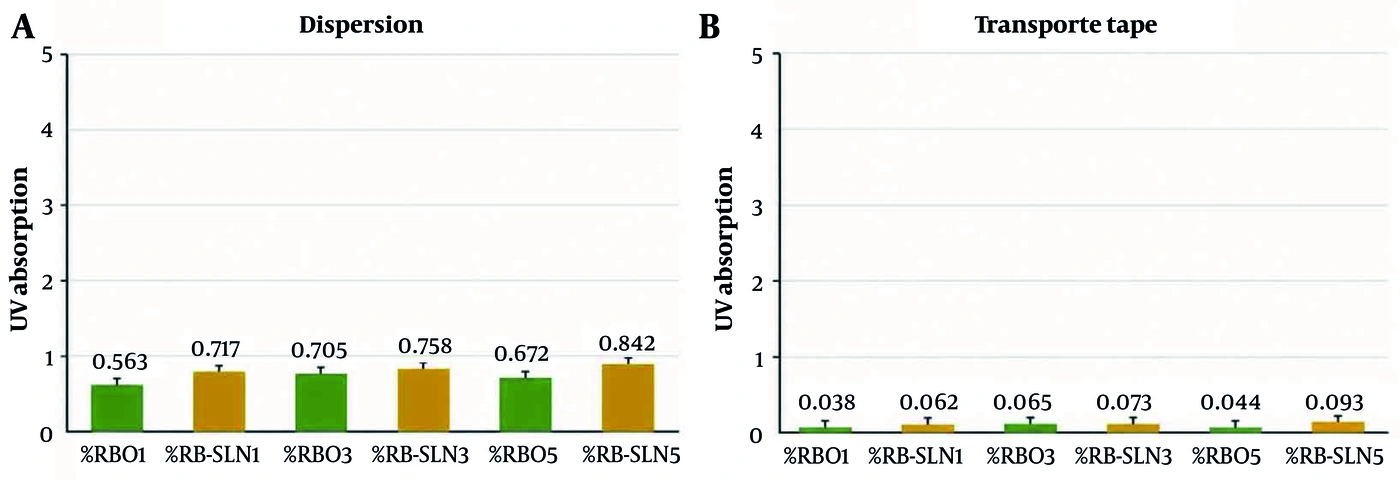
Figure 5.
UV absorbance of rice bran oil (RBO) and RBO-loaded solid lipid nanoparticles (RBO-SLNs) at 327 nm (UVA), determined using (A) the dispersion method and (B) the Transpore® tape method.
4.6.2. UVB (313 Nm)
At 313 nm, the UVB absorbance profiles of free RBO and RBO-SLNs varied according to the measurement method. Using the dispersion method, RBO-SLNs exhibited significantly higher UVB absorbance than free RBO at all tested concentrations (1%, 3%, and 5%; P ≤ 0.05), with corresponding P values of 2.49 × 10⁻⁵, 2.97 × 10⁻⁷, and 6.72 × 10⁻⁶, respectively. In contrast, although RBO-SLNs showed numerically higher UVB absorbance than free RBO when evaluated using the Transpore® tape method, the differences between the formulations were not statistically significant (Figure 6). These findings indicate that differences in UVB absorbance between free RBO and RBO-SLNs depended on the measurement method. Therefore, the higher absorbance observed with the dispersion method should not, by itself, be interpreted as evidence of superior photoprotective performance under application-relevant conditions.
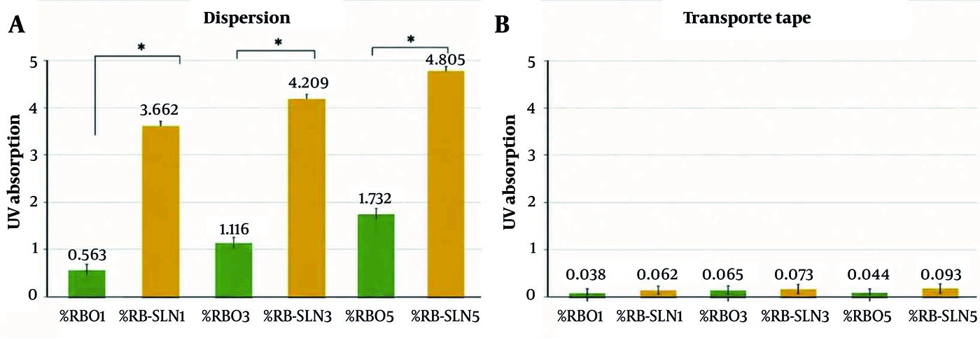
Figure 6.
UV absorbance of rice bran oil (RBO) and RBO-loaded solid lipid nanoparticles (RBO-SLNs) at 313 nm (UVB), determined using (A) the dispersion method and (B) the Transpore® tape method.
4.6.3. UVC (222 Nm)
At 222 nm, RBO-SLNs exhibited higher UVC absorbance than free RBO at all tested concentrations (1%, 3%, and 5%) when assessed using the dispersion method. These differences were statistically significant, with corresponding P values of 5.67 × 10⁻⁴, 3.79 × 10⁻³, and 4.10 × 10⁻⁴, respectively (Figure 7). The Transpore® tape measurements also showed numerically higher UVC absorbance for RBO-SLNs than for free RBO at all three concentrations. However, the statistical significance of these differences should be established independently using replicate-level data. Although the dispersion method yielded substantially higher measured absorbance values than the Transpore® tape method, this difference should be interpreted as a method-dependent variation in the measured response rather than as direct evidence of superior photoprotective efficacy. Further validation under application-relevant conditions is required to establish the photoprotective performance of the formulations.
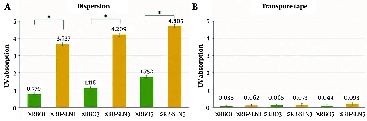
Figure 7.
UV absorbance of rice bran oil (RBO) and RBO-loaded solid lipid nanoparticles (RBO-SLNs) at 222 nm (UVC), determined using (A) the dispersion method and (B) the Transpore® tape method.
4.7. Sun Protection Factor of Rice Bran Oil, Blank Solid Lipid Nanoparticles, and Rice Bran Oil-Loaded Solid Lipid Nanoparticles
The SPF values of RBO, blank SLNs, and RBO-SLNs at 1%, 3%, and 5% were calculated using the spectrophotometric model of Mansur The SPF of both free RBO and blank SLNs increased with increasing concentration; however, the differences between these two groups were not statistically significant (P ≥ 0.05) (Table 2).
Table 2.
Sun Protection Factor (SPF) Values of Rice Bran Oil (RBO), Blank Solid Lipid Nanoparticles (SLNs), and RBO-Loaded SLNs (RBO-SLNs) at Concentrations of 1%, 3%, and 5%
| Variables | Values |
|---|---|
| RBO-SLNs 1% | 19.34 |
| RBO-SLNs 3% | 22.22 |
| RBO-SLNs 5% | 22.88 |
| SLNs 1% | 4.13 |
| SLNs 3% | 9.74 |
| SLNs 5% | 13.60 |
| RBO 1% | 3.17 |
| RBO 3% | 9.82 |
| RBO 5% | 13.57 |
Rice bran oil-loaded SLNs exhibited a significant concentration-related increase in estimated SPF (P ≤ 0.05). The highest estimated SPF was 22.88 at 5%, compared with 22.22 at 3%; however, this difference was not statistically significant. At 1% and 3%, the estimated SPF values of RBO-SLNs exceeded the sums of the corresponding values for free RBO and blank SLNs, whereas this pattern was not observed at 5%. This finding suggests a potentially nonadditive contribution of the formulation components to UV absorption; however, it does not establish synergism without appropriate statistical testing. Considering the comparable estimated SPF values at 3% and 5%, this observation, and the cell viability findings, the 3% formulation may be a promising candidate for further investigation rather than a definitively optimized formulation.
These results represent spectrophotometric SPF estimates and should not be considered equivalent to validated in vivo sunscreen protection. Further studies are required to confirm photoprotective efficacy, skin compatibility, and formulation stability under application-relevant conditions.
5. Discussion
Ultraviolet (UV) radiation is an important environmental factor that contributes to cutaneous damage (7). Excessive generation of reactive oxygen species is a major mechanism underlying UV-mediated injury, resulting in oxidative damage to cellular components and structural alterations in the skin (8, 21). Therefore, developing effective strategies to attenuate UV-induced damage remains an important objective in topical skin protection.
Rice bran oil has attracted increasing interest in cosmetic and personal-care applications because of its bioactive constituents and potential antiaging, photoprotective, and dermatological properties (10, 11). Nanotechnology-based delivery systems may further improve the physicochemical properties, stability, and biological performance of plant-derived oils (15, 22). However, the application of SLNs specifically to enhance the photoprotective properties of RBO remains insufficiently explored.
In the present study, RBO was successfully incorporated into an SLN system, yielding an EE of 85%, a mean particle size of 23.53 nm, and a highly negative zeta potential of approximately −48 mV. These characteristics indicate efficient RBO incorporation and favorable surface-charge properties. Nanoencapsulation enhanced the UV absorption of RBO, particularly in the UVB and UVC regions under the experimental conditions. This finding suggests that incorporating RBO into the lipid matrix can modify its optical behavior and potentially improve its photoprotective performance. Nevertheless, the results should be interpreted in light of the limitations of the in vitro spectrophotometric approach and should not be considered direct evidence of biological photoprotection.
The present findings are consistent with previous research on nanoencapsulated RBO. Rigo et al. (17) developed RBO-loaded lipid nanocapsules and evaluated their ability to reduce UVB-induced skin damage in a murine model. Their nanocarriers had a mean diameter of approximately 200 nm, a zeta potential of approximately −9 mV, and a PDI below 0.20 before incorporation into a topical hydrogel (17). The smaller particle size observed in the present study may reflect differences in nanocarrier composition, preparation conditions, and energy input during particle formation. Probe sonication provides high-energy cavitation that can promote droplet disruption and the formation of smaller nanoscale structures; however, its contribution should be considered alongside other formulation variables rather than as the sole determinant of particle size. Despite these physicochemical differences, both studies support the potential of nanostructured RBO systems to improve protection against UVB-associated skin damage (17).
The photoprotective potential of RBO has also been reported in nonnanostructured formulations. Syamsidi et al. demonstrated a concentration-dependent increase in SPF after incorporation of increasing amounts of RBO into compact powder formulations, supporting the oil’s intrinsic photoprotective potential (18). Similarly, Jeong Ha et al. reported that dietary rice bran attenuated several manifestations of UVB-induced skin damage in mice, including wrinkle formation, transepidermal water loss, and epidermal thickening (23). Although their administration route and experimental design differed from the topical nanocarrier approach used here, these findings further support the biological relevance of rice bran-derived constituents in mitigating UV-associated skin alterations.
Rice bran oil has also been incorporated into nanotechnology-based delivery systems for applications unrelated to topical photoprotection. A self-nanoemulsifying drug delivery system containing RBO, Kolliphor RH40 or Kolliphor EL, and Transcutol HP was developed to improve the oral delivery of poorly water-soluble drugs, including fenofibrate and itraconazole (24). These findings demonstrate the versatility of RBO as an oil-based vehicle for incorporating lipophilic compounds into advanced delivery systems and support its compatibility with nanotechnology-based formulation strategies.
Taken together, the present findings and previous studies provide complementary evidence of the potential of rice bran-derived materials for UV-related skin protection. The enhanced UV absorption after incorporating RBO into SLNs suggests that nanostructuring may improve the functional performance of this naturally derived oil. However, the absence of a blank-SLN control in the UV absorption experiments limits the ability to determine the independent contribution of the lipid nanoparticle matrix. Furthermore, the current assessment was based on in vitro spectrophotometric models. Therefore, additional studies using reconstructed skin, ex vivo, and in vivo models are required to establish whether the enhanced UV absorption translates into meaningful biological photoprotection.
5.1. Conclusions
The present study demonstrated the successful development of RBO-loaded SLNs as a nanoscale platform with potential topical photoprotective applications. The formulation exhibited favorable physicochemical characteristics, including nanoscale particle size, efficient RBO encapsulation, and a highly negative surface charge. Incorporating RBO into the SLN matrix enhanced UV absorption, particularly in the UVB and UVC regions. These findings support the potential use of RBO-SLNs in advanced skin-care and photoprotective formulations. However, further studies are required to establish their practical applicability, particularly through accelerated and real-time stability testing and evaluation in ex vivo and in vivo skin models. Such investigations will be essential to confirm the safety, stability, and photoprotective efficacy of RBO-SLNs and determine their suitability for future topical sunscreen formulations.